This website uses cookies to ensure you get the best experience on our website.


- Table of Contents
This guide uses the catalog antibody’s 1:50 dilution and HRP/DAB detection for paraffin-section IHC (datasheet M03580). Assess the nuclear pattern against the reported ubiquitous tissue staining, including high staining in bone marrow hematopoietic cells (HPA tissue IHC).

Expected localisation, validated protocols, controls and antibodies — the at-a-glance facts below, then the full design guide.
| Expected localisation | Nuclear staining in tissue (HPA tissue IHC) | |
| Staining pattern | Ubiquitous nuclear staining across tissue cell types (HPA tissue IHC) | |
| Antigen retrieval | EDTA pH 8.0 HIER, heat-mediated (datasheet M03580) | |
| Positive control | Adipose tissue+4 more · see all | |
| Negative control | None in HPA (detected in all 45 tissues); use no-primary + isotype controls |
| Fixation | Keep fixation conditions consistent across sections. (standard IHC practice; not target-specific) | |
| Caveat | Marrow peroxidase can mimic DAB staining (HPA tissue IHC; standard IHC practice) | |
| Regulation | No staining-linked regulator listed (UniProt) | |
| Isoform / epitope | No isoforms listed; no extracellular domain (UniProt) |
The catalog antibody protocol uses EDTA pH 8.0 retrieval (datasheet: M03580). The options below summarize three published paraffin-section IHC protocols (PMC9079359; PMC12618602; PMC12439426).
| Sample | Paraffin-embedded human lung cancer tissue; fixative not specified (datasheet M03580) |
| Fixation | Image fixative and duration unreported (datasheet M03580); verify before use. |
| Sectioning | 4–5 µm sections on charged slides (standard) |
| Deparaffinisation | Xylene, graded ethanol series to water (standard) |
| Antigen retrieval | Heat retrieval: EDTA pH 8.0 (datasheet M03580); 20 min, 95–100 °C (standard) |
| Peroxidase block | 3% H2O2, 10 min, room temperature (standard) |
| Blocking | 10% goat serum (datasheet M03580) |
| Primary antibody | Rabbit monoclonal (clone 22A24) anti-ALYREF, 1:50 (datasheet M03580) |
| Primary incubation | Overnight at 4 °C (datasheet M03580) |
| Detection | HRP-conjugated secondary, DAB chromogen (datasheet M03580) |
| Counterstain | Hematoxylin, blue, dehydrate and mount (standard) |
| Expected result | ALYREF-positive staining in adipocytes of adipose tissue (HPA tissue IHC: High). HPA tissue profile: Ubiquitous nuclear expression. No signal in the no-primary control. |
ALYREF is expected mainly in nuclei across tissue cell types, with high staining reported in several cell populations including bone marrow hematopoietic cells (HPA: ubiquitous nuclear expression; High in hematopoietic cells). Nuclear speckles are a documented location; cytoplasmic transit is also described (UniProt Q86V81: subcellular location). Its lack of a transmembrane segment gives no basis for a membrane pattern (UniProt Q86V81: topology). HPA rates the tissue IHC pattern Supported (HPA: tissue IHC reliability).
| Many nuclei stain in the section, including nuclei of the chosen HPA high control population (HPA: ubiquitous nuclear expression; High in bone marrow hematopoietic cells). | This fits the reported IHC distribution. Compare cells within the same section and a matched negative control before judging intensity; HPA's High labels describe observed cell populations, not a required intensity in every specimen (HPA: tissue IHC profile and levels; standard IHC practice). |
| The dominant signal outlines membranes or fills cytoplasm while nuclei are unstained. | A dominant membrane pattern conflicts with the reported nuclear IHC pattern and lack of a transmembrane segment (HPA: ubiquitous nuclear expression; UniProt Q86V81: topology). UniProt describes cytoplasmic transit, so limited cytoplasmic signal alone is inconclusive; review the nuclear signal and controls before calling an artefact (UniProt Q86V81: subcellular location; standard IHC practice). |
| Only an unexpected cell population stains while neighboring nuclei and the chosen high control population remain blank. | Check cross-reactivity or endogenous chromogen activity against the negative control (standard IHC practice). HPA reports ubiquitous nuclear expression and supplies no negative cell population here, so an unlisted cell type cannot be declared ALYREF-negative from these data (HPA: tissue IHC profile; supplied positive/negative lists). |
| Brown color spreads between cells or coats much of the section without distinct nuclear boundaries. | Diffuse background does not establish ALYREF localization. Review reagent-only and tissue controls, wash quality, antibody concentration and chromogen development under the local IHC workflow (standard IHC practice); do not score indistinct color as positive nuclei (HPA: ubiquitous nuclear expression). |
| The chosen known-positive control has no discernible nuclear signal, including its listed high population. | The run is uninterpretable until detection and tissue handling are checked (standard IHC practice). Bone marrow hematopoietic cells are one reported high IHC example, but HPA's rating does not guarantee a signal with every antibody or run condition (HPA: High in hematopoietic cells; tissue IHC reliability). |
| Which compartment should drive the IHC score? | Nuclear localization is the tissue-level anchor (HPA: ubiquitous nuclear expression). UniProt also reports cytoplasmic travel with mRNA, so minor cytoplasmic color is interpreted with controls rather than used alone as proof of specificity (UniProt Q86V81: subcellular location; standard IHC practice). |
| How much weight should the HPA antibody record carry? | HPA lists tissue IHC as Supported and two antibodies with Supported IHC entries, HPA019799 and CAB016281 (HPA: tissue IHC reliability; antibody validation list). Those labels support the reported pattern; they do not validate an unlisted antibody or establish a universal dilution (HPA: antibody validation list). |
| Can processing or isoforms explain a different compartment? | The supplied record lists no signal peptide, propeptide, transmembrane segment or isoforms, and describes a chain spanning residues 2–257 (UniProt Q86V81: processing, topology, isoforms). It provides no basis to predict a secreted or membrane-specific IHC pattern (UniProt Q86V81: processing and topology). |
| Can an epitope or retrieval setting be chosen from this record? | The RRM spans residues 106–182 and modified residues are listed, but no antibody epitope or target-specific retrieval response is supplied (UniProt Q86V81: domain and modified residues; supplied antibody data). Select retrieval conditions from the IHC-validated antibody's instructions and local controls (standard IHC practice). |
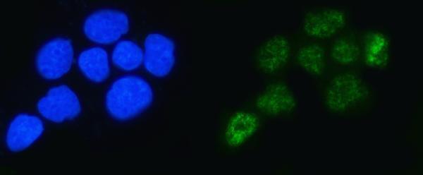
| IF/ICC Q&A: what pattern should fluorescence show? | HPA approves nucleoplasm and nuclear speckles as main locations in ICC-IF (HPA: subcellular summary). Speckles can help interpret fluorescence images; their visibility is not a requirement for chromogenic paraffin IHC (HPA: ICC-IF location; standard IHC practice). |
| Situation | Likely cause | Next action |
|---|---|---|
| No nuclear stain in the positive control. | An unsuccessful IHC run is possible; the pattern alone cannot identify whether tissue handling, retrieval, primary antibody or detection caused it (standard IHC practice). | Confirm the control contains an HPA high population, then check the catalog antibody's IHC-P instructions and each detection step (HPA: High in listed populations; standard IHC practice). |
| Weak nuclear color in the sample, but the control stains. | HPA's high examples and ubiquitous profile do not specify the intensity of every specimen or cell (HPA: tissue IHC profile and positive list). | Compare matched sections and scoring thresholds within the run; report weak nuclear signal as observed rather than forcing a high call (standard IHC practice). |
| Predominantly cytoplasmic or membrane color. | Dominant extranuclear color is discordant with tissue IHC; limited cytoplasmic transit is documented, but a membrane location is unsupported (HPA: ubiquitous nuclear expression; UniProt Q86V81: subcellular location and topology). | Inspect the negative control and nuclear counterstain; optimize the IHC workflow before assigning specificity to the extranuclear color (standard IHC practice). |
| Only one unexpected cell population appears positive. | Cross-reactivity or endogenous detection activity is possible; the supplied HPA list contains no confirmed negative cell type (standard IHC practice; HPA: supplied positive/negative lists). | Compare a primary-antibody omission control and an HPA high population in the same run; describe the finding as unresolved if controls cannot distinguish it (HPA: positive list; standard IHC practice). |
| Diffuse brown background obscures nuclei. | Excess chromogen, insufficient washing or nonspecific detection can obscure a compartment-specific result (standard IHC practice). | Review the negative control, wash and development conditions, then rescore only nuclei with clear cell boundaries (standard IHC practice; HPA: ubiquitous nuclear expression). |
| The investigator expects visible nuclear speckles in a chromogenic section. | Speckles are an approved ICC-IF location, while the tissue IHC summary reports ubiquitous nuclear expression without a speckle-resolution requirement (HPA: subcellular summary; tissue IHC profile). | Judge paraffin IHC by interpretable nuclear staining and tissue controls; use the separate IF/ICC guide when speckle localization itself is the question (HPA: tissue IHC profile; ICC-IF location; standard IHC practice). |
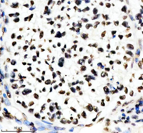
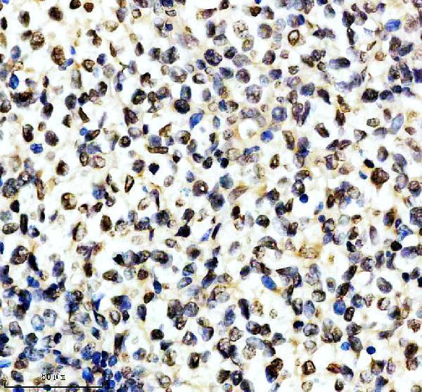
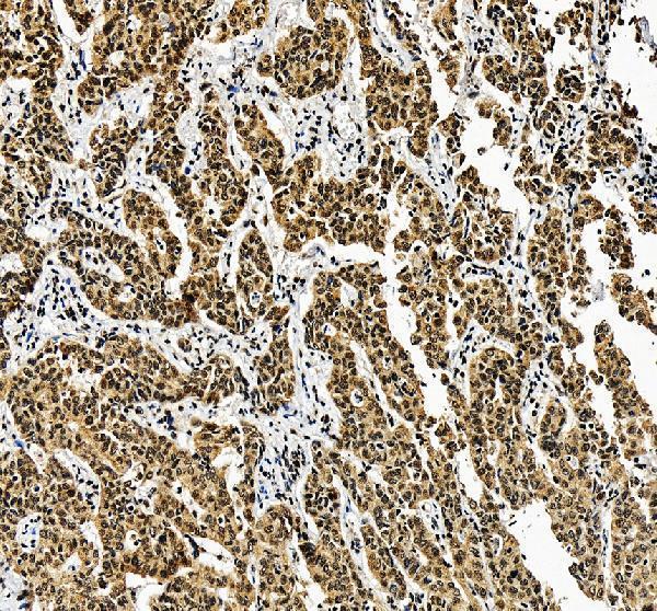
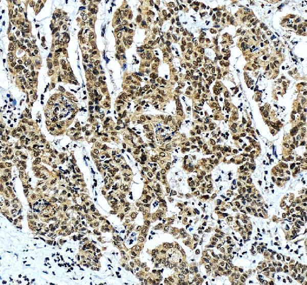
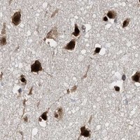


Comprehensive Human Protein Atlas IHC scoring per tissue (reliability: Supported — High consistency between antibody staining and RNA expression data.). Rows are taken directly from the HPA tissue chart — click any row's HPA link to view the source.
| Tissue | Cell type | Level | Evidence | Source |
|---|---|---|---|---|
| Adipose tissue | Adipocytes | High | Protein (IHC) | HPA → |
| Adrenal gland | Glandular cells | High | Protein (IHC) | HPA → |
| Appendix | Glandular cells | High | Protein (IHC) | HPA → |
| Bone marrow | Hematopoietic cells | High | Protein (IHC) | HPA → |
| Breast | Adipocytes | High | Protein (IHC) | HPA → |
| Tissue | Cell type | Level | Evidence | Source |
|---|---|---|---|---|
| None in HPA: ALYREF is detected in all 45 scored tissues. Use a no-primary (secondary-only) and an isotype control instead. | ||||
Troubleshoot ALYREF staining in paraffin sections by checking retrieval, nuclear localisation, controls and scoring before interpreting changes in expression.
Both catalog antibodies have IHC images from human paraffin sections (IHC captions: M03580, A03580-3); A03580-3 also has an IF image from A431 cells (IF caption: A03580-3).
M03580 has IHC images from human lung cancer, lymphoma, glioma, and testis cancer paraffin sections (M03580 IHC captions). A03580-3 has IHC images from human liver cancer and liver paraffin sections and an IF image from A431 cells (A03580-3 IHC and IF captions).
Which to pick: For tissue IHC, choose M03580 if a monoclonal antibody and its 1:50 starting dilution suit the experiment (catalog: clone 22A24; M03580 IHC caption: human paraffin section, EDTA pH 8.0 retrieval); its fixative is unreported (M03580 IHC caption). For IF/ICC, A03580-3 has a documented A431-cell IF example at 5 μg/mL (A03580-3 IF caption). Both list Human, Mouse, and Rat reactivity (catalog: M03580, A03580-3), but their IHC images document human sections only, and neither IHC caption reports the fixative (M03580 and A03580-3 IHC captions).