This website uses cookies to ensure you get the best experience on our website.


- Table of Contents
Plan paraffin section IHC for AMY2A using pancreatic exocrine cells as the expected positive population (HPA tissue IHC). This guide covers fixation, detection and interpretation of the observed cytoplasmic pattern (HPA tissue IHC).

Expected localisation, validated protocols, controls and antibodies — the at-a-glance facts below, then the full design guide.
| Expected localisation | Cytoplasmic staining in pancreatic exocrine cells (HPA tissue IHC) | |
| Staining pattern | Selective cytoplasmic staining in pancreatic exocrine cells (HPA tissue IHC) | |
| Antigen retrieval | Citrate pH 6.0 HIER, 95–98 °C, 20 min (rule: cytoplasmic / membrane antigen) | |
| Positive control | Pancreas | |
| Negative control | Adipose tissue+4 more · see all |
| Fixation | Keep fixation consistent across paraffin sections. (standard IHC practice; not target-specific) | |
| Caveat | Staining may include protein from another gene (HPA tissue IHC) | |
| Regulation | Expression regulation is not annotated (UniProt) | |
| Isoform / epitope | 2 isoforms; signal peptide 1–15 is removed; epitope impact unknown (UniProt) |
Compare the catalog antibody’s IHC-P protocol with a published AMY2A brain-section IHC workflow (PMC8028266).
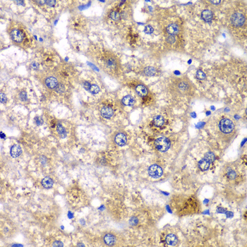
| Sample | Paraffin-embedded human liver injury tissue; fixative not specified (datasheet A06771) |
| Fixation | Image fixative and duration unreported (datasheet A06771); verify before use. |
| Sectioning | 4–5 µm sections on charged slides (standard) |
| Deparaffinisation | Xylene, graded ethanol series to water (standard) |
| Antigen retrieval | Heat-induced epitope retrieval in citrate buffer, pH 6.0, 20 min at 95–98 °C (standard rule: cytoplasmic / membrane antigen) |
| Peroxidase block | 3% H2O2, 10 min, room temperature (standard) |
| Blocking | 10% normal serum of the secondary host, 30 min, room temperature (standard) |
| Primary antibody | Rabbit anti-AMY2A, 1:50-1:200 (datasheet A06771) |
| Primary incubation | Overnight at 4 °C (standard) |
| Detection | HRP-polymer secondary, DAB chromogen 5–10 min (standard) |
| Counterstain | Hematoxylin, blue, dehydrate and mount (standard) |
| Expected result | AMY2A-positive staining in exocrine glandular cells of pancreas (HPA tissue IHC: High). HPA tissue profile: Selective cytoplasmic expression in pancreatic exocrine glandular cells. No signal in the no-primary control. |
In paraffin-section IHC, expect selective, strong cytoplasmic staining in pancreatic exocrine glandular cells (HPA: High; Enhanced tissue reliability). AMY2A is a secreted protein without a transmembrane segment, so its biology also permits protein outside cells (UniProt P04746: secreted; no transmembrane segment). HPA cautions that its antibodies can target proteins from more than one gene; this pattern alone does not establish AMY2A-specific staining (HPA: tissue IHC caution).
| Strong cytoplasmic staining in pancreatic exocrine glandular cells, with little signal elsewhere. | This matches the reported selective tissue pattern (HPA: High in pancreatic exocrine glandular cells; Enhanced tissue reliability). Record intensity and the fraction of stained cells together, since a strong signal in a few cells differs from widespread exocrine staining (general IHC practice). |
| Predominantly nuclear or sharply cell-membrane staining, with little exocrine cytoplasmic signal. | This does not match the reported IHC pattern (HPA: selective cytoplasmic expression). A membrane-only pattern also lacks support from the recorded topology (UniProt P04746: no transmembrane segment). Check morphology and detection artefacts before interpreting it as AMY2A localisation (general IHC practice). |
| Strong staining in adipocytes or other cells outside the expected pancreatic exocrine population. | Adipocytes are reported as not detected in adipose tissue (HPA: Not detected in adipocytes). Consider antibody recognition of another protein, including another gene product, or endogenous detection activity; morphology alone cannot identify the cause (HPA: multi-gene targeting caution; general IHC practice). |
| Diffuse colour across tissue, empty spaces, or section edges, without a clear cellular pattern. | The distribution is difficult to score against the selective exocrine cytoplasmic reference (HPA: tissue IHC profile). Because AMY2A is secreted, extracellular signal is biologically possible, but a diffuse deposit alone cannot establish target-specific staining (UniProt P04746: secreted; general IHC practice). |
| No staining in pancreatic exocrine glandular cells on the positive-control section. | This conflicts with the reported high signal in those cells (HPA: High in pancreatic exocrine glandular cells). Review tissue identity, section integrity, antibody and detection steps before calling the test specimen negative (general IHC practice). |
| Secretory route and processing | AMY2A has a signal peptide at residues 1–15 and a mature chain at 16–511; it is secreted (UniProt P04746: processing; subcellular location). These facts support caution when judging extracellular colour, but do not establish a specific luminal staining pattern in paraffin sections. |
| Antibody specificity and validation | HPA rates the tissue pattern Enhanced, while warning that antibodies can target products of multiple genes (HPA: tissue reliability and caution). Individual listed IHC ratings differ: CAB045960 is Enhanced; HPA045394, HPA045399 and HPA046980 are Supported (HPA: antibody validation). Match any conclusion to the antibody used. |
| IF/ICC Q: What localisation should be expected? | HPA reports Golgi apparatus localisation as approved and cytosol as uncertain in ICC-IF (HPA: subcellular profile). That evidence comes with a multi-gene antibody caution and does not replace the pancreatic exocrine cytoplasmic IHC reference (HPA: subcellular caution; tissue IHC profile). |
| Isoforms and modifications | UniProt lists two isoforms and one glycosylation site at residue 476 (UniProt P04746: isoforms; glycosylation). The supplied record gives no antibody epitope or isoform-specific staining result, so these annotations cannot explain a positive or negative section on their own. |
| Situation | Likely cause | Next action |
|---|---|---|
| Pancreatic positive control is blank. | An interrupted IHC workflow or unsuitable control section is possible; the expected exocrine signal is High (HPA: tissue IHC profile; general IHC practice). | Confirm exocrine morphology, then check primary-antibody application, detection reagents and chromogen with a working control (general IHC practice). Do not assign a target-specific fixation effect from this result. |
| Exocrine cells stain weakly despite a readable section. | The observed signal falls below the reported High reference pattern, but the stain alone does not identify which workflow step limited detection (HPA: tissue IHC profile; general IHC practice). | Check the antibody's IHC validation and compare positive-control sections processed alongside the test; adjust routine retrieval or detection conditions only with appropriate controls (HPA: antibody validation; general IHC practice). |
| Nuclei dominate the colour pattern. | Nuclear predominance is inconsistent with the reported selective cytoplasmic IHC pattern (HPA: tissue IHC profile). | Check the counterstain and chromogen distribution, then review a positive control for cytoplasmic staining before scoring the section (general IHC practice). |
| Adipocytes or broadly unrelated cells stain strongly. | Adipocytes are reported as not detected; multi-gene antibody recognition or nonspecific detection remains possible (HPA: adipose tissue; multi-gene caution; general IHC practice). | Compare with a no-primary control and the pancreatic positive control, and interpret any residual signal at the cell-type level (general IHC practice; HPA: High in pancreatic exocrine cells). |
| Colour spreads across tissue or pools outside cells. | Secreted protein may occur extracellularly, while diffuse chromogen or nonspecific background can look similar (UniProt P04746: secreted; general IHC practice). | Inspect tissue boundaries and cellular detail; compare a no-primary control before attributing diffuse signal to AMY2A (general IHC practice; HPA: multi-gene caution). |
| Two antibodies give different tissue patterns. | HPA lists different per-antibody IHC validation levels and cautions about multi-gene targeting (HPA: antibody validation; tissue IHC caution). | Record which antibody produced each pattern, compare each against the pancreatic exocrine reference, and avoid treating agreement with one antibody as proof of AMY2A specificity (HPA: tissue IHC profile and caution). |


Comprehensive Human Protein Atlas IHC scoring per tissue (reliability: Enhanced — High consistency between antibody staining and RNA expression data. Caution, targets protein from more than one gene. Secreted protein, tissue location of RNA and protein is expected to differ.). Rows are taken directly from the HPA tissue chart — click any row's HPA link to view the source.
| Tissue | Cell type | Level | Evidence | Source |
|---|---|---|---|---|
| Pancreas | Exocrine glandular cells | High | Protein (IHC) | HPA → |
| Tissue | Cell type | Level | Evidence | Source |
|---|---|---|---|---|
| Adipose tissue | Adipocytes | Not detected | Protein (IHC) | HPA → |
| Adrenal gland | Glandular cells | Not detected | Protein (IHC) | HPA → |
| Appendix | Glandular cells | Not detected | Protein (IHC) | HPA → |
| Bone marrow | Hematopoietic cells | Not detected | Protein (IHC) | HPA → |
| Breast | Adipocytes | Not detected | Protein (IHC) | HPA → |
Use pancreatic exocrine staining as the main reference while checking compartment, tissue integrity, and controls before interpreting AMY2A chromogenic IHC (HPA tissue IHC).
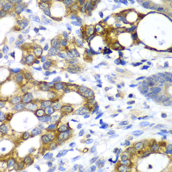
A06771 has IHC images from paraffin-embedded human liver injury and gastric cancer samples (IHC image captions). The catalog lists human, mouse and rat reactivity; it provides no IF data (catalog: reactivity and applications).
A06771 will render with an IHC figure from paraffin-embedded human liver injury at 1:100 (IHC image caption). A second caption shows paraffin-embedded human gastric cancer at 1:100; the catalog lists IHC and human, mouse and rat reactivity (IHC image caption; catalog: applications and reactivity).
Which to pick: Choose A06771 for paraffin-section IHC: its caption shows that processing, while the fixative is unreported (IHC image caption). A06771 has no listed IF/ICC application or IF figure, so there is no supported IF/ICC pick here (catalog: applications and IF image alts). For mouse or rat work, A06771 lists reactivity in both species, but its IHC captions show human samples only; clonality is unspecified (catalog: reactivity and clone; IHC image captions).