ANK1 / Ankyrin-1 · IHC design guide

Design Immunohistochemistry for ANK1

Plan ANK1 chromogenic IHC on paraffin sections with the catalog antibody at 2–5 μg/ml (datasheet A02716-1). Compare erythrocyte membranes, marrow hematopoietic cells and Purkinje cells with the reported tissue staining (HPA tissue IHC).

Evidence assembled Oct 2026 · For research use; verify linked source records and product datasheet before use
Immunohistochemistry protocol sheet for ANK1 (IHC for ANK1): expected localisation Erythrocyte membrane; Purkinje cytoplasm/membrane (HPA tissue IHC), antibody A02716-1, validated IHC image, and IHC protocol steps
Printable ANK1 IHC protocol sheet — expected localisation Erythrocyte membrane; Purkinje cytoplasm/membrane (HPA tissue IHC), antibody A02716-1, controls and protocol steps. Open the full ANK1 IHC guide →

ANK1 Immunohistochemistry Experimental Design Guide

Expected localisation, validated protocols, controls and antibodies — the at-a-glance facts below, then the full design guide.

Must know before staining
Expected localisation Erythrocyte membrane; Purkinje cytoplasm/membrane (HPA tissue IHC)
Staining pattern Membranous erythrocytes; marrow cells; Purkinje cytoplasm/membrane (HPA tissue IHC)
Antigen retrieval EDTA pH 8.0 HIER, heat-mediated (datasheet A02716-1)
Positive control ⓘ Bone marrow+1 more · see all
Negative control ⓘ Adipose tissue+4 more · see all
Important caveats
Reasons your staining may differ from the expected pattern.
Fixation Keep fixation consistent across paraffin sections and controls. (selected-SKU IHC image A02716-1)
Caveat Erythrocyte heme can mimic DAB positivity (standard IHC practice)
Regulation Marrow/Purkinje high; myocytes low (HPA tissue IHC)
Isoform / epitope 23 isoforms; verify epitope coverage across variants (UniProt)
Section 1

Recommended ANK1 IHC & IF Protocols

Use the catalog antibody’s paraffin IHC protocol (datasheet A02716-1) alongside the published pancreatic tumor microarray protocol (PMC5085203).

Recommended immunohistochemistry (IHC-P) protocol parameters
SampleParaffin-embedded human colon cancer tissue; fixative not specified (datasheet A02716-1)
FixationImage fixative and duration unreported (datasheet A02716-1); verify before use.
Sectioning4–5 µm sections on charged slides (standard)
DeparaffinisationXylene, graded ethanol series to water (standard)
Antigen retrievalHeat retrieval: EDTA pH 8.0 (datasheet A02716-1); 20 min, 95–100 °C (standard)
Peroxidase block3% H2O2, 10 min, room temperature (standard)
Blocking10% goat serum (datasheet A02716-1)
Primary antibodyRabbit anti-ANK1, 2-5 μg/ml (datasheet A02716-1)
Primary incubationOvernight at 4 °C (datasheet A02716-1)
DetectionStreptavidin-biotin complex (SABC), DAB chromogen (datasheet A02716-1)
CounterstainHematoxylin, blue, dehydrate and mount (standard)
Expected resultANK1-positive staining in hematopoietic cells of bone marrow (HPA tissue IHC: High). HPA tissue profile: Selective membranous expression in erythrocytes, cells in bone marrow and Purkinje cells. No signal in the no-primary control.
💡Decision noteStart with heat-mediated EDTA pH 8.0 retrieval (datasheet A02716-1) and include erythrocytes as a positive control (PMC5085203).
Section 2

What Is the Expected ANK1 Staining Pattern?

ANK1 is associated with the cytoskeleton and membrane, with no transmembrane segment (UniProt P16157 topology and subcellular location). In paraffin-section IHC, expect selective membranous staining of erythrocytes, strong staining in bone-marrow hematopoietic cells, and cytoplasmic or membranous staining in cerebellar Purkinje cells (HPA: tissue IHC, Supported). HPA cautions that splice or transcript discrepancies exist (HPA: tissue IHC reliability description).

What am I looking at on my slide?
Erythrocytes show a defined peripheral signal; bone-marrow hematopoietic cells and Purkinje cells stain strongly (HPA: tissue IHC).This matches the reported selective erythrocyte membrane pattern and High staining in the two listed positive cell populations (HPA: tissue IHC). Score the named cells and their compartments rather than treating every cell in a positive tissue as positive (HPA: tissue IHC cell annotations).
The dominant signal is nuclear in erythrocytes or Purkinje cells, or appears as an indiscriminate nuclear stain across the section.A nuclear pattern does not match the reported membrane or cytoplasm locations (HPA: tissue IHC; UniProt P16157 subcellular location). Review morphology and detection controls before assigning it to ANK1 (standard IHC practice).
Strong signal appears in adipocytes or respiratory epithelial cells while the expected positive cells are identifiable (HPA: tissue IHC).Those annotated cell populations were Not detected by HPA (HPA: adipose tissue and bronchus IHC). Consider cross-reactivity or endogenous detection activity; staining of erythrocytes present in the same section is a separate observation (HPA: tissue IHC; standard IHC practice).
Weak color covers cells, extracellular space, or the entire section without a discernible erythrocyte rim or positive-cell pattern.Diffuse color cannot establish the selective pattern reported by HPA (HPA: tissue IHC). Compare a detection-only control and reassess blocking, washing, and chromogen development as general IHC checks (standard IHC practice).
Bone-marrow hematopoietic cells or cerebellar Purkinje cells show no convincing signal (HPA: High in both).Treat the run as unresolved until the tissue, cell identity, antibody application, and detection controls are checked (HPA: tissue IHC; standard IHC practice). HPA's Supported rating includes a splice or transcript discrepancy caution, so one negative section alone does not establish biological absence (HPA: reliability description).
💡Expected ANK1 appearanceA convincing positive is selective erythrocyte membrane staining with High signal in bone-marrow hematopoietic cells or cytoplasmic/membranous Purkinje cells; widespread nuclear or diffuse staining fails that pattern (HPA: tissue IHC; UniProt P16157 subcellular location).
How each factor affects the staining
Membrane association and compartmentANK1 links membrane proteins to cytoskeletal elements and has no transmembrane segment (UniProt P16157 function and topology). Interpret an erythrocyte rim as membrane-associated staining; do not infer that the protein spans the membrane (HPA: tissue IHC; UniProt P16157 topology).
Cell population and expression levelHPA reports High staining in bone-marrow hematopoietic cells and Purkinje cells, but Low staining in skeletal-muscle myocytes (HPA: tissue IHC). A faint myocyte result therefore carries less weight as a positive control than the listed high-staining populations (HPA: tissue IHC).
Isoform contextUniProt lists 23 isoforms, including skeletal-muscle Mu17–Mu20 and brain Br21 (UniProt P16157 isoforms and tissue specificity). HPA also flags a splice or transcript discrepancy; do not assume every antibody recognizes every isoform or that all tissues share one staining pattern (HPA: reliability description; standard IHC interpretation).
Evidence boundary across applicationsThe tissue pattern and Supported assessment concern IHC (HPA: tissue IHC and antibody validation). HPA provides no ICC/IF image or main subcellular location for ANK1, so these IHC observations do not independently validate an IF/ICC pattern (HPA: subcellular record).
Why is my staining missing, weak or wrong?
SituationLikely causeNext action
No signal in the expected positive cells.The run or cell identification may need review; HPA reports High staining in bone-marrow hematopoietic cells and Purkinje cells (HPA: tissue IHC).Confirm the annotated cell population, use an IHC-validated antibody, and inspect the run's detection controls before scoring the section negative (HPA: antibody validation; standard IHC practice).
Most cells show nuclear color.Nuclear staining conflicts with the supplied compartment evidence (HPA: tissue IHC; UniProt P16157 subcellular location).Compare the detection-only control, verify cell morphology, and score only a reproducible expected compartment pattern (standard IHC practice; HPA: tissue IHC).
Color appears in HPA-negative cell populations.Cross-reactivity or endogenous detection activity is possible (standard IHC practice); HPA reports adipocytes and bronchial respiratory epithelium as Not detected (HPA: tissue IHC).Check a detection-only control and identify the stained cells before calling the tissue positive; erythrocyte staining can be evaluated separately (standard IHC practice; HPA: tissue IHC).
Background obscures erythrocyte outlines.Diffuse color prevents assessment of HPA's selective membranous pattern (HPA: tissue IHC); excess detection signal is a general possibility (standard IHC practice).Review blocking, washing, detection-only controls, and chromogen development; then reassess whether a distinct cell outline is visible (standard IHC practice; HPA: tissue IHC).
Skeletal muscle stains weakly despite clear positive controls.HPA lists myocytes as Low, while UniProt identifies muscle isoforms Mu17–Mu20 (HPA: tissue IHC; UniProt P16157 tissue specificity).Interpret the weak myocyte result in that context and compare the same run with a listed high-staining cell population (HPA: tissue IHC; standard IHC practice).
IF/ICC Q: Can this IHC pattern predict fluorescence staining?HPA has no ANK1 ICC/IF images or assigned main subcellular location (HPA: subcellular record).Treat the IF/ICC pattern as unconfirmed here and use the separate IF/ICC guide for application-specific design; retain the IHC tissue pattern as context only (HPA: tissue IHC and subcellular record).

Sample controls for ANK1 IHC & IF

🧪Run bone marrow first and look for staining in hematopoietic cells (HPA: High in bone marrow hematopoietic cells). Use adipose tissue as the negative tissue (HPA: Not detected in adipocytes); on the bone marrow slide, evaluate nonhematopoietic stromal cells for background staining without assuming they are ANK1-negative.
Positive control tissue: Bone marrow (Hematopoietic cells, HPA High)
Negative control tissue: Adipose tissue (HPA Not detected)
ICC-IF cell lines (HPA subcellular resource): HPA carries no ICC-IF cell line for ANK1; derive a cell-line control from the positive tissue's cell type (Hematopoietic cells) and confirm it by RNA or western blot first.
Technical controls: Include no-primary and secondary-only slides, a species- and clonality-matched isotype control (caption: rabbit primary; clonality unreported), and independently validated ANK1-knockout material as a biological negative. In bone marrow, quench endogenous peroxidase and check for endogenous biotin before using the captioned biotin–SABC/DAB detection system (caption: biotinylated secondary and SABC/DAB).
⚠️Feasibility: No target-specific fixation window or fixation effect is reported, and the selected A02716-1 tissue-IHC caption does not state a fixative. That caption uses heat retrieval in EDTA at pH 8.0 on a paraffin section, but does not establish that ANK1 staining requires this retrieval condition (caption: EDTA pH 8.0). The supplied evidence does not establish whether frozen sections or IF/ICC are easier; bone marrow erythrocytes can complicate interpretation because ANK1 is associated with the erythrocyte membrane (UniProt P16157: function and subcellular location).

HPA tissue IHC evidence for ANK1

Comprehensive Human Protein Atlas IHC scoring per tissue (reliability: Supported — External characterization data supports antibody staining. Caution, Splice and/or transcript discrepancy exists.). Rows are taken directly from the HPA tissue chart — click any row's HPA link to view the source.

Positive expression · recommended positive controls

TissueCell typeLevelEvidenceSource
Bone marrow Hematopoietic cells High Protein (IHC) HPA →
Cerebellum Purkinje cells - cytoplasm/membrane High Protein (IHC) HPA →

Undetected expression · recommended negative controls

TissueCell typeLevelEvidenceSource
Adipose tissue Adipocytes Not detected Protein (IHC) HPA →
Adrenal gland Glandular cells Not detected Protein (IHC) HPA →
Appendix Glandular cells Not detected Protein (IHC) HPA →
Breast Adipocytes Not detected Protein (IHC) HPA →
Bronchus Respiratory epithelial cells Not detected Protein (IHC) HPA →
Section 3

Advanced ANK1 IHC Tips

Troubleshoot ANK1 staining in paraffin sections by checking retrieval, cell identity and the expected membrane associated pattern before scoring (datasheet A02716-1; HPA tissue IHC).

What retrieval should I try when ANK1 staining is weak in paraffin sections?
Use heat mediated antigen retrieval in EDTA at pH 8.0 for paraffin sections (datasheet A02716-1). The selected antibody produced staining with this retrieval condition, followed by 2 μg/ml primary antibody overnight at 4°C (datasheet A02716-1). If signal is weak, first check that heating, cooling and buffer pH were consistent across sections, then compare retrieval durations on adjacent sections (standard IHC practice). Keep antibody concentration and detection conditions constant during that comparison so a change in signal can be attributed to retrieval (standard IHC practice). Score staining in the expected cells and compartments rather than judging overall section darkness (HPA tissue IHC; standard IHC practice).
Could fixation explain weak or uneven ANK1 staining?
ANK1 specific sensitivity to fixation is unknown from the supplied evidence; the selected paraffin section caption does not state a fixative (datasheet A02716-1). Record the actual fixative, fixation duration and tissue processing for each specimen before comparing staining intensity (standard IHC practice). If sections differ, compare similarly processed specimens using the same EDTA pH 8.0 retrieval and 2 μg/ml antibody conditions (datasheet A02716-1; standard IHC practice). Examine morphology alongside staining, because damaged or poorly preserved cells make compartment scoring unreliable (standard IHC practice). Do not infer a fixation effect from ANK1 localisation or its listed modifications (UniProt P16157).
Where should ANK1 staining appear in a tissue section?
Look for membrane associated staining in erythrocytes and bone marrow cells, and cytoplasmic or membranous staining in cerebellar Purkinje cells (HPA tissue IHC). ANK1 is a cytoskeletal adaptor associated with membranes and has no transmembrane segment, so a peripheral pattern need not indicate an extracellular epitope (UniProt P16157). Its recorded locations also include the myofibril M line and sarcoplasmic reticulum, which matter when interpreting muscle structures (UniProt P16157). Compare the pattern with cell morphology and an adjacent control section before calling diffuse staining specific (standard IHC practice). Assess each cell population separately, since the expected compartments differ across these settings (HPA tissue IHC; UniProt P16157).
Why might an ANK1 antibody stain some expected cells but miss others?
ANK1 has 23 listed isoforms, including erythrocyte, muscle and brain forms, so isoform coverage depends on the antibody epitope (UniProt P16157). The supplied caption identifies staining conditions but does not establish which isoforms the catalog antibody recognizes (datasheet A02716-1). Obtain its immunogen or mapped epitope and compare that sequence with isoforms expected in the cells being scored (standard IHC practice; UniProt P16157). Muscle isoforms Mu17–Mu20 are expressed in skeletal muscle, while Br21 is expressed in brain (UniProt P16157). Treat a negative cell population cautiously until epitope coverage and section quality have been checked (standard IHC practice).
How should I interpret ANK1 in a multiplex IF experiment?
For a separate IF experiment, pair ANK1 with a marker that identifies the expected cell population, then assess whether its signal follows the relevant cell boundary or cytoplasm (HPA tissue IHC; standard IF practice). Choose a fluorophore channel with low tissue autofluorescence and inspect single channel controls before interpreting overlap (standard IF practice). ANK1 has no transmembrane segment and is associated with the cytoskeletal side of membranes, so intracellular epitope access generally calls for permeabilisation after fixation (UniProt P16157; standard IF practice). Optimise that step against preservation of cell boundaries (standard IF practice). No ICC/IF images are supplied here to establish an antibody specific IF staining pattern (HPA subcellular).
How can I separate ANK1 staining from chromogenic background?
The selected paraffin section image used 10% goat serum blocking, a biotinylated secondary antibody, a streptavidin–biotin complex and DAB (datasheet A02716-1). If staining is widespread, inspect a no primary control and verify that peroxidase blocking and washes were effective (standard IHC practice). With this detection chemistry, check whether signal persists without primary antibody before assigning brown deposits to ANK1 (datasheet A02716-1; standard IHC practice). Compare suspect staining with cell morphology and the expected membrane associated or cytoplasmic pattern (HPA tissue IHC). Titrate detection conditions on matched sections while retaining a known positive compartment as a reference (standard IHC practice; HPA tissue IHC).
What should I quantify when comparing ANK1 across sections? ⚠ ANSWER MARKED FOR VERIFICATION
Define the cell population and compartment before scoring, because ANK1 staining is reported in erythrocytes, bone marrow cells and Purkinje cells (HPA tissue IHC). For nucleated cells, record the percentage positive and an intensity score of 0–3, or combine them as an H-score of 0–300 (standard IHC practice). For erythrocytes, report positive cell density per mm² or the fraction of identified erythrocytes that stain (standard IHC practice). Normalise to the counted cells or sampled tissue area, and use matched processing and imaging conditions across groups (standard IHC practice). Keep compartment specific scores separate when staining patterns differ (HPA tissue IHC; standard IHC practice).
When is an unexpected ANK1 positive area likely to be artefactual?
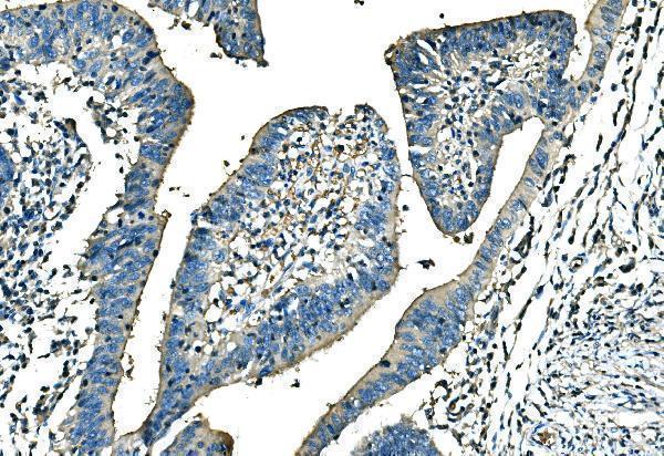
Judge a positive result against cell identity and compartment: HPA reports selective membranous staining in erythrocytes and bone marrow cells, with cytoplasmic or membranous Purkinje cell staining (HPA tissue IHC). Diffuse staining of unrelated cells or signal confined to section edges warrants review before assigning it to ANK1 (HPA tissue IHC; standard IHC practice). Exclude necrotic regions and inspect the no primary control for residual endogenous peroxidase or detection background (standard IHC practice). The selected caption establishes staining in a paraffin embedded human colon cancer section, but does not identify which cells account for that signal (datasheet A02716-1). HPA flags a splice or transcript discrepancy, so a divergent pattern needs epitope and cell identity checks (HPA tissue IHC).
Boster reagents

Best ANK1 / Ankyrin-1 IHC Antibodies

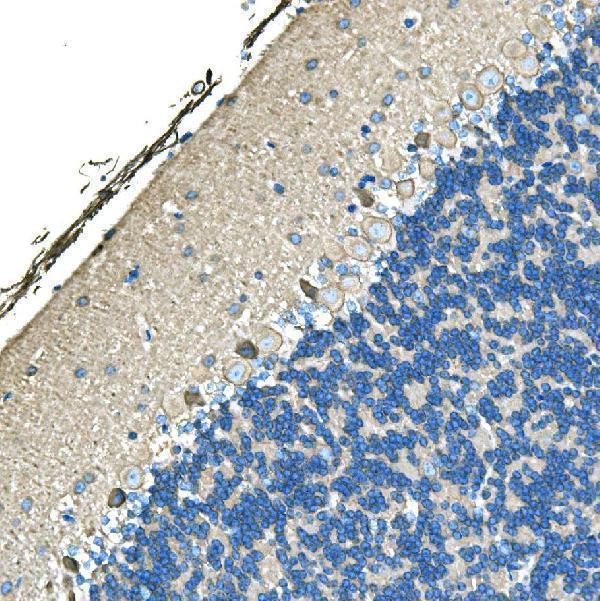
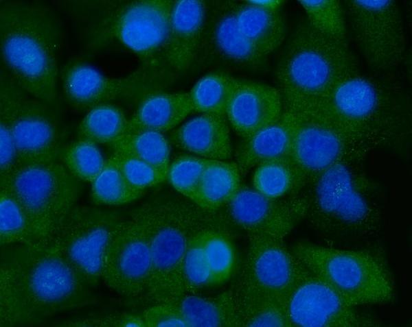
The catalog antibody has IHC images from paraffin sections of human colon cancer and mouse and rat cerebellum, plus IF/ICC data from A549 cells (catalog image captions).

Real IHC data IHC analysis of Ankyrin Erythroid/ANK/ANK1 using anti-Ankyrin Erythroid/ANK/ANK1 antibody (A02716-1). Ankyrin Erythroid/ANK/ANK1 was detected in a paraffin-embedded section of human colon cancer tissue. Heat mediated antigen retrieval was performed in EDTA buffer (pH 8.0, epitope retrieval solution). The tissue section was blocked with 10% goat serum. The tissue section was then incubated with 2 μg/ml rabbit anti-Ankyrin Erythroid/ANK/ANK1 Antibody (A02716-1) overnight at 4°C. Biotinylated goat anti-rabbit IgG was used as secondary antibody and incubated for 30 minutes at 37°C. The tissue section was developed using Strepavidin-Biotin-Complex (SABC) (Catalog # SA1022) with DAB as the chromogen.
Anti-Ankyrin erythroid/ANK/ANK1 Antibody ®
Cat # A02716-1

A02716-1 has IHC images from paraffin sections of human colon cancer and mouse and rat cerebellum (catalog IHC captions). The same SKU has an IF/ICC image from A549 cells (catalog IF caption).

Which to pick: Choose A02716-1 for paraffin section IHC; its captions document EDTA retrieval at pH 8.0 and primary antibody at 2 μg/ml (catalog IHC captions). Choose A02716-1 for IF/ICC on A549 cells, where its caption documents 5 μg/ml (catalog IF caption). It is listed as reactive with human, mouse and rat and has IHC images for all three (catalog reactivity; catalog IHC captions); clonality and the paraffin sections’ fixative are unreported (catalog clone field; catalog IHC captions).

Each figure is that product's own IHC / IF validation image from its datasheet.

References

  1. UniProt Consortium. UniProt entry P16157 (ANK1_HUMAN, Ankyrin-1).
  2. Human Protein Atlas. ANK1 tissue IHC expression (reliability: Supported).
  3. Human Protein Atlas. ANK1 subcellular location (ICC-IF): Highest expression in Rh30: 132.3 nTPM.
  4. Human Protein Atlas. ANK1 antibody validation summary (2 antibodies).
  5. Overexpression of ankyrin1 promotes pancreatic cancer cell growth. Oncotarget 2016 — PMC5085203.
  6. FGFR1 fusions as a novel molecular driver in rhabdomyosarcoma. Genes, chromosomes & cancer 2024 — PMC11385681.
  7. ANK1 inhibits malignant progression of osteosarcoma by promoting ferroptosis. BMC cancer 2024 — PMC11365275.
  8. Exploring the pathogenesis of steroid-induced osteonecrosis of the femoral head co-regulated by mesenchymal stem cells and pyroptosis-related genes with the use of transcriptome. European journal of medical research 2025 — PMC12532964.
  9. PubMed PMID:2137557 — UniProt-cited evidence.
  10. PubMed PMID:1689849 — UniProt-cited evidence.
  11. PubMed PMID:9235914 — UniProt-cited evidence.