This website uses cookies to ensure you get the best experience on our website.
- Table of Contents
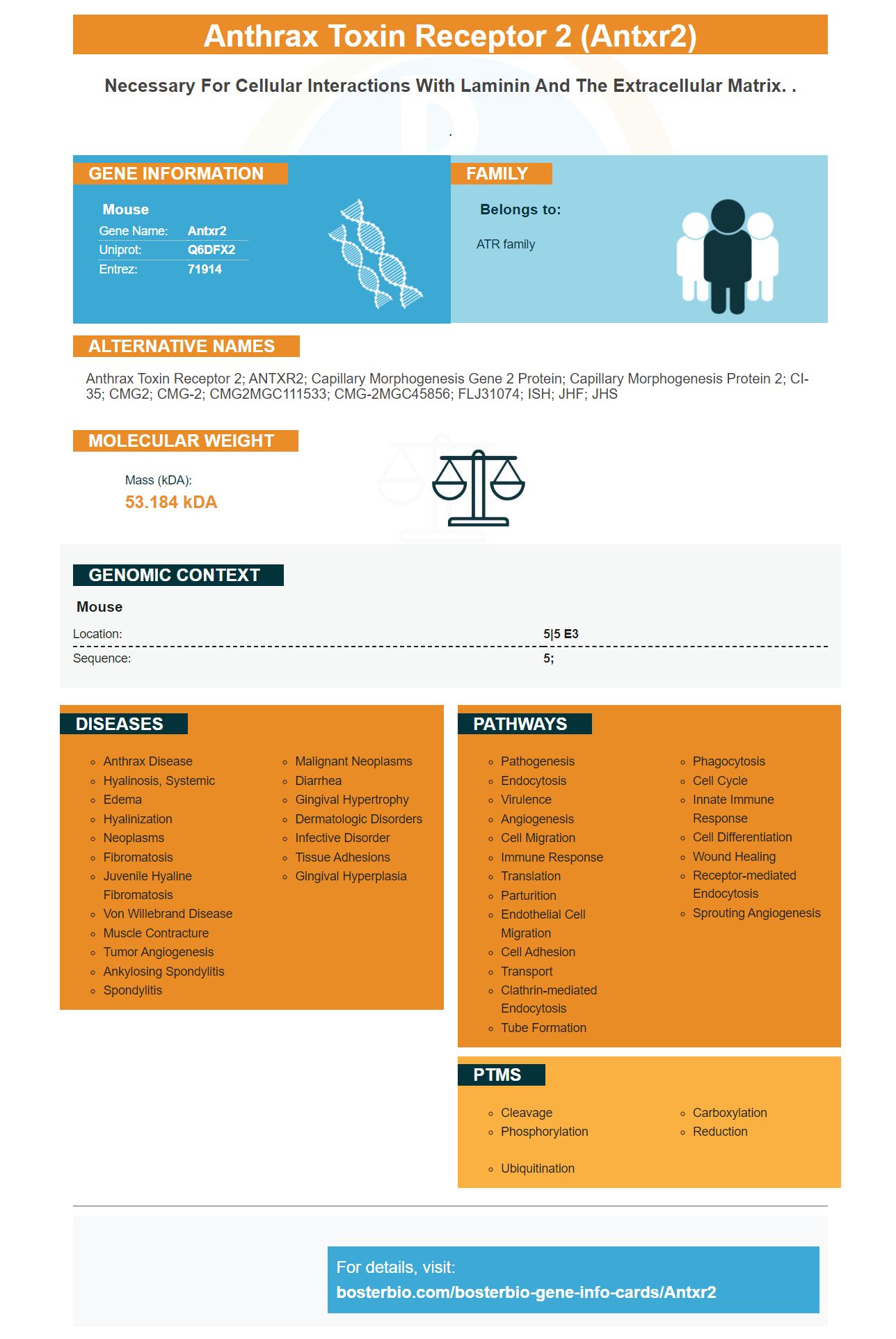
Facts about Anthrax toxin receptor 2.
.
Mouse | |
---|---|
Gene Name: | Antxr2 |
Uniprot: | Q6DFX2 |
Entrez: | 71914 |
Belongs to: |
---|
ATR family |
anthrax toxin receptor 2; ANTXR2; Capillary morphogenesis gene 2 protein; capillary morphogenesis protein 2; cI-35; CMG2; CMG-2; CMG2MGC111533; CMG-2MGC45856; FLJ31074; ISH; JHF; JHS
Mass (kDA):
53.184 kDA
Mouse | |
---|---|
Location: | 5|5 E3 |
Sequence: | 5; |
This article will discuss the Biological significance of ANTXR2 marker, High affinity primary antibodies, as well clinical applications of this antibody. We'll also talk about how ANTXR2Marker works in the body as well as in cells. We will also discuss some of the most important uses for this antibody. If you're not familiar with the ANTXR2 Marker, we strongly encourage you to read this article.
If you are a scientist looking for a new antibody, Boster Bio Anti-Anthrax Toxin Receptor 2 (#A03325) may be the perfect choice. This antibody was validated in Western Blotting, Immunohistochemistry, and ELISA and reacts with human, mouse, rat, and rat. Boster Bio has the right product for you, whether you're interested in the immune response to anthrax and/or you're looking for experiments to examine the receptor's role as a catalyst for bacterial growth.
The use of boster Bio high-affinity prim antibodies is one of many ways to develop highly targeted antibody treatments. Boster Bio uses its PCD platform for the development of high affinity clones specifically targeted at Diagnostics & Therapeutics targets. Boster prevents the B cells from secreting antigens by using proprietary chemistry. Boster then incubates splenocytes with flurochrome-conjugated antigens and plasma cells. For downstream screening, the brightest clones will be isolated.
PicoKine(tm), a company-exclusive ELISA platform, uses proprietary trade secrets for high-affinity and sensitive ELISA kits that can be used in a variety of applications. The kits contain sufficient reagents for 96 tests. The kits are suitable for use as flow cytometry, immunoassays, and other applications that require specificity.
The immunization of animals with a specific antibody produces high-affinity primaries antibodies. The antibodies are produced by selecting the best responders by performing a titration ELISA for each test-bleed. After this, spleen cell are removed and hybridoma is performed. Once the supernatants are positive, they are subjected to limiting dilution subcloning, and then further application testing.
Specific antibodies bind target antigens in enzyme-linked immunosorbent (ELISA) assays. The detection system detects the binding. Boster Bio high-affinity prima antibodies can be used to enhance the sensitivity and specificity of assays. Boster Bio high affinity primary antibodies are trusted worldwide because they are sensitive and specific. With this technology, scientists can use enzyme-linked immunosorbent assays to discover the role of a specific protein in the body.
Enzyme linked immunoassays (ELISA) can reveal the specificity or primary antibodies by determining how often they bind to the target protein. This data is used to determine whether antibodies are specific to the target protein and to identify which ones are not. ELISA results will be the final deliverable for antibody clones. Customers can also receive ELISA results, maps showing the vector, and purified antibodies.
Molecular studies revealed that the ANTXR2 marker could be used to identify ectopic uterine stromal and ectopic uterine cells. This gene is involved in the activation of YAP1. It is also associated with the cell adhesion protein CD44 and the cell-surface proteins Col5A2. YAP1 was shown to be significantly linked with the genes Col5A2 & CD44. Using ChIPPCR, we found that ANTXR2 was associated with the genes locus IL-1b/CD44.
ANTXR1 expression has been shown to be significantly associated with GC-cell invasion and migration. Knockdown of ANTXR1 reduced invasion and migration in BGC823 and MGC803 cells. Overexpression ANTXR1 effected an increase in invasion, migration, and cell proliferation in HGC27 cells. Several studies have revealed that ANTXR1 expression is involved in GC progression.
The ANTXR2 protein gene gives instructions for creating ANTXR2. This protein is found on many types of cells' surfaces. It interacts directly with components of extracellular matrix, which is a network that contains proteins outside the cell. It plays a key role in cell migration regulation and angiogenesis. While further studies are needed, this gene has already been shown to be important in clinical trials.
These results demonstrate that ANTXR1 in breast tissue is a biomarker for CSCs. This is significant because ANTXR1 expression in 3D spheres is not upregulated. The same results apply for ANTXR2, indicating that the cell does not express ANTXR1 in other tumor entities. It is important to identify other biomarkers that can help differentiate PaCSCs and other tumor entities.
Many independent studies show that the receptor does no dissociate from PA pore in prepore-to pore conversion. This observation is consistent and in line with other lines of work where ANTXR1-ANTXR2 coprecipitated with the pore or prepore. This is unlikely, however, to explain the differences between these two markers. Further research is needed to confirm that ANTXR2 is a causal gene at the 4q21 GWAS location.
The ANTXR2 genes provides instructions for the production of a protein that is found at the surface of many kinds of cells. The ANTXR2 protein interacts to the proteins in ECM, which is a complex network of proteins that surrounds the cells and supports connective tissue like skin, bones, and cartilage. The ANTXR2 protein helps the cell to regulate the correct balance of proteins, and may also be involved in the formation of tiny blood vessels.
Using ANTXR1 along with other CSC marker markers could help identify distinct subpopulations. In addition, ANTXR1+ cells exhibited elevated self-renewal capacity and enriched expression of pluripotency-associated genes. These findings support the proposed use of ANTXR1 as a PaCSC marker and could help in the development of CSC-targeted therapies.
In the present study, ANTXR1+ cell were detected in freshly digested PDXs. They were EpCAM-positive but DAPI negative. Panc215 PDX showed the highest level of ANTXR1+ cells. However, cross-reactivity of ANTXR1 & ANTXR2 is a major limitation to developing specific ligands in targeted therapy. SVV is an oncolytic agent with promising results in clinical trials. This finding also highlights that there is potential for further development of antiangiogenic antibodies therapy.
GC cell invasion is associated with metastasis and antxR1 expression. In vitro, ANTXR1 gene expression in GC is associated in a similar way with invasive as well as migration abilities. These findings suggest that ANTXR1 might be a critical tumorigenic factor in GC cell cells. Although these results are preliminary, they are useful as a diagnostic tool.
PMID: 21183079 by Huttlin E.L., et al. A tissue-specific atlas of mouse protein phosphorylation and expression.