This website uses cookies to ensure you get the best experience on our website.


- Table of Contents
Plan chromogenic ANXA13 IHC on paraffin sections with the catalog antibody at 1:50–1:200 (datasheet A11738). Use strongly stained small-intestine enterocytes as a positive reference and unstained adipocytes as a negative reference (HPA tissue IHC).

Expected localisation, validated protocols, controls and antibodies — the at-a-glance facts below, then the full design guide.
| Expected localisation | Cytoplasmic staining in gastrointestinal, biliary, renal and tubal cells (HPA tissue IHC) | |
| Staining pattern | Cytoplasmic signal in GI, bile duct, gallbladder, renal and tubal cells (HPA tissue IHC) | |
| Antigen retrieval | Citrate pH 6.0 HIER, heat-mediated (datasheet A11738) | |
| Positive control | Appendix+4 more · see all | |
| Negative control | Adipose tissue+4 more · see all |
| Fixation | Keep fixation consistent across sections (standard IHC practice; not target-specific) | |
| Caveat | Signal varies by cell type within an organ (HPA tissue IHC) | |
| Regulation | RNA is enhanced in gallbladder and intestine (HPA tissue RNA) | |
| Isoform / epitope | 2 isoforms (A, B); epitope coverage is unknown (UniProt) |
The catalog antibody protocol (datasheet A11738) is paired with published ANXA13 IHC methods for kidney sections (PMC12915081) and colorectal tumor and adjacent normal tissues (PMC5400614).
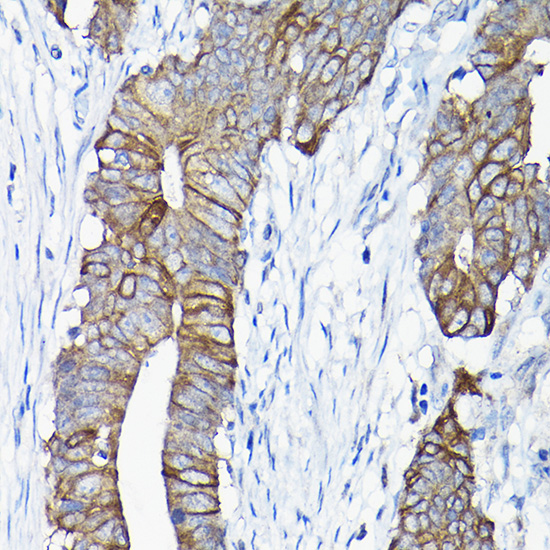
| Sample | Paraffin-embedded human colon carcinoma tissue; fixative not specified (datasheet A11738) |
| Fixation | Image fixative and duration unreported (datasheet A11738); verify before use. |
| Sectioning | 4–5 µm sections on charged slides (standard) |
| Deparaffinisation | Xylene, graded ethanol series to water (standard) |
| Antigen retrieval | Heat retrieval: Citrate pH 6.0 (datasheet A11738); 20 min, 95–100 °C (standard) |
| Peroxidase block | 3% H2O2, 10 min, room temperature (standard) |
| Blocking | 10% normal serum of the secondary host, 30 min, room temperature (standard) |
| Primary antibody | Rabbit anti-ANXA13, 1:50-1:200 (datasheet A11738) |
| Primary incubation | Overnight at 4 °C (standard) |
| Detection | HRP-polymer secondary, DAB chromogen 5–10 min (standard) |
| Counterstain | Hematoxylin, blue, dehydrate and mount (standard) |
| Expected result | ANXA13-positive staining in endocrine cells of appendix (HPA tissue IHC: High). HPA tissue profile: Cytoplasmic expression in gastrointestinal tract, bile ducts, gallbladder, renal tubules and fallopian tube. No signal in the no-primary control. |
ANXA13 is associated with the apical cell membrane and cytoplasmic vesicles; it has no transmembrane segment, and membrane association includes myristoylation and reversible calcium dependent lipid binding (UniProt P27216 topology and subcellular location). In paraffin IHC, expect epithelial staining in gastrointestinal tissue, gallbladder, bile ducts, renal tubules, and fallopian tube (HPA: tissue IHC profile). HPA rates its tissue pattern Enhanced, reflecting high consistency between staining and RNA expression (HPA: reliability).
| Apical staining in small intestinal enterocytes, particularly the microvilli, with epithelial cytoplasmic staining. | This fits the high enterocyte microvillar signal and broader gastrointestinal cytoplasmic profile observed by HPA (HPA: small intestine High; tissue IHC profile). UniProt also places ANXA13 at the apical membrane and in cytoplasmic vesicles (UniProt P27216 subcellular location). Judge the labeled cells and compartment together; uniform staining of every cell is not the expected tissue pattern (HPA: tissue IHC profile). |
| Staining is predominantly nuclear, while expected epithelial cytoplasmic or apical staining is absent. | Treat this as discordant with the tissue IHC profile and investigate staining specificity or background (HPA: tissue IHC profile; standard IHC interpretation). A nuclear signal cannot automatically be dismissed across applications: HPA reports an additional, approved nucleoplasmic location in ICC-IF, alongside its supported plasma membrane location (HPA: subcellular ICC-IF). |
| Strong signal appears chiefly in cell types outside the reported positive populations. | Compare cell identity with the documented pattern before scoring the section positive (HPA: tissue IHC positive-cell entries). For example, HPA reports high signal in duodenal endocrine cells and medium signal in colonic goblet cells, which are different populations (HPA: duodenum High; colon Medium). Unexpected staining can reflect antibody cross-reactivity or endogenous detection activity; it does not establish ANXA13 expression without supporting controls (standard IHC interpretation). |
| Diffuse color covers tissue and spaces between cells, obscuring epithelial boundaries. | This distribution is difficult to reconcile with the reported cell-associated cytoplasmic and apical pattern (HPA: tissue IHC profile; UniProt P27216 subcellular location). Consider nonspecific antibody binding, incomplete blocking, or excess chromogen development, and inspect the negative reagent control (standard IHC practice). Background intensity alone should not be scored as target-positive staining (standard IHC interpretation). |
| No signal appears in a section containing expected positive epithelial cells. | An absence in small intestinal enterocytes or appendix or duodenal endocrine cells conflicts with HPA's high staining in those specified cells (HPA: small intestine, appendix, and duodenum High). First verify that the relevant cells are present and preserved; then review the IHC-validated antibody, detection reagents, and staining conditions with a positive control (standard IHC practice). A negative slide by itself cannot establish absence of ANXA13 (standard IHC interpretation). |
| Cell type and site | Expected intensity varies by population: small intestinal enterocyte microvilli and fallopian tube cilia axonemes are High, whereas colonic goblet cells, gallbladder glandular cells, and kidney proximal tubule microvilli are Medium (HPA: tissue IHC positive-cell entries). Score the named cells, not the whole organ as one uniform compartment (standard IHC interpretation). |
| Membrane association and topology | ANXA13 lacks a transmembrane segment, yet myristoylation anchors it to membranes and calcium dependent binding can be reversible (UniProt P27216 topology and subcellular location). This supports interpreting apical or membrane-associated staining alongside cytoplasmic vesicle signal; it does not prescribe an antigen-retrieval condition for paraffin sections (UniProt P27216 subcellular location; standard IHC practice). |
| Antibody validation | HPA lists Enhanced IHC validation for HPA018535, HPA019569, HPA019650, and CAB025135; its tissue reliability is also Enhanced (HPA: antibodies; tissue IHC reliability). These summaries support the reported pattern but do not make an unexpected compartment or cell population positive without controls (standard IHC interpretation). |
| IF/ICC Q: What localization should an IF result show? | A: HPA reports mainly plasma membrane localization, supported in ICC-IF, with additional approved nucleoplasmic localization; image-bearing cell lines are A-549 and U-251MG (HPA: subcellular ICC-IF). That ICC-IF observation is separate from the paraffin tissue IHC pattern and supplies no IHC protocol condition (HPA: subcellular ICC-IF; tissue IHC profile). |
| Situation | Likely cause | Next action |
|---|---|---|
| Expected-positive epithelium is unstained. | The relevant cells may be absent from the section, or the IHC detection run may have failed; HPA reports high staining only for specified positive cells, such as small intestinal enterocyte microvilli (HPA: small intestine High; standard IHC practice). | Confirm the cells on the counterstained section, then compare a known-positive section processed in the same run; review the catalog antibody's IHC-P instructions and reagent performance (standard IHC practice). |
| Color is diffuse across cells and extracellular areas. | Nonspecific binding, inadequate blocking, or excessive development can obscure the cell-associated pattern (standard IHC practice; HPA: tissue IHC profile). | Inspect a negative reagent control; optimize blocking, antibody dilution, washing, and chromogen development according to the IHC workflow (standard IHC practice). |
| Staining is mainly nuclear in tissue IHC. | The tissue IHC pattern is described as cytoplasmic, although HPA separately approves an additional nucleoplasmic ICC-IF location (HPA: tissue IHC profile; subcellular ICC-IF). | Check whether epithelial cytoplasmic or apical staining is also present, inspect controls, and avoid assigning isolated nuclear color to ANXA13 without corroboration (standard IHC interpretation). |
| Strong signal appears in an unexpected cell population. | Cross-reactivity or endogenous detection activity is possible when staining does not follow the documented cell-specific pattern (HPA: tissue IHC positive-cell entries; standard IHC interpretation). | Identify the stained cells with the counterstain, inspect reagent controls, and compare with the named HPA positive and not-detected populations before scoring (HPA: tissue IHC positive and negative entries; standard IHC practice). |
| A low-signal tissue is being used to judge assay failure. | HPA records Low staining in nasopharyngeal and bronchial ciliated rootlets and stomach glandular cells, so faint signal there has limited value as a positive-run check (HPA: tissue IHC low entries). | Use a section containing a documented High population, such as small intestinal enterocyte microvilli, as the positive comparison; score the specified cells (HPA: small intestine High; standard IHC practice). |
| A not-detected tissue shows convincing-looking color. | HPA reports Not detected in specified cells, including adipocytes in adipose tissue and hematopoietic cells in bone marrow; apparent color warrants specificity review (HPA: tissue IHC negative entries). | Verify cell identity and inspect negative reagent controls, then compare staining with a documented positive cell population in the same run (HPA: tissue IHC positive and negative entries; standard IHC practice). |


Comprehensive Human Protein Atlas IHC scoring per tissue (reliability: Enhanced — High consistency between antibody staining and RNA expression data.). Rows are taken directly from the HPA tissue chart — click any row's HPA link to view the source.
| Tissue | Cell type | Level | Evidence | Source |
|---|---|---|---|---|
| Appendix | Endocrine cells | High | Protein (IHC) | HPA → |
| Duodenum | Endocrine cells | High | Protein (IHC) | HPA → |
| Fallopian tube | Ciliated cells (cilia axoneme) | High | Protein (IHC) | HPA → |
| Small intestine | Enterocytes - Microvilli | High | Protein (IHC) | HPA → |
| Testis | Sertoli cells | High | Protein (IHC) | HPA → |
| Tissue | Cell type | Level | Evidence | Source |
|---|---|---|---|---|
| Adipose tissue | Adipocytes | Not detected | Protein (IHC) | HPA → |
| Adrenal gland | Glandular cells | Not detected | Protein (IHC) | HPA → |
| Bone marrow | Hematopoietic cells | Not detected | Protein (IHC) | HPA → |
| Breast | Adipocytes | Not detected | Protein (IHC) | HPA → |
| Caudate | Glial cells | Not detected | Protein (IHC) | HPA → |
Troubleshoot ANXA13 staining in paraffin sections by checking retrieval, cell identity and subcellular pattern before comparing staining intensity.
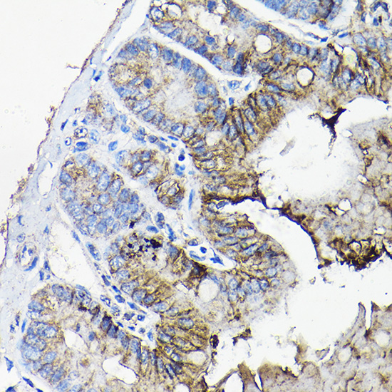
A11738 has IHC images from paraffin-embedded human colon carcinoma and mouse jejunum (catalog IHC captions). Human, mouse and rat reactivity is listed; no IF image is supplied (catalog reactivity; catalog images).
A11738 is listed for IHC and has paraffin-section images from human colon carcinoma and mouse jejunum, each stained at 1:50 after high-pressure retrieval in 10 mM citrate, pH 6.0 (catalog applications; catalog IHC captions). Its listed reactivity is human, mouse and rat; the payload supplies no IF image (catalog reactivity; catalog images).
Which to pick: Choose A11738 for paraffin-section IHC: its own captions document staining in human colon carcinoma and mouse jejunum, but do not report the fixative (catalog IHC captions). For cross-species work, A11738 lists human, mouse and rat reactivity; the supplied images document human and mouse tissue only (catalog reactivity; catalog IHC captions). For IF/ICC, A11738 has no listed IF application or IF image, although its catalog text mentions immunofluorescence microscopy testing; optimize and verify it for the intended sample (catalog applications; catalog text; catalog images).