This website uses cookies to ensure you get the best experience on our website.


- Table of Contents
Plan AOX1 staining in paraffin sections using its cytoplasmic tissue pattern and an IHC-validated antibody (HPA tissue IHC; datasheet). Compare positive hepatocytes with an appropriate negative control, and score staining by cell type (HPA tissue IHC; standard IHC practice).

Expected localisation, validated protocols, controls and antibodies — the at-a-glance facts below, then the full design guide.
| Expected localisation | Cytoplasmic staining (HPA tissue IHC) | |
| Staining pattern | Cytoplasm in hepatocytes, renal tubules, Leydig and adrenal cells (HPA tissue IHC) | |
| Antigen retrieval | Citrate pH 6.0 HIER, 95–98 °C, 20 min (rule: cytoplasmic / membrane antigen) | |
| Positive control | Adrenal gland+2 more · see all | |
| Negative control | Adipose tissue+4 more · see all |
| Fixation | Keep formalin fixation consistent across sections (standard IHC practice; not target-specific) | |
| Caveat | Adipocytes are unstained despite adipose tissue expression (HPA tissue IHC; UniProt) | |
| Regulation | No significant human sex effect (UniProt) | |
| Isoform / epitope | 0 annotated isoforms; one cytoplasmic chain (UniProt) |
The catalog antibody’s IHC-P protocol is accompanied by four published AOX1 chromogenic IHC protocols (PMC3415238; PMC8287305; PMC13304650; PMC8058741).
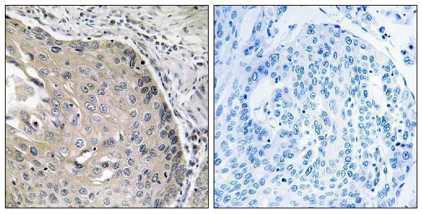
| Sample | Paraffin-embedded human lung carcinoma tissue; fixative not specified (datasheet A02144-1) |
| Fixation | Image fixative and duration unreported (datasheet A02144-1); verify before use. |
| Sectioning | 4–5 µm sections on charged slides (standard) |
| Deparaffinisation | Xylene, graded ethanol series to water (standard) |
| Antigen retrieval | Heat-induced epitope retrieval in citrate buffer, pH 6.0, 20 min at 95–98 °C (standard rule: cytoplasmic / membrane antigen) |
| Peroxidase block | 3% H2O2, 10 min, room temperature (standard) |
| Blocking | 10% normal serum of the secondary host, 30 min, room temperature (standard) |
| Primary antibody | Rabbit anti-AOX1, 1:100-1:300 (datasheet A02144-1) |
| Primary incubation | Overnight at 4 °C (standard) |
| Detection | HRP-polymer secondary, DAB chromogen 5–10 min (standard) |
| Counterstain | Hematoxylin, blue, dehydrate and mount (standard) |
| Expected result | AOX1-positive staining in glandular cells of adrenal gland (HPA tissue IHC: High). HPA tissue profile: Cytoplasmic expression most abundant in hepatocytes, renal tubules, Leydig cells and adrenal glandular cells. No signal in the no-primary control. |
AOX1 should show cytoplasmic staining in hepatocytes, adrenal glandular cells and Leydig cells, where HPA reports high staining (HPA tissue IHC). Its reported location is cytoplasmic, consistent with a protein lacking a transmembrane segment (UniProt Q06278). HPA rates the tissue IHC pattern “Enhanced,” citing high consistency between antibody staining and RNA expression data (HPA tissue IHC).
| Strong cytoplasmic staining in hepatocytes, adrenal glandular cells or Leydig cells. | This fits the reported high staining in those cell populations (HPA tissue IHC) and AOX1’s cytoplasmic location (UniProt Q06278). Judge the signal in the named cells, since staining across an entire tissue section is less informative than its cellular distribution (general IHC practice). |
| Predominantly nuclear or sharply membrane-bound staining. | These compartments depart from the expected cytoplasmic IHC pattern (HPA tissue IHC; UniProt Q06278 topology). Review morphology and controls before interpreting such signal as AOX1; compartment mismatch raises concern for nonspecific staining or an imaging or scoring artefact (general IHC practice). |
| Strong staining in adipocytes or bronchial respiratory epithelial cells. | HPA reports AOX1 as not detected in these specific cell types (HPA tissue IHC). Check whether the stained cells were identified correctly, then assess background and detection controls; unexpected signal may reflect cross-reactivity or endogenous detection activity (general IHC practice). |
| Diffuse colour across cells and tissue spaces, with little cellular distinction. | A uniform haze does not reproduce the cell-restricted cytoplasmic pattern reported for AOX1 (HPA tissue IHC). Compare with a no-primary control and inspect washing, blocking and detection conditions for background that could obscure genuine staining (general IHC practice). |
| No convincing signal in hepatocytes in a liver section. | This conflicts with HPA’s high hepatocyte staining and UniProt’s report of abundant liver expression (HPA tissue IHC; UniProt Q06278). Confirm that tissue and cells are suitable, then check the antibody’s IHC-P instructions, retrieval, detection reagents and run controls (general IHC practice). |
| Which tissue and cells provide a positive reference? | Liver hepatocytes, adrenal glandular cells and testicular Leydig cells are reported as high (HPA tissue IHC). HPA also describes cytoplasmic expression in renal tubules, while its listed proximal-tubule cell-body level is low; use that qualified record when judging kidney (HPA tissue IHC). |
| Can a whole tissue be treated as uniformly negative? | Interpret the named cell type: HPA reports no detection in adipocytes from adipose tissue or breast, among other listed cells (HPA tissue IHC). UniProt reports expression in adipose tissue without specifying those cells (UniProt Q06278); these records do not establish that every adipose-tissue cell is negative. |
| How strong is the tissue-pattern evidence? | HPA assigns tissue IHC “Enhanced” reliability and lists two rabbit polyclonal antibodies with Enhanced IHC status, HPA040199 and HPA040215 (HPA tissue IHC; HPA antibodies). That supports the reported pattern, while an individual staining run still needs appropriate controls (general IHC practice). |
| Does AOX1 processing suggest a separate secreted staining pattern? | UniProt describes one chain spanning residues 1–1338, no signal peptide or propeptide, and no transmembrane segment (UniProt Q06278). Its reported cytoplasmic location supports cytoplasmic interpretation; these annotations alone do not predict retrieval requirements or fixation sensitivity (UniProt Q06278). |
| What should IF/ICC show? | Mainly cytosolic signal is supported by HPA’s ICC-IF record; nuclear bodies, microtubules and the cytokinetic bridge are additional uncertain locations (HPA subcellular). Treat those additional patterns cautiously. HPA lists HPA040199 as ICC Supported; this IHC section supplies no IF/ICC protocol (HPA antibodies). |
| Situation | Likely cause | Next action |
|---|---|---|
| Liver positive control is blank or very faint. | High hepatocyte staining is expected, so a blank result may indicate a failed staining run or unsuitable specimen (HPA tissue IHC; general IHC practice). | Verify hepatocytes are present; review the catalog antibody’s IHC-P instructions, retrieval conditions, dilution, detection and run controls (general IHC practice). No AOX1-specific fixation sensitivity is established by the supplied sources. |
| Nuclei dominate the signal. | Dominant nuclear staining conflicts with AOX1’s cytoplasmic location and the reported tissue IHC pattern (UniProt Q06278; HPA tissue IHC). | Recheck compartment boundaries with the counterstain, compare the no-primary control and review antibody and detection conditions before scoring the nuclear signal as specific (general IHC practice). |
| Adipocytes or bronchial respiratory epithelium stain strongly. | HPA reports AOX1 as not detected in those cell types; the apparent signal may reflect cell-identification error, cross-reactivity or endogenous detection activity (HPA tissue IHC; general IHC practice). | Confirm cell identity on the section and compare negative and no-primary controls; inspect the detection system for background before assigning AOX1 positivity (general IHC practice). |
| Brown colour spreads beyond cell boundaries. | Diffuse colour lacks the cellular and cytoplasmic definition expected from HPA’s AOX1 pattern (HPA tissue IHC). Background from detection or incomplete washing is a general possibility (general IHC practice). | Compare a no-primary control, inspect tissue morphology and review blocking, washes and detection development using standard IHC controls (general IHC practice). |
| Kidney looks weaker than liver. | The HPA profile mentions renal tubules, but its listed proximal-tubule cell-body level is low, whereas hepatocytes are high (HPA tissue IHC). | Score each identified cell population against its own reported level; do not require kidney intensity to equal liver intensity (HPA tissue IHC). |
| IF/ICC shows nuclear-body or microtubule signal. | HPA labels these additional locations uncertain, while its main cytosolic location is supported (HPA subcellular). | Prioritise cytosolic localisation when interpreting AOX1 and verify unusual structures with suitable imaging controls (HPA subcellular; general IF practice). Consult the separate IF/ICC guide for that application. |


Comprehensive Human Protein Atlas IHC scoring per tissue (reliability: Enhanced — High consistency between antibody staining and RNA expression data.). Rows are taken directly from the HPA tissue chart — click any row's HPA link to view the source.
| Tissue | Cell type | Level | Evidence | Source |
|---|---|---|---|---|
| Adrenal gland | Glandular cells | High | Protein (IHC) | HPA → |
| Liver | Hepatocytes | High | Protein (IHC) | HPA → |
| Testis | Leydig cells | High | Protein (IHC) | HPA → |
| Tissue | Cell type | Level | Evidence | Source |
|---|---|---|---|---|
| Adipose tissue | Adipocytes | Not detected | Protein (IHC) | HPA → |
| Appendix | Glandular cells | Not detected | Protein (IHC) | HPA → |
| Bone marrow | Hematopoietic cells | Not detected | Protein (IHC) | HPA → |
| Breast | Adipocytes | Not detected | Protein (IHC) | HPA → |
| Bronchus | Respiratory epithelial cells | Not detected | Protein (IHC) | HPA → |
Troubleshoot AOX1 staining by checking retrieval, cytoplasmic localisation, tissue context and control behaviour in paraffin section IHC.
The catalog includes human-reactive AOX1 antibodies for IHC and IF (catalog: A02144-1, A02144-2); one has a paraffin-section IHC image from human lung carcinoma (A02144-1 image caption).
The rendered card is A02144-1: its IHC image shows paraffin-embedded human lung carcinoma tissue with a peptide-blocked comparison (A02144-1 image caption). IF is listed as an application for A02144-1, with human and rat reactivity, but no IF image is supplied (catalog: A02144-1).
Which to pick: Choose A02144-1 for paraffin-section IHC when a matching image matters; its caption does not report the fixative (A02144-1 image caption). For IF/ICC, A02144-1 is the only listed IF option, though the catalog supplies no IF image or ICC validation (catalog: A02144-1, A02144-2). For mouse tissue IHC, consider A02144-2 because it lists mouse reactivity and IHC, while noting that no IHC image is supplied for that SKU (catalog: A02144-2).