This website uses cookies to ensure you get the best experience on our website.
- Table of Contents
1 Citations


Facts about DNA-(apurinic or apyrimidinic site) lyase 2.

Displays also double-stranded DNA 3'-5' exonuclease, 3'-phosphodiesterase activities. Shows robust 3'-5' exonuclease activity on 3'-recessed heteroduplex DNA and is able to remove mismatched nucleotides preferentially.
| Human | |
|---|---|
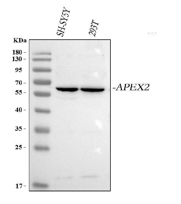
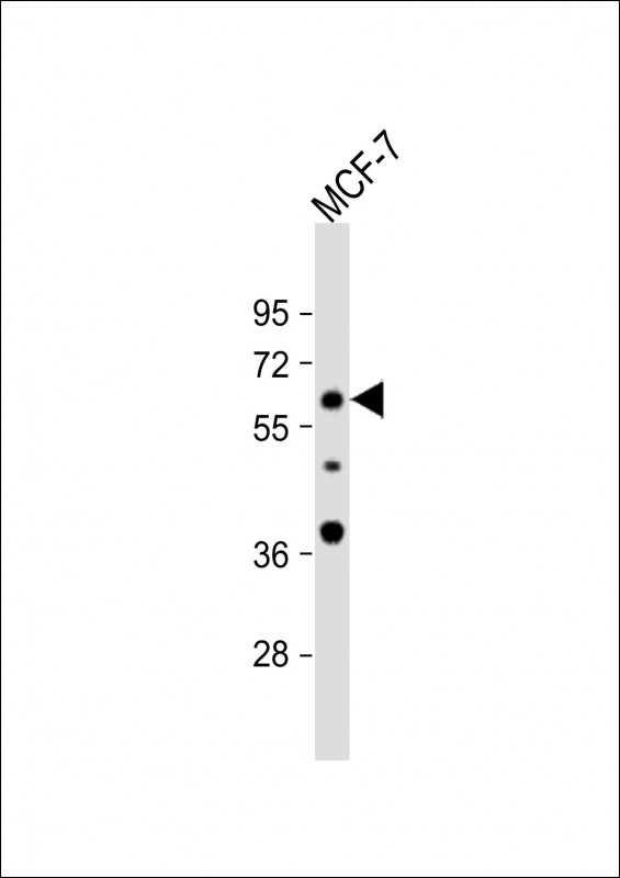
| Gene Name: | APEX2 |
| Uniprot: | Q9UBZ4 |
| Entrez: | 27301 |

| Belongs to: |
|---|
| DNA repair enzymes AP/ExoA family |

AP endonuclease 2; APE2; APEX nuclease (apurinic/apyrimidinic endonuclease) 2; APEX nuclease 2; APEXL2; Apurinic-apyrimidinic endonuclease 2; DNA-(apurinic or apyrimidinic site) lyase 2; DNA-apurinic or apyrimidinic site lyase 2; EC 3.1; EC 4.2.99.18; XTH2
Mass (kDA):
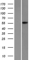
57.401 kDA

| Human | |
|---|---|
| Location: | Xp11.21 |
| Sequence: | X; NC_000023.11 (55000363..55009057) |
Highly expressed in brain and kidney. Weakly expressed in the fetal brain.
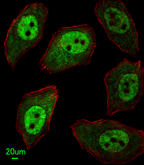
Nucleus. Cytoplasm. Mitochondrion. Together with PCNA, is redistributed in discrete nuclear foci in presence of oxidative DNA damaging agents.


PMID: 11376153 by Tsuchimoto D., et al. Human APE2 protein is mostly localized in the nuclei and to some extent in the mitochondria, while nuclear APE2 is partly associated with proliferating cell nuclear antigen.
PMID: 16687656 by Burkovics P., et al. Human Ape2 protein has a 3'-5' exonuclease activity that acts preferentially on mismatched base pairs.