This website uses cookies to ensure you get the best experience on our website.


- Table of Contents
Plan chromogenic APOC3 IHC on paraffin sections with the IHC-validated antibody (datasheet M01416). Compare cellular staining with plasma positivity and extracellular deposits, and use consistent fixation and controls to interpret the pattern (HPA tissue IHC; standard IHC practice).

Expected localisation, validated protocols, controls and antibodies — the at-a-glance facts below, then the full design guide.
| Expected localisation | Plasma and extracellular deposits (HPA tissue IHC) | |
| Staining pattern | Plasma/deposits; enterocytes and Paneth cells high, hepatocytes medium (HPA tissue IHC) | |
| Antigen retrieval | EDTA pH 8.0 HIER, heat-mediated (datasheet M01416) | |
| Positive control | Duodenum+2 more · see all | |
| Negative control | Adipose tissue+4 more · see all |
| Fixation | Keep fixation consistent across paraffin sections. (standard IHC practice; not target-specific) | |
| Caveat | Secreted APOC3 may stain away from its source cells (UniProt; HPA tissue IHC) | |
| Regulation | Staining regulation is not established (UniProt; HPA tissue IHC) | |
| Isoform / epitope | No isoforms annotated; mature chain spans residues 21–99 (UniProt) |
The catalog antibody protocol uses EDTA pH 8.0 heat retrieval (datasheet M01416). The published APOC3 IHC protocols below cover liver, ovarian, aortic sinus and arterial arch sections (PMCs: 13113833, 10493025, 11383354, 13080510).
| Sample | Paraffin-embedded human melanoma tissue; fixative not specified (datasheet M01416) |
| Fixation | Image fixative and duration unreported (datasheet M01416); verify before use. |
| Sectioning | 4–5 µm sections on charged slides (standard) |
| Deparaffinisation | Xylene, graded ethanol series to water (standard) |
| Antigen retrieval | Heat retrieval: EDTA pH 8.0 (datasheet M01416); 20 min, 95–100 °C (standard) |
| Peroxidase block | 3% H2O2, 10 min, room temperature (standard) |
| Blocking | 10% goat serum (datasheet M01416) |
| Primary antibody | Rabbit monoclonal (clone 17A47) anti-APOC3, 1:50-1:200 (datasheet M01416) |
| Primary incubation | Overnight at 4 °C (datasheet M01416) |
| Detection | HRP-conjugated secondary, DAB chromogen (datasheet M01416) |
| Counterstain | Hematoxylin, blue, dehydrate and mount (standard) |
| Expected result | APOC3-positive staining in enterocytes of duodenum (HPA tissue IHC: High). HPA tissue profile: Plasma positivity and extracellular deposits. Expressed in hepatocytes and small intestines. No signal in the no-primary control. |
APOC3 is a secreted protein with no transmembrane segment (UniProt P02656 topology). In paraffin section IHC, expect staining associated with hepatocytes and intestinal epithelial cells, plus plasma positivity or extracellular deposits (HPA tissue IHC profile). HPA reports high staining in duodenal enterocytes and small intestinal Paneth cells, and medium staining in hepatocytes (HPA tissue IHC). Tissue IHC reliability is Supported (HPA tissue IHC).
| Enterocytes or Paneth cells stain strongly; hepatocytes stain more moderately, with plasma or extracellular staining (HPA tissue IHC). | This matches the reported tissue pattern: high in duodenal enterocytes and small intestinal Paneth cells, medium in hepatocytes (HPA tissue IHC). Plasma positivity and extracellular deposits also fit a secreted protein (HPA tissue IHC profile; UniProt P02656 subcellular location). Score cell-associated and extracellular signal separately when comparing sections (general IHC practice). |
| Only nuclei stain in a paraffin section, without the expected cellular or extracellular pattern (HPA tissue IHC profile). | Treat isolated nuclear IHC as discordant with the reported plasma and extracellular tissue pattern and secreted location (HPA tissue IHC profile; UniProt P02656 subcellular location). HPA separately reports nucleoplasm, nucleoli and cell junctions in ICC-IF, so nuclear staining alone cannot prove an artefact; verify the IHC signal with controls and an independently validated antibody (HPA subcellular ICC-IF; general IHC practice). |
| Strong chromogen appears in adipocytes, adrenal glandular cells or respiratory epithelium (HPA tissue IHC negatives). | These listed cell types were not detected in HPA tissue IHC (HPA tissue IHC). If they stain while appropriate positive tissue also stains, consider antibody cross-reactivity or endogenous detection activity as general IHC possibilities. Check a no-primary control and compare staining with the expected positive cell types (general IHC practice). |
| Broad haze covers many compartments and obscures cell boundaries or extracellular deposits (general IHC practice). | The result cannot be scored confidently against HPA’s cell-specific and extracellular profile (HPA tissue IHC profile; general IHC practice). Possible general IHC causes include insufficient blocking, concentrated primary antibody, inadequate washing or detection background. A no-primary control can identify signal arising from the detection system, but cannot establish antibody specificity (general IHC practice). |
| No signal appears in duodenal enterocytes, small intestinal Paneth cells or hepatocytes (HPA tissue IHC positives). | A blank result in these reported positive cells makes a technical false negative plausible, especially when all tissue compartments are blank (HPA tissue IHC; general IHC practice). Check tissue integrity, primary antibody use, retrieval conditions and detection controls before concluding APOC3 is absent (general IHC practice). HPA’s Supported tissue rating does not guarantee staining in every specimen (HPA tissue IHC reliability). |
| Secretion and processing (UniProt P02656 topology and processing) | APOC3 has a signal peptide at residues 1–20, a mature chain at 21–99, and no transmembrane segment (UniProt P02656). Secreted material can therefore contribute to plasma or extracellular staining alongside positive cells (UniProt P02656 subcellular location; HPA tissue IHC profile). |
| Tissue source versus deposited protein (UniProt P02656; HPA tissue IHC) | UniProt lists liver tissue specificity, while HPA reports positive intestinal cells and extracellular deposits (UniProt P02656 tissue specificity; HPA tissue IHC). Tissue staining alone does not establish which local cell made extracellular APOC3 (HPA tissue IHC reliability note). |
| Tissue IHC antibody evidence (HPA antibodies) | HPA073918 has Supported IHC validation; HPA065365 has Approved ICC validation but no listed IHC status (HPA antibodies). Keep evidence for the two applications separate when interpreting a paraffin section (HPA antibodies). |
| IF/ICC Q: Should nuclear IF define a positive IHC result? (HPA subcellular ICC-IF) | No. HPA reports mainly nucleoplasmic ICC-IF signal, with nucleoli and cell junctions also observed, whereas tissue IHC reports plasma positivity and extracellular deposits (HPA subcellular ICC-IF; HPA tissue IHC profile). Interpret each application against its own evidence (HPA antibodies). |
| Target-specific fixation effects are not established by the supplied assay evidence. Verify with a matched IHC source before attributing a result to fixation. | Target-specific fixation effects are not established by the supplied assay evidence. Verify with a matched IHC source before attributing a result to fixation. |
| Situation | Likely cause | Next action |
|---|---|---|
| Expected positive cells are blank, but the slide has intact tissue (HPA tissue IHC positives; general IHC practice). | Primary antibody, retrieval or detection may have failed; the pattern alone cannot identify which step failed (general IHC practice). | Confirm the antibody is IHC validated, then inspect the positive control, retrieval record and detection controls; repeat with the validated IHC procedure if a step failed (HPA antibodies; general IHC practice). |
| Nuclear-only IHC appears in otherwise negative tissue (HPA tissue IHC profile). | The IHC pattern conflicts with HPA’s reported plasma and extracellular tissue pattern, although nuclear ICC-IF is separately reported (HPA tissue IHC profile; HPA subcellular ICC-IF). | Review morphology and no-primary staining; confirm with an independently validated IHC antibody before calling the nuclear pattern APOC3-specific (general IHC practice; HPA antibodies). |
| Chromogen appears in HPA-negative cell types across several tissues (HPA tissue IHC negatives). | Cross-reactivity or endogenous detection activity are possible general IHC explanations; HPA lists those cell types as not detected (HPA tissue IHC; general IHC practice). | Compare a no-primary control and known positive tissue; address detection background and reassess antibody specificity if staining persists only with primary antibody (general IHC practice; HPA tissue IHC positives). |
| Diffuse background obscures enterocytes, Paneth cells or hepatocytes (HPA tissue IHC positives). | Blocking, primary concentration, washing or detection background may be contributing; appearance alone cannot distinguish them (general IHC practice). | Inspect the no-primary control, then optimize blocking, dilution and washes one variable at a time while retaining positive tissue for comparison (general IHC practice; HPA tissue IHC positives). |
| Extracellular signal dominates, leaving cell-associated staining hard to score (HPA tissue IHC profile). | Plasma positivity and extracellular deposits are part of the reported profile of secreted APOC3; their presence alone does not identify the local producing cell (HPA tissue IHC profile; UniProt P02656 subcellular location). | Record extracellular and cell-associated staining separately, and base cell-type calls on identifiable enterocytes, Paneth cells or hepatocytes (general IHC practice; HPA tissue IHC positives). |
| A result differs between paraffin IHC and ICC-IF (HPA tissue IHC; HPA subcellular ICC-IF). | HPA reports extracellular tissue IHC and mainly nucleoplasmic ICC-IF; the supplied antibody validations also cover different applications (HPA tissue IHC profile; HPA subcellular ICC-IF; HPA antibodies). | Use the tissue IHC profile and IHC-supported antibody evidence to judge the paraffin section; document the ICC-IF result as a separate observation (HPA tissue IHC; HPA antibodies). |


Comprehensive Human Protein Atlas IHC scoring per tissue (reliability: Supported — No tissue staining or RNA expression data available for comparison. Secreted protein, tissue location of RNA and protein is expected to differ.). Rows are taken directly from the HPA tissue chart — click any row's HPA link to view the source.
| Tissue | Cell type | Level | Evidence | Source |
|---|---|---|---|---|
| Duodenum | Enterocytes | High | Protein (IHC) | HPA → |
| Small intestine | Paneth cells | High | Protein (IHC) | HPA → |
| Liver | Hepatocytes | Medium | Protein (IHC) | HPA → |
| Tissue | Cell type | Level | Evidence | Source |
|---|---|---|---|---|
| Adipose tissue | Adipocytes | Not detected | Protein (IHC) | HPA → |
| Adrenal gland | Glandular cells | Not detected | Protein (IHC) | HPA → |
| Appendix | Endocrine cells | Not detected | Protein (IHC) | HPA → |
| Bone marrow | Hematopoietic cells | Not detected | Protein (IHC) | HPA → |
| Breast | Adipocytes | Not detected | Protein (IHC) | HPA → |
Troubleshoot APOC3 staining in paraffin sections by checking retrieval, compartment, controls and scoring against the available tissue evidence.
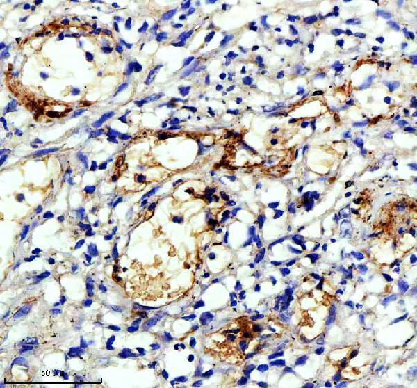
One human-reactive APOC3 antibody has a real IHC image from a paraffin-embedded human melanoma section (M01416 image caption; catalog: M01416 reactivity). No IF image is supplied (catalog: M01416).
M01416 is listed for human IHC and WB (catalog: M01416 applications and reactivity). Its IHC image shows staining in a paraffin-embedded human melanoma section following EDTA pH 8 retrieval and DAB detection (M01416 image caption).
Which to pick: For tissue IHC, choose M01416: it is a rabbit monoclonal with a human paraffin-section IHC example and a listed dilution of 1:50–1:200 (catalog: M01416; M01416 image caption). No SKU in this payload is listed for IF/ICC or nonhuman reactivity (catalog: M01416 applications and reactivity). The M01416 caption does not report the fixative, so its paraffin-section image does not establish tissue processing (M01416 image caption). The selected M01416 tissue-IHC caption documents paraffin sections, but does not specify the fixative (selected-SKU IHC image M01416).