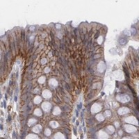
ARF6 / ADP-ribosylation factor 6 · IHC design guide

Design Immunohistochemistry for ARF6

Plan chromogenic ARF6 IHC in paraffin sections around the cytoplasmic and membranous pattern seen in most tissues (HPA tissue IHC). Start with catalog antibody PB9987 at 0.5–1 μg/mL (datasheet: PB9987), and account for the absence of detectable cardiomyocyte staining (HPA tissue IHC).

Evidence assembled Oct 2026 · For research use; verify linked source records and product datasheet before use
Immunohistochemistry protocol sheet for ARF6 (IHC for ARF6): expected localisation Cytoplasmic and membranous in most tissues (HPA tissue IHC), antibody PB9987, validated IHC image, and IHC protocol steps
Printable ARF6 IHC protocol sheet — expected localisation Cytoplasmic and membranous in most tissues (HPA tissue IHC), antibody PB9987, controls and protocol steps. Open the full ARF6 IHC guide →

ARF6 Immunohistochemistry Experimental Design Guide

Expected localisation, validated protocols, controls and antibodies — the at-a-glance facts below, then the full design guide.

Must know before staining
Expected localisation Cytoplasmic and membranous in most tissues (HPA tissue IHC)
Staining pattern Most tissues show cytoplasmic and membranous staining (HPA tissue IHC)
Antigen retrieval EDTA pH 8.0 HIER, heat-mediated (datasheet PB9987)
Positive control ⓘ Adrenal gland+4 more · see all
Negative control ⓘ Bone marrow+4 more · see all
Important caveats
Reasons your staining may differ from the expected pattern.
Fixation Keep fixation conditions consistent across sections. (standard IHC practice; not target-specific)
Caveat Cardiomyocytes were unstained despite reported higher heart levels (HPA tissue IHC; UniProt)
Regulation No stimulus-dependent regulation reported (UniProt)
Isoform / epitope No isoforms annotated; chain 2–175 has no extracellular domain (UniProt)
Section 1

Recommended ARF6 IHC & IF Protocols

The catalog antibody protocol uses EDTA pH 8.0 retrieval (datasheet PB9987). The published IHC options cover prostate, zebrafish, mouse allograft, and breast specimens (PMC4499334; PMC10054615; PMC4336811).

Recommended immunohistochemistry (IHC-P) protocol parameters
SampleParaffin-embedded human melanoma tissue; fixative not specified (datasheet PB9987)
FixationImage fixative and duration unreported (datasheet PB9987); verify before use.
Sectioning4–5 µm sections on charged slides (standard)
DeparaffinisationXylene, graded ethanol series to water (standard)
Antigen retrievalHeat retrieval: EDTA pH 8.0 (datasheet PB9987); 20 min, 95–100 °C (standard)
Peroxidase block3% H2O2, 10 min, room temperature (standard)
Blocking10% goat serum (datasheet PB9987)
Primary antibodyRabbit anti-ARF6, 0.5-1μg/ml (datasheet PB9987)
Primary incubationOvernight at 4 °C (datasheet PB9987)
DetectionStreptavidin-biotin complex (SABC), DAB chromogen (datasheet PB9987)
CounterstainHematoxylin, blue, dehydrate and mount (standard)
Expected resultARF6-positive staining in glandular cells of adrenal gland (HPA tissue IHC: Medium). HPA tissue profile: Cytoplasmic and membranous expression in most tissues. No signal in the no-primary control.
💡Decision noteStart with EDTA pH 8.0 HIER for the catalog antibody (datasheet PB9987); use the cited retrieval conditions when reproducing a published protocol (PMC10054615; PMC4336811).
Section 2

What Is the Expected ARF6 Staining Pattern?

ARF6 is found in the cytosol and at cell, endosomal, and recycling endosomal membranes; it has no transmembrane segment (UniProt P62330). In tissue IHC, expect cytoplasmic and membranous staining in many cell types, including medium staining in glandular cells of colon, breast, and appendix (HPA tissue IHC). HPA rates its tissue staining Approved but reports low consistency with RNA expression (HPA tissue IHC).

What am I looking at on my slide?
Cytoplasmic staining with a membranous component in glandular cells (HPA tissue IHC).This fits the reported tissue pattern and ARF6 localisation (HPA tissue IHC; UniProt P62330). Compare cells within the same section: the strongest interpretable result is staining in the expected compartment of the expected cell type, with a clean background (general IHC practice).
Predominantly nuclear staining, without a credible cytoplasmic or membranous component (HPA tissue IHC; UniProt P62330).A nuclear-only pattern is outside the supplied ARF6 localisation evidence (UniProt P62330; HPA tissue IHC). Check the no-primary control, counterstain, and detection conditions before interpreting it as target signal (general IHC practice).
Strong staining of bone marrow hematopoietic cells or heart cardiomyocytes (HPA tissue IHC).HPA reports ARF6 as not detected in those cells by tissue IHC (HPA tissue IHC). Review cell identification and controls for cross-reactivity or endogenous detection activity (general IHC practice); HPA's low antibody–RNA consistency limits a definitive negative call (HPA tissue IHC).
Diffuse color across cells and extracellular spaces, obscuring cell borders (general IHC practice).This distribution cannot establish ARF6's reported cytoplasmic and membranous localisation (HPA tissue IHC; UniProt P62330). Examine the no-primary control and background suppression before scoring cells as positive (general IHC practice).
No staining in colon glandular cells despite an intact section (HPA tissue IHC).HPA records medium staining in this cell population, making it a useful comparison tissue (HPA tissue IHC). A blank result warrants checks of antibody, retrieval, detection, and control performance; it does not alone establish absence of ARF6 (general IHC practice).
💡Expected ARF6 appearanceCall a convincing positive when identifiable glandular cells show medium cytoplasmic and membranous staining, as reported for colon (HPA tissue IHC); nuclear-only color or diffuse, cell-independent deposit needs control review (UniProt P62330; general IHC practice).
How each factor affects the staining
Compartment and cell identityARF6's cytosolic and membrane associations support a combined pattern, while HPA describes cytoplasmic and membranous tissue staining (UniProt P62330; HPA tissue IHC). Score the named cell population separately from surrounding tissue (general IHC practice).
Choice of comparison tissueColon, breast, and appendix glandular cells have medium HPA staining; bone marrow hematopoietic cells are listed as not detected (HPA tissue IHC). These are observed IHC comparisons, not absolute positive or negative standards (HPA tissue IHC).
Evidence and antibody validationHPA lists CAB002778 as IHC Approved, while its tissue profile notes low consistency between antibody staining and RNA expression (HPA antibodies; HPA tissue IHC). Treat unexpected staining as a finding to validate with controls (general IHC practice).
Topology and processingUniProt reports no transmembrane segment, signal peptide, propeptide, or annotated isoforms; its listed chain spans residues 2–175 (UniProt P62330). Those annotations do not identify this antibody's epitope or predict its retrieval requirements (UniProt P62330).
IF/ICC Q&A: what localisation should I expect?HPA supports a cytosolic main location in ICC-IF; UniProt also describes membrane-associated pools (HPA subcellular; UniProt P62330). Use the separate IF/ICC guide for imaging decisions; tissue IHC intensity should not be assumed to match ICC-IF (HPA tissue IHC; HPA subcellular).
Why is my staining missing, weak or wrong?
SituationLikely causeNext action
No signal in an HPA medium-staining glandular tissue, such as colon (HPA tissue IHC).The run may have failed at retrieval, primary incubation, or detection (general IHC practice).Check section identity and a positive control, then review the validated antibody's IHC-P instructions and each detection step (general IHC practice).
Color appears in the no-primary control (general IHC practice).Endogenous detection activity or nonspecific reagent deposition may contribute (general IHC practice).Address the detection system's background controls and repeat the comparison before assigning ARF6 positivity (general IHC practice).
Broad haze hides the reported cytoplasmic and membranous pattern (HPA tissue IHC).Excess background can obscure cell boundaries and compartment assignment (general IHC practice).Review blocking, primary antibody concentration, washes, and chromogen development against controls (general IHC practice).
Only nuclei stain in an otherwise readable section (UniProt P62330; HPA tissue IHC).The pattern conflicts with the supplied cytosolic and membrane localisation (UniProt P62330; HPA tissue IHC).Check counterstain and no-primary control; withhold a positive call until cytoplasmic or membranous signal is credible (general IHC practice).
Unexpected strong signal appears in HPA not-detected cell populations (HPA tissue IHC).Cell misidentification, cross-reactivity, or endogenous detection activity are possibilities (general IHC practice).Confirm morphology and controls, and report the HPA discrepancy rather than treating its not-detected category as proof of absence (HPA tissue IHC; general IHC practice).
Staining varies markedly between cells in one section (general IHC practice).Cell populations can differ in HPA staining level; uneven assay performance is another possibility (HPA tissue IHC; general IHC practice).Score identified cell types separately, compare the section's control staining, and avoid pooling unrelated compartments into one intensity score (general IHC practice).

Sample controls for ARF6 IHC & IF

🧪Run colon first: glandular cells should show ARF6 staining (HPA: Colon glandular cells, Medium). Use parathyroid gland as the negative tissue; its glandular cells are listed as not detected (HPA: Parathyroid gland glandular cells, Not detected). On the colon slide, assess neighboring nonglandular cells as candidate internal negatives only if independently verified; they should retain counterstain without specific chromogenic signal (standard IHC practice).
Positive control tissue: Adrenal gland (Glandular cells, HPA Medium)
Negative control tissue: Bone marrow (HPA Not detected)
ICC-IF cell lines (HPA subcellular resource): HPA ICC-IF images show ARF6 in A-431, U-251MG, U2OS, with annotated localisation: Cytosol (supported) (HPA subcellular).
Technical controls: Include a no-primary, secondary-only control, an isotype-matched rabbit IgG control, and ARF6 knockout tissue or a validated immunizing-peptide competition control (catalog caption: rabbit primary; standard IHC practice). Quench endogenous peroxidase and, if using the caption’s streptavidin–biotin detection, control endogenous biotin in colon sections (catalog caption: SABC with DAB; standard IHC practice).
⚠️Feasibility: A target-specific fixation window or fixation effect is unreported in the supplied evidence, and the selected PB9987 paraffin-section caption does not state a fixative (PB9987 caption). Heat retrieval in EDTA at pH 8.0 was used for the catalog antibody’s paraffin-section example; whether retrieval is required has not been established here (PB9987 caption). The evidence does not establish that frozen sections or IF are easier for ARF6; assess colon-section endogenous biotin when using the caption’s biotin-based detection (PB9987 caption; standard IHC practice).

HPA tissue IHC evidence for ARF6

Comprehensive Human Protein Atlas IHC scoring per tissue (reliability: Approved — Low consistency between antibody staining and RNA expression data.). Rows are taken directly from the HPA tissue chart — click any row's HPA link to view the source.

Positive expression · recommended positive controls

TissueCell typeLevelEvidenceSource
Adrenal gland Glandular cells Medium Protein (IHC) HPA →
Appendix Glandular cells Medium Protein (IHC) HPA →
Breast Glandular cells Medium Protein (IHC) HPA →
Colon Glandular cells Medium Protein (IHC) HPA →
Duodenum Glandular cells Medium Protein (IHC) HPA →

Undetected expression · recommended negative controls

TissueCell typeLevelEvidenceSource
Bone marrow Hematopoietic cells Not detected Protein (IHC) HPA →
Heart muscle Cardiomyocytes Not detected Protein (IHC) HPA →
Parathyroid gland Glandular cells Not detected Protein (IHC) HPA →
Skeletal muscle Myocytes Not detected Protein (IHC) HPA →
Smooth muscle Smooth muscle cells Not detected Protein (IHC) HPA →
Section 3

Advanced ARF6 IHC Tips

Use the catalog antibody’s paraffin section workflow as the IHC starting point, then judge staining against ARF6’s expected cellular distribution (datasheet PB9987; UniProt P62330).

Which retrieval condition should I start with for weak ARF6 staining in paraffin sections?
Start with heat-mediated antigen retrieval in EDTA pH 8.0 for paraffin sections (datasheet PB9987). If staining remains weak, compare retrieval duration on adjacent sections while keeping antibody concentration and detection conditions constant; include a section processed without primary antibody to reveal background. The documented example used 10% goat serum blocking and 1 µg/mL primary antibody overnight at 4°C (datasheet PB9987). Evaluate both cytoplasmic and membranous staining, since both occur in tissue IHC (HPA: cytoplasmic and membranous expression in most tissues), and avoid treating increased diffuse staining alone as improved retrieval.
How should I assess whether fixation is masking the ARF6 epitope?
Target-specific sensitivity to fixation is unknown: the selected paraffin section caption does not report a fixative (datasheet PB9987). Compare matched sections prepared under your available fixation conditions, using the same EDTA pH 8.0 retrieval and detection workflow for each (datasheet PB9987). Record fixation duration and section handling, then compare signal in equivalent cell populations alongside morphology and the no-primary control; poor morphology can make apparent staining gains misleading. Do not infer fixation tolerance from ARF6’s membrane association or from tissue staining patterns (UniProt P62330: subcellular location; HPA: tissue IHC profile), because neither tests this antibody across fixation conditions.
Should ARF6 stain the cytoplasm, the cell membrane, or both?
Score cytoplasmic and membranous compartments separately: tissue IHC reports both in most tissues (HPA: tissue IHC profile), while the supported subcellular location in the cell imaging record is cytosol (HPA: subcellular summary). ARF6 is also annotated at the cell membrane and several endosomal membranes, without a transmembrane segment (UniProt P62330: subcellular location and topology). A peripheral rim or intracellular puncta may therefore be plausible, but a rim alone does not prove plasma membrane localisation in a thin section. Compare adjacent cells and section planes, and use an appropriate compartment marker if precise membrane assignment drives the conclusion.
Can I assign a membrane signal to a specific ARF6 isoform or epitope?
Do not assign a stained compartment to an isoform: the supplied record annotates 0 isoforms and no transmembrane segment for ARF6 (UniProt P62330: isoforms and topology). Membrane association is consistent with its annotated myristoylation requirement, but the catalog caption does not specify the antibody’s binding epitope (UniProt P62330: subcellular annotation; datasheet PB9987). Ask for epitope mapping or independent validation before making claims about recognition of a modified terminus. For IHC optimisation, compare the same cells across retrieval conditions and keep the EDTA pH 8.0 method as the reference (datasheet PB9987).
How can IF help check an ambiguous ARF6 IHC pattern?
Use IF as a separate localisation check, rather than assuming that the paraffin section IHC conditions transfer to it (datasheet PB9987: paraffin section IHC). Multiplex ARF6 with a marker for the cell type you expect to stain, and choose a fluorophore channel away from the specimen’s strongest autofluorescence; inspect unstained and single-label controls. If testing intracellular epitopes, compare gentle permeabilisation with an unpermeabilised condition, because ARF6 is annotated in cytosol and on the cytoplasmic face of membrane-associated compartments (UniProt P62330: subcellular location and topology). The reported cell imaging localisation is cytosol, providing a comparison point rather than validation of this catalog antibody in IF (HPA: subcellular summary).
What controls distinguish ARF6 staining from chromogenic background?
Run a no-primary control through the full secondary and chromogen workflow, and check whether pigment or reaction product persists without primary antibody. The selected IHC example used a biotinylated goat anti-rabbit secondary, streptavidin–biotin complex and DAB (datasheet PB9987); assess endogenous biotin and peroxidase contributions when using that detection scheme. Apply a peroxidase block as a general chromogenic IHC step, then compare control and test sections at the same development endpoint. If background obscures cells, review blocking, washes, primary concentration and edge staining before calling diffuse brown signal ARF6; the documented starting concentration is 1 µg/mL (datasheet PB9987).
How should I quantify ARF6 IHC when staining is both cytoplasmic and membranous? ⚠ ANSWER MARKED FOR VERIFICATION
Define the cell population and compartment before scoring, because the reported tissue pattern includes cytoplasmic and membranous expression (HPA: tissue IHC profile). For each compartment, record the percentage of positive cells and an intensity category, then calculate an H-score if intensity is reproducible across the staining run. Alternatively, report positive-cell density per mm² of viable tissue when cell counts are the primary endpoint. Normalise comparisons to the same tissue area or eligible cell count, apply identical chromogen development and image settings, and exclude folds, necrosis and section edges using rules set before reviewing group labels.
When is an apparent ARF6-positive IHC result biologically credible?
A credible result follows cell boundaries or cytoplasmic structures and is reproducible in equivalent cells on adjacent sections; cytoplasmic and membranous staining are reported in tissue IHC (HPA: tissue IHC profile). Interpret an isolated nuclear signal cautiously, because the supplied localisation annotations emphasise cytosol and membranes (UniProt P62330: subcellular location; HPA: subcellular summary). Reject patterns confined to section edges, folds or necrotic areas, and investigate staining retained in the no-primary control for endogenous enzyme or detection background. Treat a positive field as suggestive rather than definitive when antibody staining and RNA data disagree, since the tissue IHC record reports low consistency between them (HPA: reliability description).
Boster reagents

Best ARF6 / ADP-ribosylation factor 6 IHC Antibodies

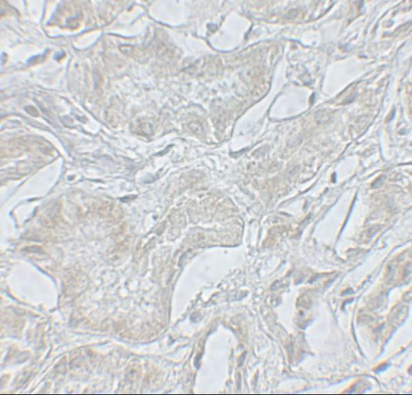
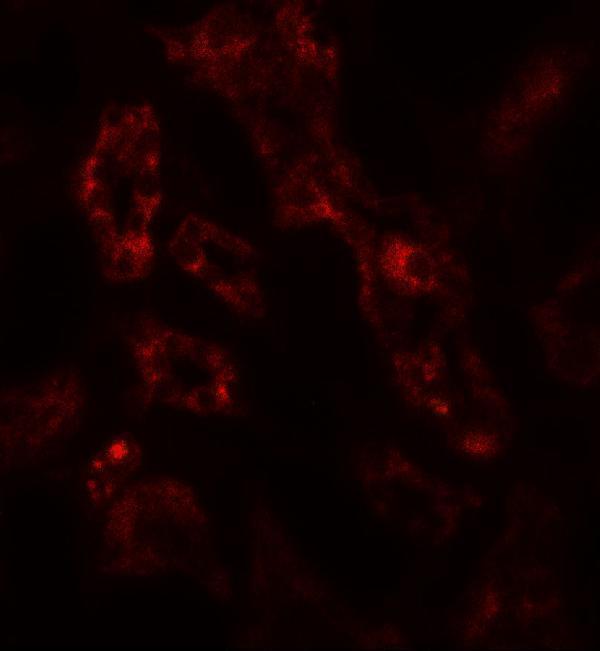
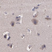
Anti-ARF6 antibodies have IHC images from human melanoma, human kidney, and rat cardiac muscle tissue (catalog IHC captions), plus IF images from human kidney tissue and A431 cells (catalog IF captions).

Real IHC data IHC analysis of ARF6 using anti-ARF6 antibody (PB9987). ARF6 was detected in paraffin-embedded section of human melanoma tissue. Heat mediated antigen retrieval was performed in EDTA buffer (pH8.0, epitope retrieval solution). The tissue section was blocked with 10% goat serum. The tissue section was then incubated with 1μg/ml rabbit anti-ARF6 Antibody (PB9987) overnight at 4°C. Biotinylated goat anti-rabbit IgG was used as secondary antibody and incubated for 30 minutes at 37°C. The tissue section was developed using Strepavidin-Biotin-Complex (SABC) (Catalog # SA1022) with DAB as the chromogen.
Anti-ARF6 Antibody ®
Cat # PB9987
Real IHC data Immunohistochemistry of ARF6 in human kidney tissue with ARF6 antibody at 5 μg/mL.
Anti-ADP-ribosylation factor 6 ARF6 Antibody
Cat # A00927
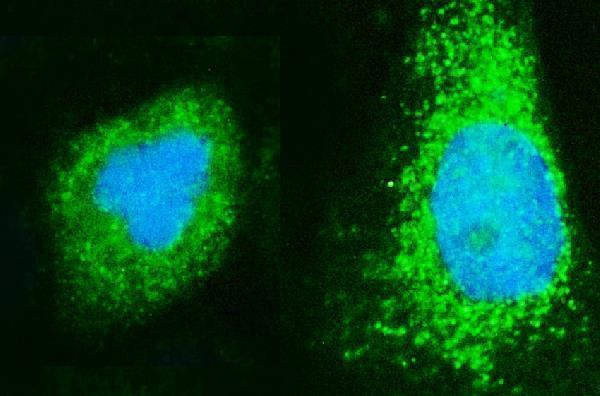
Real IF data IF analysis of ARF6 using anti-ARF6 antibody (A00927-2). ARF6 was detected in immunocytochemical section of A431 cells. Enzyme antigen retrieval was performed using IHC enzyme antigen retrieval reagent (AR0022) for 15 mins. The cells were blocked with 10% goat serum. And then incubated with 5μg/mL rabbit anti-ARF6 Antibody (A00927-2) overnight at 4°C. DyLight®488 Conjugated Goat Anti-Rabbit IgG (BA1127) was used as secondary antibody at 1:100 dilution and incubated for 30 minutes at 37°C. The section was counterstained with DAPI. Visualize using a fluorescence microscope and filter sets appropriate for the label used.
Anti-ARF6 Antibody ®
Cat # A00927-2

PB9987 shows IHC in paraffin sections of human melanoma and rat cardiac muscle (PB9987 IHC captions); A00927 shows IHC and IF in human kidney tissue (A00927 image captions). A00927-2 shows IF/ICC in A431 cells (A00927-2 IF caption).

Which to pick: For paraffin-section tissue IHC, choose PB9987 at 0.5–1 μg/mL (PB9987 catalog dilution); its IHC captions show paraffin sections, but do not report the fixative (PB9987 IHC captions). For IF/ICC in cells, choose A00927-2 at 5 μg/mL (A00927-2 IF caption); its application list includes IF and ICC, and its IHC dilution is unreported (A00927-2 catalog). For work involving human and rat tissue, PB9987 has IHC images in both species (PB9987 IHC captions); all three SKUs list human, mouse, and rat reactivity, while clonality is unreported (catalog).

Each figure is that product's own IHC / IF validation image from its datasheet.

References

  1. UniProt Consortium. UniProt entry P62330 (ARF6_HUMAN, ADP-ribosylation factor 6).
  2. Human Protein Atlas. ARF6 tissue IHC expression (reliability: Approved).
  3. Human Protein Atlas. ARF6 subcellular location (ICC-IF): Localized to the cytosol..
  4. Human Protein Atlas. ARF6 antibody validation summary (2 antibodies).
  5. Increased expression of ARF GTPases in prostate cancer tissue. SpringerPlus 2015 — PMC4499334.
  6. VGLL2-NCOA2 leverages developmental programs for pediatric sarcomagenesis. Cell reports 2023 — PMC10054615.
  7. ARF6 mediates nephrin tyrosine phosphorylation-induced podocyte cellular dynamics. PloS one 2017 — PMC5589247.
  8. lincRNA-RoR and miR-145 regulate invasion in triple-negative breast cancer via targeting ARF6. Molecular cancer research : MCR 2015 — PMC4336811.
  9. PubMed PMID:1993656 — UniProt-cited evidence.
  10. PubMed PMID:14659046 — UniProt-cited evidence.
  11. PubMed PMID:9653160 — UniProt-cited evidence.