This website uses cookies to ensure you get the best experience on our website.


- Table of Contents
Plan ARFGAP3 chromogenic IHC in paraffin sections using cytoplasmic staining in glandular cells as the tissue reference (HPA tissue IHC). Compare staining cautiously with RNA data because their reported consistency is low (HPA tissue IHC).

Expected localisation, validated protocols, controls and antibodies — the at-a-glance facts below, then the full design guide.
| Expected localisation | Cytoplasmic tissue staining (HPA tissue IHC) | |
| Staining pattern | Cytoplasmic staining, most abundant in glandular cells (HPA tissue IHC) | |
| Antigen retrieval | Citrate pH 6.0 HIER, 95–98 °C, 20 min (rule: cytoplasmic / membrane antigen) | |
| Positive control | Pancreas+4 more · see all | |
| Negative control | Adipose tissue+4 more · see all |
| Fixation | Keep fixation consistent across paraffin sections. (standard IHC practice; not target-specific) | |
| Caveat | Antibody staining has low consistency with RNA data (HPA tissue IHC) | |
| Regulation | Highest expression in endocrine glands and testis (UniProt) | |
| Isoform / epitope | 2 isoforms; check epitope coverage for each (UniProt) |
The catalog antibody’s IHC-P protocol is paired with one published ARFGAP3 anterior vaginal wall IHC study (PMC7774809).
| Sample | Paraffin-embedded mouse stomach tissue; fixative not specified (datasheet A06839-1) |
| Fixation | Image fixative and duration unreported (datasheet A06839-1); verify before use. |
| Sectioning | 4–5 µm sections on charged slides (standard) |
| Deparaffinisation | Xylene, graded ethanol series to water (standard) |
| Antigen retrieval | Heat-induced epitope retrieval in citrate buffer, pH 6.0, 20 min at 95–98 °C (standard rule: cytoplasmic / membrane antigen) |
| Peroxidase block | 3% H2O2, 10 min, room temperature (standard) |
| Blocking | 10% normal serum of the secondary host, 30 min, room temperature (standard) |
| Primary antibody | Rabbit anti-ARFGAP3, 1:50-1:200 (datasheet A06839-1) |
| Primary incubation | Overnight at 4 °C (standard) |
| Detection | HRP-polymer secondary, DAB chromogen 5–10 min (standard) |
| Counterstain | Hematoxylin, blue, dehydrate and mount (standard) |
| Expected result | ARFGAP3-positive staining in exocrine glandular cells of pancreas (HPA tissue IHC: Medium). HPA tissue profile: Cytoplasmic expression in several different tissue types, most abundant in glandular cells. No signal in the no-primary control. |
ARFGAP3 should appear primarily in the cytoplasm, with a Golgi component and possible peripheral puncta (HPA subcellular; UniProt Q9NP61 localization). Glandular cells in several tissues show medium IHC staining, including pancreatic exocrine cells and prostate glandular cells (HPA tissue IHC). ARFGAP3 has no transmembrane segment (UniProt Q9NP61 topology). The tissue IHC profile is Approved, although antibody staining and RNA expression have low consistency (HPA tissue IHC).
| Cytoplasmic staining in pancreatic exocrine glandular cells, with a concentrated perinuclear component. | This fits medium staining in those cells (HPA tissue IHC) and the cytosolic and Golgi locations (HPA subcellular). A Golgi-like concentration may be easier to judge in IF; chromogenic IHC can show the broader cytoplasmic pattern without resolving individual structures (general IHC practice). |
| Predominantly nuclear staining, with little cytoplasmic signal in glandular cells. | A nuclear-dominant pattern conflicts with the reported cytosol and Golgi locations (HPA subcellular; UniProt Q9NP61 localization). Treat it as suspect and compare the antibody-stained section with its negative control before assigning ARFGAP3 localization (general IHC practice). |
| Strong staining in adipocytes while the expected glandular-cell pattern is absent. | Adipocytes were not detected in the HPA tissue IHC profile, whereas several glandular-cell populations stained at medium levels (HPA tissue IHC). Consider cross-reactivity or endogenous chromogenic activity; the adipocyte result does not establish that every cell in adipose tissue is negative (general IHC practice; HPA tissue IHC). |
| Broad, fairly even colour across cells and surrounding section, obscuring cell boundaries. | This is less consistent with a cell-associated cytoplasmic pattern (HPA tissue IHC; HPA subcellular). Background from detection reagents or insufficient blocking may obscure a real signal; assess the negative control and background before scoring cells (general IHC practice). |
| No staining in pancreatic exocrine or prostate glandular cells. | Both are medium-staining populations in the HPA tissue IHC profile (HPA tissue IHC). Check tissue integrity, retrieval, antibody and detection controls before calling the sample negative (general IHC practice). An HPA reference pattern is a comparator, not a guarantee for every specimen (HPA tissue IHC). |
| Cellular location and topology | ARFGAP3 is reported at the Golgi membrane, in cytoplasm and on peripheral puncta likely associated with the ER–Golgi intermediate compartment; it has no transmembrane segment (UniProt Q9NP61 localization and topology). Judge cytoplasmic signal in context rather than requiring a crisp membrane outline (general IHC practice). |
| Tissue and cell selection | Pancreatic exocrine, prostate, salivary, rectal, stomach and small-intestinal glandular cells show medium staining, as do placental decidual cells and seminal-vesicle glandular cells (HPA tissue IHC). Choose an annotated cell population as the positive comparator; adjacent cells need not stain equally (general IHC practice). |
| Strength of tissue evidence | The tissue IHC profile is Approved but notes low consistency between antibody staining and RNA expression; RNA specificity is low (HPA tissue IHC). Use the observed cell pattern as a reference and interpret an unexpected result with controls, without treating RNA abundance as an exact IHC intensity prediction (general IHC practice). |
| Antibody validation | HPA000638 is Approved for IHC and Supported for ICC (HPA antibodies). These application labels describe the supplied antibody validation; they do not validate every staining condition or resolve an unexpected result in a new specimen (HPA antibodies; general IHC practice). |
| IF/ICC Q: What location should fluorescence show? | A: Mainly cytosol, with an additional Golgi signal (HPA subcellular). HPA lists ICC-IF images in A-431, U-251MG and U2OS cells (HPA subcellular). IF/ICC conditions belong to its separate guide; this location summary does not specify an IF protocol. |
| Situation | Likely cause | Next action |
|---|---|---|
| The positive-comparator glandular cells have no visible signal. | The tissue may have inadequate antigen exposure, or the antibody or chromogenic detection step may have failed (general IHC practice). Pancreatic exocrine and prostate glandular cells are medium-staining HPA comparators (HPA tissue IHC). | Confirm the annotated cell population is present, inspect a positive control, and check the established retrieval and detection workflow (general IHC practice). Do not infer target-specific fixation sensitivity; none is supplied. |
| Nearly every structure develops diffuse chromogen. | Broad staining is difficult to reconcile with the reported cytoplasmic cell pattern (HPA tissue IHC; HPA subcellular). Detection background, incomplete blocking or inadequate washing can cause diffuse colour (general IHC practice). | Review the negative control, blocking, washing and detection steps; score ARFGAP3 only where cell-associated signal remains distinguishable from background (general IHC practice). |
| The main signal appears nuclear. | Neither HPA nor UniProt lists the nucleus as an ARFGAP3 location in the supplied records (HPA subcellular; UniProt Q9NP61 localization). Nuclear colour may reflect nonspecific staining or a detection artefact (general IHC practice). | Compare with the negative control and a positive tissue showing the expected cytoplasmic pattern before interpreting nuclear colour as ARFGAP3 (HPA tissue IHC; general IHC practice). |
| Unexpected cells stain strongly while annotated glandular cells are weak. | The HPA reference includes medium glandular-cell staining but also records cell populations with low or undetected staining (HPA tissue IHC). Cross-reactivity or endogenous detection activity is possible (general IHC practice). | Check the exact cell annotation, compare positive and negative controls, and assess endogenous chromogenic activity with the appropriate detection control (HPA tissue IHC; general IHC practice). |
| A positive section looks weaker than another tissue or run. | HPA assigns medium staining to several specified populations, not a fixed intensity for every specimen (HPA tissue IHC). Section quality and staining conditions can change apparent intensity (general IHC practice). | Compare equivalent cell populations under the same scoring criteria, then review section quality and run controls before changing the interpretation (general IHC practice). |
| Fine Golgi or peripheral puncta are hard to distinguish in chromogenic IHC. | UniProt reports Golgi membrane and peripheral punctate localization, while HPA reports a mainly cytosolic ICC-IF signal with an additional Golgi signal (UniProt Q9NP61 localization; HPA subcellular). Fine structures may be unresolved in tissue IHC (general IHC practice). | Assess the broader cytoplasmic glandular-cell pattern against the HPA tissue reference; use the separate IF/ICC guide if the subcellular distribution itself must be resolved (HPA tissue IHC; general IHC practice). |
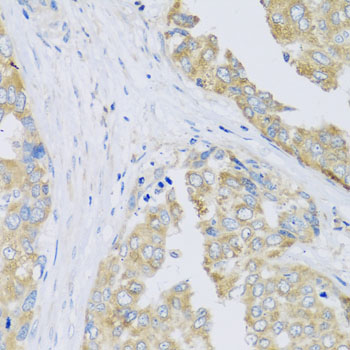
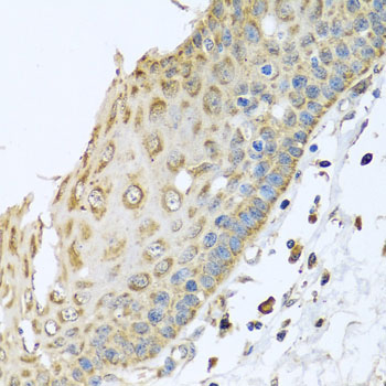
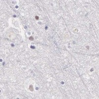


Comprehensive Human Protein Atlas IHC scoring per tissue (reliability: Approved — Low consistency between antibody staining and RNA expression data.). Rows are taken directly from the HPA tissue chart — click any row's HPA link to view the source.
| Tissue | Cell type | Level | Evidence | Source |
|---|---|---|---|---|
| Pancreas | Exocrine glandular cells | Medium | Protein (IHC) | HPA → |
| Placenta | Decidual cells | Medium | Protein (IHC) | HPA → |
| Prostate | Glandular cells | Medium | Protein (IHC) | HPA → |
| Rectum | Glandular cells | Medium | Protein (IHC) | HPA → |
| Salivary gland | Glandular cells | Medium | Protein (IHC) | HPA → |
| Tissue | Cell type | Level | Evidence | Source |
|---|---|---|---|---|
| Adipose tissue | Adipocytes | Not detected | Protein (IHC) | HPA → |
| Caudate | Glial cells | Not detected | Protein (IHC) | HPA → |
| Cerebellum | Cells in granular layer | Not detected | Protein (IHC) | HPA → |
| Cerebral cortex | Endothelial cells | Not detected | Protein (IHC) | HPA → |
| Cervix | Glandular cells | Not detected | Protein (IHC) | HPA → |
Troubleshoot ARFGAP3 staining in paraffin sections by checking retrieval, controls and whether the signal fits its reported cytosolic and Golgi localisation.
The anti-ARFGAP3 catalog antibody A06839-1 has real IHC images from paraffin-embedded mouse stomach and human liver cancer; no IF data are provided (A06839-1 image captions; catalog applications).
A06839-1 lists IHC and Human and Mouse reactivity (catalog applications and reactivity). Its IHC images show paraffin-embedded mouse stomach and human liver cancer, each at 1:100 (A06839-1 image captions).
Which to pick: Choose A06839-1 for tissue IHC in paraffin sections, as shown in its own mouse stomach and human liver cancer images (A06839-1 image captions). It is the listed choice for Human and Mouse samples (catalog reactivity). No IF/ICC choice is supported here because A06839-1 has no listed IF application or IF image; the fixative used for its paraffin sections is unreported (catalog applications; A06839-1 image captions).