ARHGAP18 / Rho GTPase-activating protein 18 · IHC design guide

Design Immunohistochemistry for ARHGAP18

Plan chromogenic ARHGAP18 IHC in paraffin sections around the predominantly cytoplasmic tissue pattern (HPA tissue IHC). The IHC-validated antibody has a starting dilution range of 1:100–1:300 (datasheet: A08418).

Evidence assembled Oct 2026 · For research use; verify linked source records and product datasheet before use
Immunohistochemistry protocol sheet for ARHGAP18 (IHC for ARHGAP18): expected localisation Cytoplasmic; membranous in a few tissues (HPA tissue IHC), antibody A08418, validated IHC image, and IHC protocol steps
Printable ARHGAP18 IHC protocol sheet — expected localisation Cytoplasmic; membranous in a few tissues (HPA tissue IHC), antibody A08418, controls and protocol steps. Open the full ARHGAP18 IHC guide →

ARHGAP18 Immunohistochemistry Experimental Design Guide

Expected localisation, validated protocols, controls and antibodies — the at-a-glance facts below, then the full design guide.

Must know before staining
Expected localisation Cytoplasmic; membranous in a few tissues (HPA tissue IHC)
Staining pattern General cytoplasmic staining with some membranous staining (HPA tissue IHC)
Antigen retrieval Citrate pH 6.0 HIER, 95–98 °C, 20 min (rule: cytoplasmic / membrane antigen)
Positive control ⓘ Adrenal gland+4 more · see all
Negative control ⓘ Adipose tissue+4 more · see all
Important caveats
Reasons your staining may differ from the expected pattern.
Fixation Keep formalin fixation consistent between sections. (standard IHC practice; not target-specific)
Caveat Staining varies by cell type; adipocytes are undetected (HPA tissue IHC)
Regulation No expression regulator annotated (UniProt)
Isoform / epitope 2 isoforms; no ectodomain; isoform epitopes unknown (UniProt)
Section 1

Recommended ARHGAP18 IHC & IF Protocols

The catalog antibody’s IHC-P protocol is paired with published ARHGAP18 protocols for human breast carcinoma (PMC5674094) and neuroendocrine tumor tissue (PMC3485222).

Recommended immunohistochemistry (IHC-P) protocol parameters
SampleParaffin-embedded human brain tissue; fixative not specified (datasheet A08418)
FixationImage fixative and duration unreported (datasheet A08418); verify before use.
Sectioning4–5 µm sections on charged slides (standard)
DeparaffinisationXylene, graded ethanol series to water (standard)
Antigen retrievalHeat-induced epitope retrieval in citrate buffer, pH 6.0, 20 min at 95–98 °C (standard rule: cytoplasmic / membrane antigen)
Peroxidase block3% H2O2, 10 min, room temperature (standard)
Blocking10% normal serum of the secondary host, 30 min, room temperature (standard)
Primary antibodyRabbit anti-ARHGAP18, 1:100-1:300 (datasheet A08418)
Primary incubationOvernight at 4 °C (standard)
DetectionHRP-polymer secondary, DAB chromogen 5–10 min (standard)
CounterstainHematoxylin, blue, dehydrate and mount (standard)
Expected resultARHGAP18-positive staining in glandular cells of adrenal gland (HPA tissue IHC: High). HPA tissue profile: General cytoplasmic expression with additional membranous expression in a few tissues. No signal in the no-primary control.
💡Decision noteStart with citrate pH 6.0 retrieval at 95–98 °C for 20 min (page antigen retrieval); the breast carcinoma protocol also specifies microwave retrieval (PMC5674094).
Section 2

What Is the Expected ARHGAP18 Staining Pattern?

ARHGAP18 is cytoplasmic and has no transmembrane segment (UniProt Q8N392). Expect predominantly cytoplasmic staining, with some membranous staining in a few tissues (HPA tissue IHC). High staining is reported in selected glandular, basal, ciliated and exocrine glandular cells (HPA tissue IHC). Treat the pattern as a guide: HPA rates its tissue IHC data Approved, with medium agreement between staining and RNA expression, pending external verification (HPA tissue IHC).

What am I looking at on my slide?
Strong cytoplasmic staining in pancreatic exocrine glandular cells or bronchial basal cells.This fits two reported high-staining cell populations (HPA tissue IHC) and the cytoplasmic location (UniProt Q8N392). Judge the signal in the named cells rather than treating every cell in the section as an expected positive.
Some cell-edge staining accompanies a predominantly cytoplasmic pattern.Additional membranous expression occurs in a few tissues (HPA tissue IHC). A cell-edge signal can therefore be compatible with the reported pattern, but the payload does not identify which tissues should show it (HPA tissue IHC); assess it alongside the cytoplasmic signal.
Nuclear staining dominates the section, with little cytoplasmic signal.That is discordant with cytoplasmic localization (UniProt Q8N392) and mainly cytosolic localization in ICC-IF (HPA subcellular). Nuclear speckles are an uncertain additional ICC-IF location (HPA subcellular), so they do not establish dominant nuclear staining as an expected IHC result.
Strong staining appears in adipocytes or parathyroid glandular cells.Both are reported as not detected (HPA tissue IHC). Consider antibody cross-reactivity or endogenous detection activity (standard IHC practice), especially if the signal persists where the primary antibody is omitted. A negative reference is a comparison, not proof of specificity by itself.
The whole section has diffuse colour, or a known-positive cell population has no visible signal.Diffuse colour that obscures cell boundaries limits compartment scoring; background from detection reagents or inadequate blocking is possible (standard IHC practice). No signal in pancreatic exocrine glandular cells, reported high by HPA, calls for a run-level check before interpreting a test section as negative (HPA tissue IHC; standard IHC practice).
💡Expected ARHGAP18 appearanceCall a result consistent when the named high-staining cells show a clear, predominantly cytoplasmic signal, with possible membranous staining in a few tissues (HPA tissue IHC; UniProt Q8N392); dominant nuclear signal or strong staining in HPA not-detected cell populations warrants investigation (HPA subcellular; HPA tissue IHC).
How each factor affects the staining
Choice of reference cellsPancreatic exocrine glandular cells and bronchial basal cells are high-staining references; adipocytes and parathyroid glandular cells are reported not detected (HPA tissue IHC). Score those cell populations specifically: a mixed tissue cannot be assigned one expected intensity from these entries.
Compartment used for scoringCytoplasm is the annotated location (UniProt Q8N392), and cytosol is the supported main ICC-IF location (HPA subcellular). Nuclear speckles and plasma membrane are uncertain additional ICC-IF locations (HPA subcellular); IHC reports additional membranous expression in a few tissues (HPA tissue IHC).
Strength of the reference evidenceThe tissue pattern is Approved, with medium consistency between antibody staining and RNA data and external verification pending (HPA tissue IHC). HPA031595 is Approved for IHC and Supported for ICC (HPA antibodies); these labels guide interpretation without establishing specificity in every specimen.
Isoforms and antibody coverageUniProt lists two ARHGAP18 isoforms (UniProt Q8N392). No antibody epitope or isoform-specific staining result is supplied, so these records cannot establish whether HPA031595 detects both isoforms (UniProt Q8N392; HPA antibodies).
Detection chemistryEndogenous activity or residual detection-reagent background can create chromogenic colour without target binding (standard IHC practice). A primary-omission control helps identify that contribution, while a matched positive reference helps assess whether the staining run worked (standard IHC practice).
Why is my staining missing, weak or wrong?
SituationLikely causeNext action
Expected high-staining reference cells have no signal.The staining run or detection step may have failed (standard IHC practice); pancreatic exocrine glandular cells are reported high (HPA tissue IHC).Check the positive reference and detection controls, then review the IHC-validated antibody's stated IHC-P workflow (standard IHC practice). Avoid calling test sections negative until the reference works.
Dominant nuclear colour appears across cells.The pattern conflicts with cytoplasmic localization (UniProt Q8N392). An uncertain nuclear-speckle observation from ICC-IF does not establish this IHC pattern (HPA subcellular).Compare the nuclear colour with a primary-omission control and inspect whether a distinct cytoplasmic signal remains (standard IHC practice).
Strong colour appears in adipocytes or parathyroid glandular cells.Those cells are reported not detected (HPA tissue IHC); cross-reactivity or endogenous detection activity is possible (standard IHC practice).Compare the same cell populations with primary-omission and positive-reference sections (standard IHC practice). Interpret persistent unexpected colour cautiously.
Diffuse colour obscures cell boundaries.Detection background or insufficient blocking may prevent reliable localization (standard IHC practice).Inspect the primary-omission control; review blocking, washing and detection steps before scoring cytoplasmic staining (standard IHC practice).
Only a cell-edge signal is visible.Some membranous expression is reported, but the expected dominant location is cytoplasmic (HPA tissue IHC; UniProt Q8N392).Check for cytoplasmic signal in a high-staining reference cell population and compare edge staining with the primary-omission control (HPA tissue IHC; standard IHC practice).
IF/ICC shows nuclear speckles or plasma-membrane signal.Both are uncertain additional locations; cytosol is the supported main ICC-IF location (HPA subcellular).Interpret them as secondary observations and compare with the cytosolic signal and appropriate imaging controls (HPA subcellular; standard IF practice). Use the separate IF/ICC guide for its workflow.

Sample controls for ARHGAP18 IHC & IF

🧪Run adrenal gland first: glandular cells should stain (HPA: High in adrenal gland glandular cells); use adipose tissue as the negative comparator (HPA: adipocytes Not detected). On the positive slide, assess morphologically distinct non-glandular cells for background-level staining, but do not designate them confirmed ARHGAP18-negative cells without independent validation.
Positive control tissue: Adrenal gland (Glandular cells, HPA High)
Negative control tissue: Adipose tissue (HPA Not detected)
ICC-IF cell lines (HPA subcellular resource): HPA ICC-IF images show ARHGAP18 in Hep-G2, U-251MG, U2OS, RPTEC/TERT1, with annotated localisation: Cytosol (supported) (HPA subcellular).
Technical controls: Include a no-primary, secondary-only control, a concentration-matched isotype control appropriate to the primary antibody’s host species and clonality, and a knockout sample or peptide-blocked section; the selected antibody’s human brain paraffin-section caption describes a peptide-blocked comparison (catalog caption: A08418). For chromogenic IHC in adrenal gland, quench endogenous peroxidase, block endogenous biotin if using avidin–biotin detection, and check endogenous pigment against the no-primary section.
⚠️Feasibility: No target-specific fixation window, fixation effect, or antigen-retrieval dependency is reported in the supplied evidence; optimize retrieval empirically for the IHC-validated antibody. The selected-SKU paraffin-section caption does not state a fixative (catalog caption: A08418), so fixation cannot be assumed. The evidence does not establish whether frozen sections or IF are easier; adrenal pigment can complicate interpretation of brown chromogen, so compare with the no-primary section. The selected A08418 tissue-IHC caption documents paraffin sections, but does not specify the fixative (selected-SKU IHC image A08418).

HPA tissue IHC evidence for ARHGAP18

Comprehensive Human Protein Atlas IHC scoring per tissue (reliability: Approved — Medium consistency between antibody staining and RNA expression data. Pending external verification.). Rows are taken directly from the HPA tissue chart — click any row's HPA link to view the source.

Positive expression · recommended positive controls

TissueCell typeLevelEvidenceSource
Adrenal gland Glandular cells High Protein (IHC) HPA →
Bronchus Basal cells High Protein (IHC) HPA →
Cervix Glandular cells High Protein (IHC) HPA →
Duodenum Glandular cells High Protein (IHC) HPA →
Epididymis Glandular cells High Protein (IHC) HPA →

Undetected expression · recommended negative controls

TissueCell typeLevelEvidenceSource
Adipose tissue Adipocytes Not detected Protein (IHC) HPA →
Cerebellum Cells in granular layer Not detected Protein (IHC) HPA →
Hippocampus Glial cells Not detected Protein (IHC) HPA →
Liver Cholangiocytes Not detected Protein (IHC) HPA →
Ovary Ovarian stroma cells Not detected Protein (IHC) HPA →
Section 3

Advanced ARHGAP18 IHC Tips

Troubleshoot ARHGAP18 staining in paraffin sections by checking retrieval, cellular distribution, antibody specificity, and cell type before scoring chromogenic signal.

What retrieval should I start with for weak ARHGAP18 staining in paraffin sections?
Start with citrate pH 6.0 heat-induced epitope retrieval at 95–98 °C for 20 min (page protocol). Keep section thickness, cooling, and detection conditions matched while comparing retrieval runs, so a change in staining can be attributed to retrieval (standard IHC practice). If staining remains weak, test a longer retrieval time on adjacent sections, watching for tissue damage or increased background (standard IHC practice). The A08418 image documents staining in paraffin-embedded human brain with peptide blocking, but its caption does not report a fixative or retrieval method (A08418 caption). Judge any improvement by cellular staining and a matched negative control, rather than overall DAB darkness (standard IHC practice).
Could fixation explain variable ARHGAP18 staining between paraffin blocks?
ARHGAP18-specific sensitivity to fixation is unknown from the supplied evidence; the A08418 paraffin-section caption does not state its fixative (A08418 caption). Record each block’s fixative and fixation duration before comparing staining intensity, because processing differences can alter antigen accessibility (standard IHC practice). Apply the same citrate pH 6.0, 95–98 °C, 20 min retrieval and detection run to adjacent sections when assessing block-to-block variation (page protocol; standard IHC practice). Include a consistently processed reference section in each run, and inspect tissue preservation alongside DAB intensity (standard IHC practice). Do not interpret differences between blocks as an ARHGAP18 fixation effect without a controlled comparison (standard IHC practice).
Should ARHGAP18 appear at the membrane or inside cells?
Expect predominantly cytoplasmic staining: UniProt assigns ARHGAP18 to the cytoplasm, and HPA supports cytosol as its main location (UniProt Q8N392; HPA subcellular). HPA also reports membranous expression in a few tissues, while its plasma-membrane subcellular assignment is uncertain (HPA tissue IHC; HPA subcellular). Because ARHGAP18 has no transmembrane segment, a crisp membrane-only DAB outline needs independent validation before being called its expected pattern (UniProt Q8N392 topology; standard IHC practice). Compare the same cell populations on adjacent sections with and without primary antibody, using a light counterstain to keep cell boundaries visible (standard IHC practice). Score cytoplasmic and membrane-associated signal separately if both persist under controlled staining conditions (standard IHC practice).
Can this IHC result distinguish ARHGAP18 isoforms or modified epitopes?
ARHGAP18 has 2 annotated isoforms, while the supplied antibody evidence does not map this reagent’s epitope to an isoform-specific sequence (UniProt Q8N392; A08418 caption). Its Rho-GAP domain spans residues 324–523, and annotated phosphorylation sites include residues 66, 69, 158, 263, and 610 (UniProt Q8N392). Do not assign a DAB pattern to one isoform or phosphorylation state without epitope mapping and a suitable independent control (standard IHC practice). The A08418 human-brain image includes a peptide-blocked comparison, which supports assessment of peptide-competitive staining for that antibody and image (A08418 caption). It does not establish isoform discrimination or modification specificity in other sections (A08418 caption; standard IHC practice).
How should I cross-check an IHC pattern with multiplex IF?
For a secondary IF check, pair ARHGAP18 with a marker of the expected cell population, such as a basal-cell marker when examining bronchial basal cells (HPA tissue IHC; standard IF practice). Choose a far-red fluorophore for a weak ARHGAP18 channel when shorter-wavelength tissue autofluorescence is prominent, and acquire single-label controls to check bleed-through (standard IF practice). ARHGAP18 is mainly cytosolic and lacks a transmembrane segment, so test mild permeabilisation for an intracellular epitope rather than assuming surface accessibility (HPA subcellular; UniProt Q8N392 topology; standard IF practice). Keep fixation and permeabilisation matched across IF controls, and compare cell-level distribution with the chromogenic section (standard IF practice). Treat IF signal as corroboration only after its own antibody and channel controls pass (standard IF practice).
What should I check when ARHGAP18 DAB staining is diffuse?
First inspect the no-primary control and a peptide-blocked section when using the A08418 antibody; its paraffin-brain caption documents a peptide-blocked comparison (A08418 caption; standard IHC practice). Block endogenous peroxidase before chromogenic detection, and shorten DAB development if background rises in the no-primary control (standard IHC practice). Check whether diffuse signal survives antibody dilution titration and washing, while keeping citrate pH 6.0 retrieval at 95–98 °C for 20 min fixed during that comparison (page protocol; standard IHC practice). ARHGAP18 is principally cytoplasmic, so uniform extracellular deposit or stain over tissue-free regions should trigger a technical review (UniProt Q8N392; standard IHC practice). Assess a high-staining reference population, such as pancreatic exocrine glandular cells, in the same run (HPA: High in pancreatic exocrine glandular cells; standard IHC practice).
How can I score ARHGAP18 while accounting for different cell mixtures? ⚠ ANSWER MARKED FOR VERIFICATION
Define the cell population and compartment before scoring: HPA reports high staining in pancreatic exocrine glandular cells and no detection in adipocytes (HPA tissue IHC). Within viable target cells, record percent positive and intensity grades 0–3, then calculate an H-score from 0–300 if intensity matters (standard IHC scoring). Normalise positive-cell counts to the number of eligible cells, or report positive-cell density per mm² of viable tissue when cell numbers cannot be counted reliably (standard IHC scoring). Exclude folds, necrosis, and section edges using the same prespecified rule across samples (standard IHC practice). Report cytoplasmic and membrane-associated scores separately because HPA describes general cytoplasmic staining with membranous staining in a few tissues (HPA tissue IHC).
Which ARHGAP18 staining patterns are convincing, and which suggest artefact?
A convincing result has cellular cytoplasmic staining in an appropriate population, consistent with UniProt’s cytoplasmic assignment and HPA’s supported cytosolic location (UniProt Q8N392; HPA subcellular). Compare suspected positives with relevant cell-type references: HPA reports high staining in bronchial basal cells but no detection in adipocytes (HPA tissue IHC). Treat nuclear-speckle-only staining cautiously because HPA marks that additional location uncertain; inspect edge-heavy signal, necrotic areas, and no-primary controls for artefact (HPA subcellular; standard IHC practice). Endogenous peroxidase can contribute DAB signal, so confirm the peroxidase block and review a matched negative control (standard IHC practice). HPA rates tissue staining Approved with medium RNA concordance and pending external verification, so retain independent antibody or peptide-block evidence when interpreting unexpected patterns (HPA tissue IHC; A08418 caption).
Boster reagents

Best ARHGAP18 / Rho GTPase-activating protein 18 IHC Antibodies

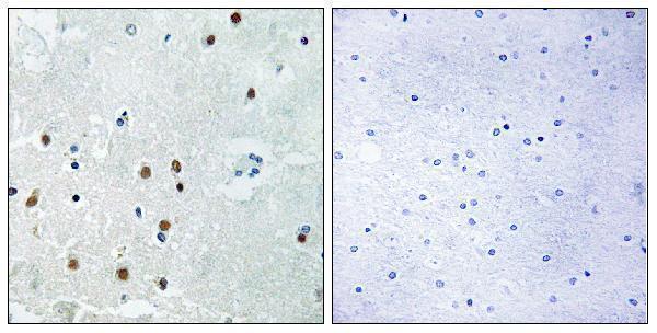
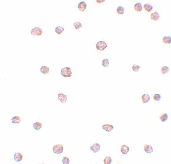
ARHGAP18 antibodies include paraffin-section IHC in human brain (A08418 image caption) and IF/ICC in 3T3 cells (A08418-1 image captions); listed reactivity spans human, mouse and rat (catalog).

Real IHC data Immunohistochemistryt analysis of paraffin-embedded human brain, using RHG18 Antibody. The lane on the right is blocked with the RHG18 peptide.
Anti-ARHGAP18 Antibody
Cat # A08418
Real IF data Immunocytochemistry of ARHGAP18 in 3T3 cells with ARHGAP18 antibody at 2.5 μg/mL.
Anti-ARHGAP18 Antibody
Cat # A08418-1

A08418 lists IHC and IF for human and mouse (catalog: applications/reactivity), with an IHC image of paraffin-embedded human brain (A08418 image caption). A08418-1 lists IF and ICC for human, mouse and rat (catalog: applications/reactivity), with both images showing 3T3 cells (A08418-1 image captions).

Which to pick: Choose A08418 for tissue IHC because IHC is listed and its image shows paraffin-embedded human brain (A08418: applications/image caption); the caption does not report the fixative (A08418 image caption). Choose A08418-1 for IF/ICC in 3T3 cells because both images use those cells (A08418-1 image captions). A08418-1 also lists rat reactivity, while A08418 lists human and mouse; neither entry reports a clone, so clonality cannot guide selection (catalog: reactivity/clone).

Each figure is that product's own IHC / IF validation image from its datasheet.

References

  1. UniProt Consortium. UniProt entry Q8N392 (RHG18_HUMAN, Rho GTPase-activating protein 18).
  2. Human Protein Atlas. ARHGAP18 tissue IHC expression (reliability: Approved).
  3. Human Protein Atlas. ARHGAP18 subcellular location (ICC-IF): Mainly localized to the cytosol. In addition localized to the nuclear speckles and plasma membrane..
  4. Human Protein Atlas. ARHGAP18 antibody validation summary (1 antibodies).
  5. Rho-GTPase activating-protein 18: a biomarker associated with good prognosis in invasive breast cancer. British journal of cancer 2017 — PMC5674094.
  6. The somatostatin analogue octreotide inhibits growth of small intestine neuroendocrine tumour cells. PloS one 2012 — PMC3485222.
  7. Thiopurines correct the effects of autophagy impairment on intestinal healing - a potential role for ARHGAP18/RhoA. Disease models & mechanisms 2021 — PMC8084572.
  8. Tenofovir Modulates Semaphorin 4D Signaling and Regulates Bone Homeostasis, Which Can Be Counteracted by Dipyridamole and Adenosine A2A Receptor. International journal of molecular sciences 2021 — PMC8583797.
  9. PubMed PMID:17974005 — UniProt-cited evidence.
  10. PubMed PMID:14574404 — UniProt-cited evidence.
  11. PubMed PMID:15489334 — UniProt-cited evidence.