ARHGEF12 / Rho guanine nucleotide exchange factor 12 · IHC design guide

Design Immunohistochemistry for ARHGEF12

Plan chromogenic ARHGEF12 IHC in paraffin sections using the catalog antibody’s documented conditions (datasheet A06802-3). Assess mainly cytoplasmic staining across cell types and interpret rare nuclear staining in context (HPA tissue IHC).

Evidence assembled Oct 2026 · For research use; verify linked source records and product datasheet before use
Immunohistochemistry protocol sheet for ARHGEF12 (IHC for ARHGEF12): expected localisation Mainly cytoplasmic, with rare nuclear staining (HPA tissue IHC), antibody A06802-3, validated IHC image, and IHC protocol steps
Printable ARHGEF12 IHC protocol sheet — expected localisation Mainly cytoplasmic, with rare nuclear staining (HPA tissue IHC), antibody A06802-3, controls and protocol steps. Open the full ARHGEF12 IHC guide →

ARHGEF12 Immunohistochemistry Experimental Design Guide

Expected localisation, validated protocols, controls and antibodies — the at-a-glance facts below, then the full design guide.

Must know before staining
Expected localisation Mainly cytoplasmic, with rare nuclear staining (HPA tissue IHC)
Staining pattern Several cell types show mainly cytoplasmic, rarely nuclear staining (HPA tissue IHC)
Antigen retrieval EDTA pH 8.0 HIER, heat-mediated (datasheet A06802-3)
Positive control ⓘ Adrenal gland+4 more · see all
Negative control ⓘ Adipose tissue+4 more · see all
Important caveats
Reasons your staining may differ from the expected pattern.
Fixation Keep fixation consistent across sections. (standard IHC practice; not target-specific)
Caveat Stimulation can shift ARHGEF12 toward the membrane (UniProt)
Regulation Ubiquitously expressed (UniProt)
Isoform / epitope 2 isoforms; check epitope coverage for each (UniProt)
Section 1

Recommended ARHGEF12 IHC & IF Protocols

The catalog antibody’s IHC-P protocol (datasheet A06802-3) appears alongside two published ARHGEF12 IHC methods (PMC12027205; PMC12374580).

Recommended immunohistochemistry (IHC-P) protocol parameters
SampleParaffin-embedded human colorectal adenocarcinoma tissue; fixative not specified (datasheet A06802-3)
FixationImage fixative and duration unreported (datasheet A06802-3); verify before use.
Sectioning4–5 µm sections on charged slides (standard)
DeparaffinisationXylene, graded ethanol series to water (standard)
Antigen retrievalHeat retrieval: EDTA pH 8.0 (datasheet A06802-3); 20 min, 95–100 °C (standard)
Peroxidase block3% H2O2, 10 min, room temperature (standard)
Blocking10% goat serum (datasheet A06802-3)
Primary antibodyRabbit anti-ARHGEF12, 2-5 μg/ml (datasheet A06802-3)
Primary incubationOvernight at 4 °C (datasheet A06802-3)
DetectionHRP-conjugated secondary, DAB chromogen (datasheet A06802-3)
CounterstainHematoxylin, blue, dehydrate and mount (standard)
Expected resultARHGEF12-positive staining in glandular cells of adrenal gland (HPA tissue IHC: Medium). HPA tissue profile: Mainly cytoplasmic expression in several different cell types, including rare cases of nuclear expression. No signal in the no-primary control.
💡Decision noteStart with heat-mediated EDTA pH 8.0 retrieval (datasheet A06802-3); sodium citrate retrieval is a published alternative (PMC12027205).
Section 2

What Is the Expected ARHGEF12 Staining Pattern?

ARHGEF12 staining should be mainly cytoplasmic, with occasional nuclear staining in tissue sections (HPA tissue IHC). Medium staining is reported in glandular cells of several organs, respiratory epithelial cells in bronchus, and endothelial cells in colon (HPA tissue IHC). HPA rates the tissue staining Approved, with medium consistency against RNA data (HPA tissue IHC). ARHGEF12 has no transmembrane segment; UniProt reports cytoplasmic localization and membrane recruitment upon stimulation (UniProt Q9NZN5 topology and subcellular location).

What am I looking at on my slide?
Cytoplasmic staining in glandular cells of adrenal gland, appendix, or duodenum; or in bronchial respiratory epithelium or colon endothelium.These are reported cell-specific patterns at Medium intensity (HPA tissue IHC). Interpret the stained cell population, rather than calling an entire tissue uniformly positive. HPA rates its tissue profile Approved, with medium RNA–staining consistency (HPA tissue IHC).
Predominantly strong nuclear staining, with little cytoplasmic signal.This differs from the mainly cytoplasmic tissue profile; HPA reports only rare nuclear expression (HPA tissue IHC). Check morphology, counterstain, and a no-primary control before assigning nuclear staining to ARHGEF12 (general IHC practice).
Strong staining in adipocytes, bone-marrow hematopoietic cells, or smooth muscle cells.These specific cell populations were Not detected by HPA tissue IHC (HPA tissue IHC). Consider cross-reactivity or endogenous chromogenic activity, especially if a no-primary control also develops color; check the actual cell type before judging the whole specimen (general IHC practice).
Diffuse brown haze across cells and extracellular spaces, obscuring boundaries.A widespread deposit does not resolve the cell-specific, mainly cytoplasmic profile (HPA tissue IHC). Assess no-primary staining, blocking, washing, antibody concentration, and chromogen development as general IHC troubleshooting steps; the supplied sources do not identify an ARHGEF12-specific cause.
No signal in glandular cells of adrenal gland or appendix while the counterstain and tissue morphology remain visible.Both are reported at Medium staining, making them useful positive comparators, though neither guarantees signal in every section (HPA tissue IHC). Check detection reagents and antibody conditions before interpreting absence as biology (general IHC practice).
💡Expected ARHGEF12 appearanceA convincing positive shows mainly cytoplasmic, approximately Medium staining in an HPA-reported cell population (HPA tissue IHC); diffuse deposit or dominant nuclear color warrants control-based review, since tissue nuclear expression is rare (HPA tissue IHC).
How each factor affects the staining
Compartment and topologyUniProt places ARHGEF12 in cytoplasm and at membranes after stimulation, without a transmembrane segment (UniProt Q9NZN5). Membrane-associated staining can therefore be plausible, but tissue IHC is described as mainly cytoplasmic (HPA tissue IHC). Do not require a crisp membrane rim in every paraffin section.
Cell-type variationHPA reports Medium staining in several glandular and endothelial populations, Low staining in lung alveolar cells, and Not detected staining in adipocytes (HPA tissue IHC). A negative field is interpretable only after identifying which cells are present; UniProt describes broad expression but does not predict equal staining in every cell (UniProt Q9NZN5 tissue specificity).
Antibody evidence and isoformsTwo listed antibodies have Approved IHC status; the supplied validation does not state Enhanced IHC evidence (HPA antibodies). UniProt lists two isoforms and reports isoform 2 in jejunum and testis (UniProt Q9NZN5). Without an epitope map, do not infer whether the IHC-validated antibody distinguishes isoforms.
IF/ICC: where should signal appear?HPA reports approved plasma-membrane and cytosol localization, with additional approved nucleoplasmic localization in ICC-IF (HPA subcellular). This answers the compartment question for IF/ICC; use its separate guide for that application. The tissue IHC profile remains mainly cytoplasmic, with rare nuclear expression (HPA tissue IHC).
Why is my staining missing, weak or wrong?
SituationLikely causeNext action
Reported positive cells appear blank.Detection or antibody conditions may have failed; HPA reports Medium staining in adrenal and appendix glandular cells (HPA tissue IHC).Run a reported positive tissue alongside the sample and check antibody dilution, retrieval conditions, detection reagents, and chromogen development (general IHC practice). No target-specific fixation sensitivity is reported in the supplied sources.
Brown signal appears even without primary antibody.The result points to endogenous detection activity or a detection-reagent background source (general IHC practice).Review endogenous-enzyme quenching, blocking, and detection controls before scoring ARHGEF12. Compare cell boundaries with the counterstain; HPA describes mainly cytoplasmic tissue staining (HPA tissue IHC).
Signal is diffuse throughout the section.Excess antibody, insufficient washing, or prolonged chromogen development can obscure specific staining (general IHC practice).Adjust those general IHC conditions and compare with a no-primary control and an HPA-reported positive cell population (HPA tissue IHC). Score only cell-associated signal with interpretable morphology.
Nuclear staining dominates the reported positive cells.This is atypical for the mainly cytoplasmic tissue pattern, although rare nuclear expression is reported (HPA tissue IHC).Check that the counterstain is not being read as chromogen, then compare no-primary and positive-control sections (general IHC practice). Do not discard isolated nuclear staining solely because it differs from the dominant pattern.
Unexpected cells stain while the expected population does not.Misidentified cells, nonspecific binding, or uneven detection are possible (general IHC practice); HPA records distinct staining levels by cell type (HPA tissue IHC).Recheck morphology and cell identity, then compare reported positive and Not detected populations on controlled sections (HPA tissue IHC). Treat the discrepancy as unresolved until controls support the cell-specific interpretation.

Sample controls for ARHGEF12 IHC & IF

🧪Run bronchus first: respiratory epithelial cells should stain (HPA: Medium in bronchus respiratory epithelial cells). Use adipose tissue as the negative tissue, focusing on adipocytes (HPA: Not detected in adipocytes); on the bronchus slide, use cells without visible chromogen as an internal background reference, without assuming a particular cell type is ARHGEF12-negative.
Positive control tissue: Adrenal gland (Glandular cells, HPA Medium)
Negative control tissue: Adipose tissue (HPA Not detected)
ICC-IF cell lines (HPA subcellular resource): HPA ICC-IF images show ARHGEF12 in A-431, U-251MG, U2OS, with annotated localisation: Plasma membrane (approved), Cytosol (approved) (HPA subcellular).
Technical controls: Include no-primary (secondary-only), concentration-matched rabbit IgG isotype, and a confirmed ARHGEF12 knockout specimen or cognate-peptide competition control (selected-SKU caption: rabbit primary antibody). For chromogenic IHC, block endogenous peroxidase and check the bronchus section for background chromogen; for IF, assess tissue autofluorescence with a no-primary control (standard IHC/IF practice).
⚠️Feasibility: A target-specific fixation window or fixation effect is unreported, and the selected-SKU paraffin-section caption does not state the fixative (selected-SKU tissue-IHC caption). That caption uses heat retrieval in EDTA at pH 8.0, so test retrieval under the chosen specimen-processing conditions (selected-SKU tissue-IHC caption). The supplied evidence does not establish that frozen sections or IF are easier; IF/ICC images exist for A-431, U-251MG and U2OS, with plasma membrane and cytosol as the main reported locations (HPA subcellular).

HPA tissue IHC evidence for ARHGEF12

Comprehensive Human Protein Atlas IHC scoring per tissue (reliability: Approved — Medium consistency between antibody staining and RNA expression data.). Rows are taken directly from the HPA tissue chart — click any row's HPA link to view the source.

Positive expression · recommended positive controls

TissueCell typeLevelEvidenceSource
Adrenal gland Glandular cells Medium Protein (IHC) HPA →
Appendix Glandular cells Medium Protein (IHC) HPA →
Bronchus Respiratory epithelial cells Medium Protein (IHC) HPA →
Colon Endothelial cells Medium Protein (IHC) HPA →
Duodenum Glandular cells Medium Protein (IHC) HPA →

Undetected expression · recommended negative controls

TissueCell typeLevelEvidenceSource
Adipose tissue Adipocytes Not detected Protein (IHC) HPA →
Bone marrow Hematopoietic cells Not detected Protein (IHC) HPA →
Caudate Glial cells Not detected Protein (IHC) HPA →
Liver Cholangiocytes Not detected Protein (IHC) HPA →
Lymph node Germinal center cells Not detected Protein (IHC) HPA →
Section 3

Advanced ARHGEF12 IHC Tips

Troubleshoot ARHGEF12 staining in paraffin sections by checking retrieval, cell type and subcellular pattern before changing how the signal is scored.

What retrieval conditions should I try when ARHGEF12 staining is weak?
Start with heat-mediated antigen retrieval in EDTA at pH 8.0 for paraffin sections (datasheet A06802-3). The selected example used this retrieval before applying 2 μg/ml catalog antibody overnight at 4°C and developing with DAB (caption A06802-3). Keep heating and cooling conditions consistent between slides, and include a no-retrieval section to assess whether retrieval improves signal or raises background (standard IHC practice). If staining remains weak, titrate heating time around 10–20 minutes; consider citrate at pH 6.0 only as a fallback and compare tissue preservation alongside signal (standard IHC practice).
How can I troubleshoot inconsistent ARHGEF12 staining across differently fixed sections?
Target-specific fixation sensitivity is unknown: the selected paraffin-section caption does not report a fixative, and tissue staining patterns do not establish a fixation response (caption A06802-3; HPA tissue IHC). Compare sections processed with different fixation intervals only while holding section thickness, embedding and retrieval conditions constant (standard IHC practice). Apply EDTA retrieval at pH 8.0 and the same 2 μg/ml antibody concentration to each comparison section (datasheet A06802-3; caption A06802-3). Assess both staining in the expected cell compartment and preservation of morphology; a change in either alone cannot establish a target-specific fixation effect (UniProt Q9NZN5 localisation; standard IHC practice).
Should a membrane rim or nuclear ARHGEF12 signal count as positive?
Score cytoplasmic staining as the primary tissue pattern, while recording membrane and nuclear staining separately (HPA tissue IHC; UniProt Q9NZN5 localisation). ARHGEF12 can translocate to the membrane upon stimulation and has no transmembrane segment, so a membrane-associated pattern is biologically plausible but needs cell-level review (UniProt Q9NZN5 localisation and topology). The subcellular atlas reports approved plasma-membrane and cytosol localisation, with nucleoplasm as an additional location; tissue IHC reports only rare nuclear expression (HPA subcellular; HPA tissue IHC). Check any rim or nuclear signal against neighboring cells, a no-primary control and the counterstain before assigning it to ARHGEF12 (standard IHC practice).
Can this IHC stain distinguish ARHGEF12 isoforms or phosphorylation states?
Do not assign an isoform from chromogenic staining alone: ARHGEF12 has 2 reported isoforms, but the supplied antibody caption does not map its epitope (UniProt Q9NZN5 isoforms; caption A06802-3). Isoform 2 is reported in jejunum and testis, which does not show that this antibody detects it selectively (UniProt Q9NZN5 tissue specificity; caption A06802-3). The protein contains PDZ, RGSL, DH and PH domains and reported phosphoserines, including positions 41 and 309; none identifies the antibody's binding site (UniProt Q9NZN5 domains and modified residues; caption A06802-3). Establish epitope coverage or modification sensitivity with mapped antibody information and an appropriate independent control before making either claim (standard IHC validation practice).
How should I investigate ARHGEF12 localisation by multiplex immunofluorescence?
For a separate IF experiment, pair ARHGEF12 with a marker identifying the expected glandular or endothelial cell population, then inspect colocalisation within individual cells (HPA tissue IHC; standard IF practice). Favor a far-red fluorophore for the weaker channel and measure an unstained section in each channel, because tissue autofluorescence can mimic diffuse signal (standard IF practice). ARHGEF12 has no transmembrane segment and is reported in the cytosol and at the plasma membrane, so include a brief permeabilisation trial, such as 0.1% Triton X-100 for 5 minutes, to access intracellular epitopes (UniProt Q9NZN5 topology; HPA subcellular; standard IF practice). Validate antibody performance and compartment patterns in IF independently of the paraffin-section IHC example (caption A06802-3; standard IF validation practice).
What controls help identify nonspecific ARHGEF12 DAB staining?
Run a no-primary section and inspect staining in cells expected to be low or undetected, such as adipocytes or bone-marrow hematopoietic cells, while retaining a plausible positive compartment on the same run (HPA tissue IHC; standard IHC practice). The selected example used 10% goat serum blocking and 2 μg/ml primary antibody overnight at 4°C (caption A06802-3). If diffuse DAB persists, titrate the primary antibody and use a peroxidase block such as 3% hydrogen peroxide for 10 minutes, checking the detection reagent's instructions (standard IHC practice). Compare background after each adjustment with preserved cytoplasmic signal and tissue morphology, rather than judging overall brown intensity alone (HPA tissue IHC; standard IHC practice).
How should I quantify ARHGEF12 staining across paraffin sections? ⚠ ANSWER MARKED FOR VERIFICATION
Define the cell population and compartment before scoring, because tissue IHC reports mainly cytoplasmic expression across several cell types (HPA tissue IHC). For a consistent chromogenic run, record the percentage of positive viable cells and an H-score from 0–300 using intensity grades 0–3; report membrane or nuclear signal separately (standard IHC scoring practice; HPA tissue IHC). Normalize cell counts to the eligible cell population, or report positive-cell density per mm² of viable tissue when comparing uneven regions (standard IHC quantification practice). Keep retrieval, exposure and threshold settings constant, and exclude folds and necrotic areas before comparing sections (standard IHC practice).
How can I distinguish genuine ARHGEF12 positivity from tissue artefact?
A convincing result follows the expected cytoplasmic pattern in identifiable cells, with membrane-associated signal assessed separately and rare nuclear staining interpreted cautiously (HPA tissue IHC; HPA subcellular). Compare the stained cell population with the atlas: colon endothelial cells and several glandular populations show medium staining, whereas adipocytes and bone-marrow hematopoietic cells are reported as undetected (HPA tissue IHC). Disregard staining confined to cut edges, folds or necrotic areas, and investigate DAB signal reproduced in a no-primary section as possible endogenous enzyme or detection background (standard IHC practice). The atlas calls tissue IHC reliability Approved with medium staining–RNA consistency, so an unexpected compartment or cell type needs independent validation before a biological conclusion (HPA tissue IHC).
Boster reagents

Best ARHGEF12 / Rho guanine nucleotide exchange factor 12 IHC Antibodies

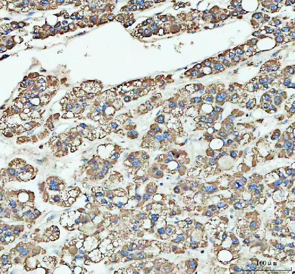
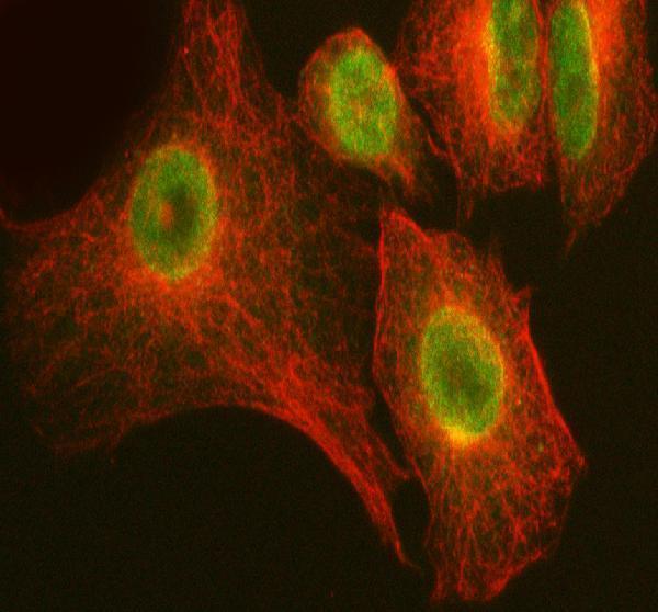
The catalog antibody has paraffin-section IHC images from human tumors and an IF/ICC image from A549 cells (catalog image captions). Its listed reactivity is human, mouse and rat (catalog reactivity).

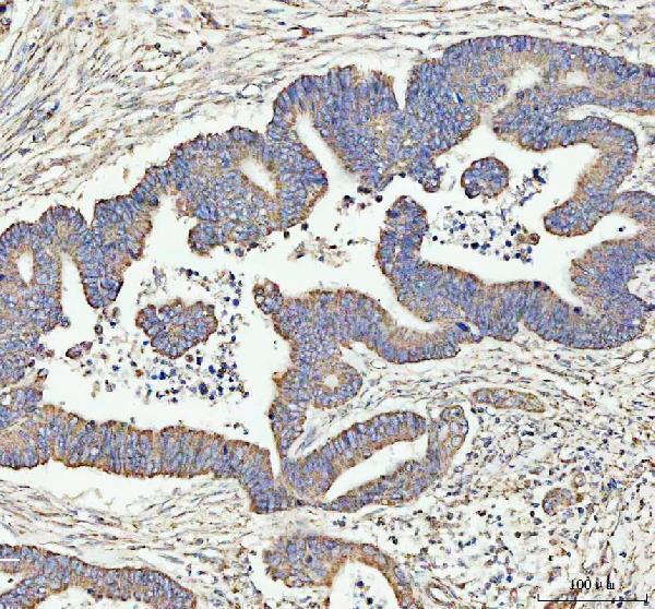
Real IHC data IHC analysis of ARHGEF12L using anti-ARHGEF12L antibody (A06802-3). ARHGEF12L was detected in a paraffin-embedded section of human colorectal adenocarcinoma tissue. Heat mediated antigen retrieval was performed in EDTA buffer (pH 8.0, epitope retrieval solution). The tissue section was blocked with 10% goat serum. The tissue section was then incubated with 2 μg/ml rabbit anti-ARHGEF12L Antibody (A06802-3) overnight at 4°C. Peroxidase Conjugated Goat Anti-rabbit IgG was used as secondary antibody and incubated for 30 minutes at 37°C. The tissue section was developed using HRP Conjugated Rabbit IgG Super Vision Assay Kit (Catalog # SV0002) with DAB as the chromogen.
Anti-LARG/ARHGEF12 Antibody ®
Cat # A06802-3

A06802-3 has IHC images from paraffin sections of human colorectal adenocarcinoma, liver cancer, parotid acinar cell carcinoma and cervical squamous cell carcinoma (catalog IHC captions). The same SKU has an IF/ICC image from A549 cells and lists human, mouse and rat reactivity (catalog IF caption; catalog reactivity).

Which to pick: Choose A06802-3 for paraffin-section IHC: its caption documents EDTA retrieval at pH 8.0 and 2 μg/ml primary antibody; the fixative is unreported (catalog IHC caption). For IF/ICC, the same SKU has an A549 cell image at 5 μg/ml (catalog IF caption). It lists human, mouse and rat reactivity overall, while its IHC dilution listing specifies human and rat; clonality is unreported (catalog reactivity; catalog IHC dilution listing; catalog clone field).

Each figure is that product's own IHC / IF validation image from its datasheet.

References

  1. UniProt Consortium. UniProt entry Q9NZN5 (ARHGC_HUMAN, Rho guanine nucleotide exchange factor 12).
  2. Human Protein Atlas. ARHGEF12 tissue IHC expression (reliability: Approved).
  3. Human Protein Atlas. ARHGEF12 subcellular location (ICC-IF): Mainly localized to the plasma membrane and cytosol. In addition localized to the nucleoplasm..
  4. Human Protein Atlas. ARHGEF12 antibody validation summary (2 antibodies).
  5. METTL3 Promotes Cutaneous T-Cell Lymphoma Progression by Regulating ARHGEF12 Expression. International journal of molecular sciences 2025 — PMC12027205.
  6. Comprehensive exome profiling identifies ARHGEF12 mutation as a driver in gastric cancer with ovarian metastasis. Theranostics 2025 — PMC12374580.
  7. Identification of Arhgef12 and Prkci as genetic modifiers of retinal dysplasia in the Crb1rd8 mouse model. PLoS genetics 2022 — PMC9212170.
  8. Trans-Synaptic Signaling through the Glutamate Receptor Delta-1 Mediates Inhibitory Synapse Formation in Cortical Pyramidal Neurons. Neuron 2019 — PMC6926483.
  9. PubMed PMID:10681437 — UniProt-cited evidence.
  10. PubMed PMID:15489334 — UniProt-cited evidence.
  11. PubMed PMID:9205841 — UniProt-cited evidence.