This website uses cookies to ensure you get the best experience on our website.


- Table of Contents
Plan chromogenic IHC for ARHGEF2 in paraffin sections using its variable cytoplasmic tissue staining as a reference (HPA tissue IHC). High staining in bone marrow hematopoietic cells and germinal center cells offers positive-control options (HPA tissue IHC).

Expected localisation, validated protocols, controls and antibodies — the at-a-glance facts below, then the full design guide.
| Expected localisation | Variable cytoplasmic tissue staining (HPA tissue IHC) | |
| Staining pattern | Cytoplasmic staining of varying intensity across tissues (HPA tissue IHC) | |
| Antigen retrieval | EDTA pH 8.0 HIER, heat-mediated (datasheet A02572-3) | |
| Positive control | Bone marrow+4 more · see all | |
| Negative control | Adipose tissue+4 more · see all |
| Fixation | Keep fixation consistent across sections (standard IHC practice; not target-specific) | |
| Caveat | Bone marrow peroxidase may mimic DAB staining (HPA tissue IHC; standard IHC practice) | |
| Regulation | Expression regulation not established (UniProt) | |
| Isoform / epitope | 3 isoforms; epitope coverage is unspecified (UniProt) |
The catalog antibody protocol is paired with one published ARHGEF2 tissue IHC protocol (PMC9637107 methods).
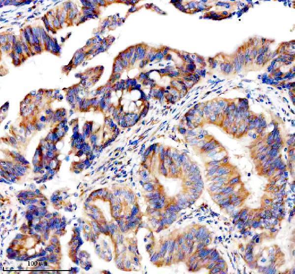
| Sample | Paraffin-embedded human colon cancer tissue; fixative not specified (datasheet A02572-3) |
| Fixation | Image fixative and duration unreported (datasheet A02572-3); verify before use. |
| Sectioning | 4–5 µm sections on charged slides (standard) |
| Deparaffinisation | Xylene, graded ethanol series to water (standard) |
| Antigen retrieval | Heat retrieval: EDTA pH 8.0 (datasheet A02572-3); 20 min, 95–100 °C (standard) |
| Peroxidase block | 3% H2O2, 10 min, room temperature (standard) |
| Blocking | 10% goat serum (datasheet A02572-3) |
| Primary antibody | Rabbit anti-ARHGEF2, 2-5 μg/ml (datasheet A02572-3) |
| Primary incubation | Overnight at 4 °C (datasheet A02572-3) |
| Detection | HRP-conjugated secondary, DAB chromogen (datasheet A02572-3) |
| Counterstain | Hematoxylin, blue, dehydrate and mount (standard) |
| Expected result | ARHGEF2-positive staining in hematopoietic cells of bone marrow (HPA tissue IHC: High). HPA tissue profile: Cytoplasmic expression of varying intensities in several different tissue types. No signal in the no-primary control. |
ARHGEF2 is a cytoplasmic protein associated with the cytoskeleton, junctions and other intracellular structures; it has no transmembrane segment (UniProt Q92974). In paraffin sections, expect cytoplasmic staining in selected cell populations, especially bone marrow hematopoietic cells and lymph node or tonsil germinal center cells (HPA: High). HPA rates the tissue IHC profile Approved, with medium agreement between staining and RNA data and external verification pending (HPA).
| Clear cytoplasmic staining in bone marrow hematopoietic cells or lymph node and tonsil germinal center cells, with adjacent structures still readable. | This fits the reported High staining in those populations (HPA) and the intracellular localization of ARHGEF2 (UniProt Q92974). Intensity can vary among tissues (HPA); score the named cell population rather than the whole section. |
| A sharply nuclear, exclusively extracellular, or uniform cell-surface signal dominates the section. | Those compartments do not match the reported cytoplasmic tissue profile (HPA) or the protein's intracellular locations and lack of a transmembrane segment (UniProt Q92974). Check morphology and controls before attributing the signal to ARHGEF2. |
| Strong staining appears in adipocytes, esophageal squamous epithelial cells, or heart cardiomyocytes. | These specific cell populations are reported as Not detected (HPA). Review tissue identification and control staining; cross-reactivity or endogenous detection activity is possible (general IHC practice). A negative HPA observation is not proof of absolute protein absence. |
| Diffuse color covers cells, stroma, and empty spaces, obscuring intracellular boundaries. | This cannot be scored as the reported cytoplasmic cell staining (HPA). Background from detection reagents, inadequate blocking, or excessive antibody concentration is possible (general IHC practice); compare a matched control section. |
| No staining is visible in an adequately sampled bone marrow hematopoietic population or lymph node germinal center. | These are reported High populations (HPA), so an entirely blank result raises an assay-performance question. Check section identity, tissue preservation, retrieval, antibody exposure, and detection controls (general IHC practice) before interpreting the specimen as negative. |
| Cell population and tissue context | HPA reports High staining in bone marrow hematopoietic cells, lymph node and tonsil germinal center cells, and late spermatids; several other populations are Medium, Low, or Not detected (HPA). Compare like cell populations when selecting controls or scoring. |
| Intracellular distribution | UniProt places ARHGEF2 in cytoplasm, cytoskeleton, tight junctions, Golgi, spindle-associated structures, ruffles, and cytoplasmic vesicles (UniProt Q92974). The HPA tissue profile summarizes variable cytoplasmic staining (HPA); routine chromogenic sections need not resolve every listed subcellular site. |
| Antibody validation and protein forms | The listed antibody HPA043437 is IHC Approved, while HPA describes medium staining–RNA agreement and pending external verification (HPA). UniProt lists 3 isoforms and a chain spanning residues 1–986 (UniProt Q92974); the payload gives no epitope, so isoform coverage cannot be inferred. |
| IF/ICC question: what localization should be expected? | Intracellular localization is biologically plausible from UniProt Q92974, but HPA provides no main ICC-IF location or cell-line images for this record (HPA subcellular). Use the separate IF/ICC guide for assay design; this IHC evidence does not establish an IF staining pattern. |
| Situation | Likely cause | Next action |
|---|---|---|
| The presumed positive control has no chromogenic signal. | The sampled section may lack the relevant cells, or a preparation or detection step may have failed (general IHC practice); HPA's High label applies to named cell populations, not every area of a tissue (HPA). | Confirm hematopoietic cells or germinal centers are present, then inspect reagent and detection controls and review the laboratory's retrieval conditions (general IHC practice). Do not assume a target-specific retrieval requirement from HPA. |
| Staining is widespread across tissue types but lacks a clear cytoplasmic pattern. | Diffuse background or nonspecific binding may be obscuring interpretable staining (general IHC practice). HPA describes cytoplasmic expression of varying intensity across tissues (HPA), which does not justify uniform color over all structures. | Compare a matched control and tissue morphology; review blocking, washing, antibody concentration, and detection exposure using the validated assay workflow (general IHC practice). Reassess the named cell populations after background is controlled. |
| Strong color appears where HPA reports Not detected staining. | Cell-type misidentification, antibody cross-reactivity, or endogenous detection activity are possibilities (general IHC practice). HPA reports Not detected adipocytes, esophageal squamous epithelial cells, and cardiomyocytes (HPA). | Verify the cell type on a counterstained section and compare reagent controls (general IHC practice). Treat the HPA entry as an observed reference pattern, with its medium agreement and pending external verification (HPA). |
| Signal seems confined to nuclei or extracellular material. | The pattern conflicts with the HPA cytoplasmic profile and UniProt intracellular localization (HPA; UniProt Q92974). Morphologic overlap, precipitate, or nonspecific signal could account for it (general IHC practice). | Check focus and cellular boundaries, inspect a matched control, and repeat scoring in a named HPA High population (general IHC practice; HPA). Do not call the unexpected compartment ARHGEF2 solely from color. |
| Two positive tissues show different staining strengths. | HPA explicitly reports variable cytoplasmic intensity: bone marrow hematopoietic cells are High, while breast glandular cells are Medium (HPA). Tissue-wide averages can also conceal differences among cell types (general IHC practice). | Score the specified cell population in each tissue and compare it with its own HPA reference level (HPA). Keep section quality and detection conditions comparable when interpreting an intensity difference (general IHC practice). |
| A result is being used to claim a specific isoform or an IF/ICC pattern. | UniProt lists three isoforms, but the supplied record does not map the antibody epitope to them (UniProt Q92974; HPA antibodies). HPA supplies no ICC-IF main location or images for this record (HPA subcellular). | Report the result as IHC staining with the catalog antibody and describe only the observed compartment and cell type (HPA; general IHC practice). Leave isoform identity and an IF/ICC pattern unresolved without separate evidence. |


Comprehensive Human Protein Atlas IHC scoring per tissue (reliability: Approved — Medium consistency between antibody staining and RNA expression data. Pending external verification.). Rows are taken directly from the HPA tissue chart — click any row's HPA link to view the source.
| Tissue | Cell type | Level | Evidence | Source |
|---|---|---|---|---|
| Bone marrow | Hematopoietic cells | High | Protein (IHC) | HPA → |
| Lymph node | Germinal center cells | High | Protein (IHC) | HPA → |
| Testis | Elongated or late spermatids | High | Protein (IHC) | HPA → |
| Tonsil | Germinal center cells | High | Protein (IHC) | HPA → |
| Adrenal gland | Glandular cells | Medium | Protein (IHC) | HPA → |
| Tissue | Cell type | Level | Evidence | Source |
|---|---|---|---|---|
| Adipose tissue | Adipocytes | Not detected | Protein (IHC) | HPA → |
| Bronchus | Basal cells | Not detected | Protein (IHC) | HPA → |
| Cervix | Glandular cells | Not detected | Protein (IHC) | HPA → |
| Epididymis | Glandular cells | Not detected | Protein (IHC) | HPA → |
| Esophagus | Squamous epithelial cells | Not detected | Protein (IHC) | HPA → |
Troubleshoot ARHGEF2 staining in paraffin sections by checking retrieval, cell type, cytoplasmic localisation and controls before scoring signal.
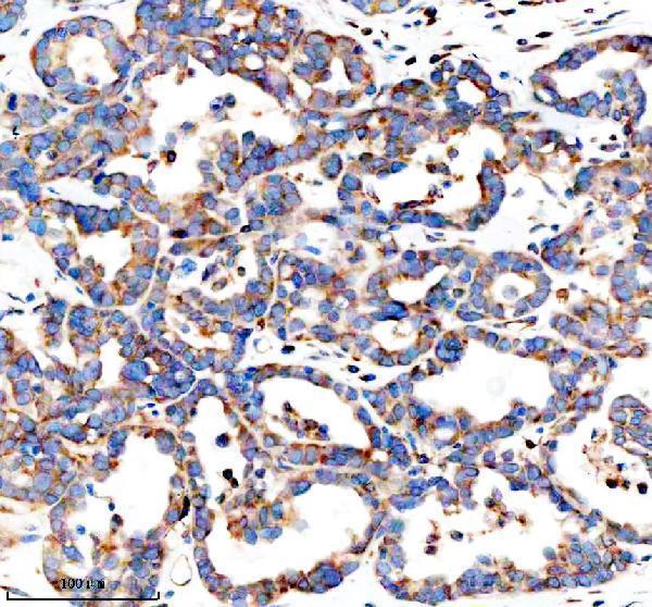
ARHGEF2 tissue IHC has a human paraffin-section image (A02572-3 image caption); IF/ICC is listed for human, mouse and rat, with no IF image supplied (M02572-1 catalog).
A02572-3 has IHC images from paraffin-embedded human colon and ovarian cancer sections (A02572-3 image captions). M02572-1 lists ICC/IF for human, mouse and rat, but supplies no IF image (M02572-1 catalog).
Which to pick: Choose A02572-3 for tissue IHC: its own captions show paraffin sections, and the fixative is unreported (A02572-3 image captions). Choose monoclonal M02572-1 for IF/ICC because that application is listed for it; it has no listed IHC validation (M02572-1 catalog). Both list human, mouse and rat reactivity, but the supplied IHC images show human tissue only (A02572-3 catalog and image captions; M02572-1 catalog).