This website uses cookies to ensure you get the best experience on our website.


- Table of Contents
Plan ARHGEF9 chromogenic IHC in paraffin sections using the reported cytoplasmic staining in CNS tissue, including Purkinje cells and neuropil (HPA tissue IHC). This guide covers tissue selection, staining controls and interpretation of cell-specific signals.

Expected localisation, validated protocols, controls and antibodies — the at-a-glance facts below, then the full design guide.
| Expected localisation | Cytoplasmic tissue staining (HPA tissue IHC) | |
| Staining pattern | Cytoplasmic CNS staining in Purkinje cells and neuropil (HPA tissue IHC) | |
| Antigen retrieval | Citrate pH 6.0 HIER, 95–98 °C, 20 min (rule: cytoplasmic / membrane antigen) | |
| Positive control | Adrenal gland+4 more · see all | |
| Negative control | Adipose tissue+4 more · see all |
| Fixation | Keep fixation consistent across sections. (standard IHC practice; not target-specific) | |
| Caveat | Hippocampal glial cells may be unstained (HPA tissue IHC) | |
| Regulation | No staining-relevant regulation specified (UniProt) | |
| Isoform / epitope | 3 isoforms; epitope coverage is unspecified (UniProt; datasheet) |
Compare the catalog antibody’s IHC-P protocol (datasheet) with a published mouse brain paraffin-section protocol (PMC6066644).
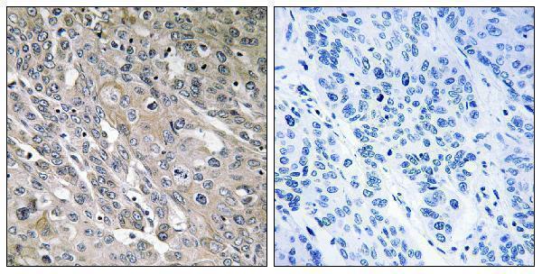
| Sample | Paraffin-embedded human lung carcinoma tissue; fixative not specified (datasheet A06931-1) |
| Fixation | Image fixative and duration unreported (datasheet A06931-1); verify before use. |
| Sectioning | 4–5 µm sections on charged slides (standard) |
| Deparaffinisation | Xylene, graded ethanol series to water (standard) |
| Antigen retrieval | Heat-induced epitope retrieval in citrate buffer, pH 6.0, 20 min at 95–98 °C (standard rule: cytoplasmic / membrane antigen) |
| Peroxidase block | 3% H2O2, 10 min, room temperature (standard) |
| Blocking | 10% normal serum of the secondary host, 30 min, room temperature (standard) |
| Primary antibody | Rabbit anti-ARHGEF9, 1:100 - 1:300 (datasheet A06931-1) |
| Primary incubation | Overnight at 4 °C (standard) |
| Detection | HRP-polymer secondary, DAB chromogen 5–10 min (standard) |
| Counterstain | Hematoxylin, blue, dehydrate and mount (standard) |
| Expected result | ARHGEF9-positive staining in glandular cells of adrenal gland (HPA tissue IHC: Medium). HPA tissue profile: Cytoplasmic expression in several tissue, most abundant CNS. No signal in the no-primary control. |
ARHGEF9 is cytoplasmic and associated with the postsynaptic density (UniProt O43307); it has no transmembrane segment (UniProt O43307 topology). In paraffin section IHC, expect cytoplasmic staining in Purkinje cells and staining of cerebral cortical neuropil, both reported at medium intensity (HPA tissue IHC). HPA rates its tissue staining Approved, with external verification pending (HPA tissue IHC).
| Cytoplasmic signal in cerebellar Purkinje cells and staining in cortical neuropil. | This matches two medium intensity observations in CNS tissue (HPA tissue IHC). Interpret neuropil staining by region and structure; it does not identify a particular cell type on its own (general IHC practice). |
| Predominantly nuclear staining, with little cytoplasmic signal. | A predominantly nuclear pattern conflicts with the reported cytoplasmic location and postsynaptic density association (UniProt O43307; HPA subcellular ICC-IF). Check the no-primary control and detection background before assigning this pattern to ARHGEF9 (general IHC practice). |
| Strong signal in a cell population recorded as not detected. | Alveolar cells in lung and germinal center cells in lymph node are reported as not detected (HPA tissue IHC). Check the exact cell population: staining outside it does not contradict that observation. Within it, consider cross-reactivity or endogenous chromogenic activity (general IHC practice). |
| Diffuse color across cells, stroma, or the whole section. | This lacks the cell and compartment pattern reported for ARHGEF9 (HPA tissue IHC; UniProt O43307). Background from detection reagents, inadequate blocking, or excessive primary antibody is possible; a no-primary section helps distinguish detection background (general IHC practice). |
| No signal in Purkinje cells of an interpretable cerebellar section. | Purkinje cells show medium staining in the HPA reference (HPA tissue IHC). First verify that these cells are present, then review the antibody's IHC validation, detection controls, and run conditions; a blank section alone cannot establish absent protein (general IHC practice). |
| Cell and tissue choice | Cerebellar Purkinje cells and cortical neuropil offer documented medium signal; hippocampal glial cells are listed as not detected (HPA tissue IHC). Compare the specified cells, not whole tissues, when choosing controls (general IHC practice). |
| Antibody validation | One listed rabbit antibody has Approved IHC status; its external verification remains pending (HPA HPA035419; HPA tissue IHC). This supports a reference pattern but does not make every stain specific (general IHC practice). |
| Isoforms and epitope | UniProt lists three ARHGEF9 isoforms (UniProt O43307). The supplied evidence gives no antibody epitope or isoform coverage, so staining cannot be assigned to a particular isoform from this pattern alone. |
| Topology and processing | ARHGEF9 has no transmembrane segment, signal peptide, propeptide, or reported processing into shorter chains in this record (UniProt O43307). A membrane outline or extracellular deposit is therefore not the expected reference pattern (UniProt O43307; HPA tissue IHC). |
| IF/ICC: where should signal localize? | HPA reports supported cytosolic localization in ICC-IF, while UniProt also lists the postsynaptic density (HPA subcellular ICC-IF; UniProt O43307). This is a localization answer only; the supplied evidence gives no IF/ICC protocol option. |
| Antigen retrieval evidence | Target-specific fixation effects are not established by the supplied assay evidence. Verify with a matched IHC source before attributing a result to fixation. |
| Situation | Likely cause | Next action |
|---|---|---|
| Cerebellar positive control is blank. | The reference reports medium Purkinje cell staining (HPA tissue IHC); absent signal may reflect a failed staining run or unavailable antigen, without identifying which (general IHC practice). | Confirm Purkinje cells are present, then check the antibody's IHC validation, detection reagents, and run controls (general IHC practice). |
| Nuclei dominate the stain. | Nuclear enrichment conflicts with the cytoplasmic reference localization (UniProt O43307; HPA subcellular ICC-IF). Nonspecific staining or detection background is possible (general IHC practice). | Review a no-primary section and whether cytoplasmic signal follows the expected cells and structures (general IHC practice; HPA tissue IHC). |
| Color appears in alveolar cells or lymph node germinal center cells. | Both populations are listed as not detected (HPA tissue IHC). Signal there may represent cross-reactivity or endogenous detection activity (general IHC practice). | Compare the same cell populations with no-primary and appropriate detection controls; avoid calling the whole tissue positive from unrelated cells (general IHC practice). |
| Background obscures the cytoplasm. | Diffuse color can arise from detection background, inadequate blocking, or excessive primary antibody (general IHC practice); HPA describes a cytoplasmic tissue pattern (HPA tissue IHC). | Inspect a no-primary control, then review blocking, washes, and the dilution specified for the IHC-validated antibody (general IHC practice). |
| A strong membrane outline replaces cytoplasmic staining. | ARHGEF9 lacks a transmembrane segment and is annotated in cytoplasm and postsynaptic density (UniProt O43307); an outline is discordant with the supplied localization. | Check whether the outline persists without primary antibody and compare it with cytoplasmic Purkinje cell staining in the same run (general IHC practice; HPA tissue IHC). |
| Two tissue sections give different apparent intensity. | HPA records medium staining in selected cells and not-detected or low staining in others (HPA tissue IHC). Differences may reflect which cells were sampled, alongside run variation (general IHC practice). | Score the named cell population and compartment in each section; compare staining only after checking controls and run conditions (general IHC practice; HPA tissue IHC). |


Comprehensive Human Protein Atlas IHC scoring per tissue (reliability: Approved — Pending external verification.). Rows are taken directly from the HPA tissue chart — click any row's HPA link to view the source.
| Tissue | Cell type | Level | Evidence | Source |
|---|---|---|---|---|
| Adrenal gland | Glandular cells | Medium | Protein (IHC) | HPA → |
| Bronchus | Respiratory epithelial cells | Medium | Protein (IHC) | HPA → |
| Cerebellum | Purkinje cells | Medium | Protein (IHC) | HPA → |
| Cerebral cortex | Neuropil | Medium | Protein (IHC) | HPA → |
| Colon | Glandular cells | Medium | Protein (IHC) | HPA → |
| Tissue | Cell type | Level | Evidence | Source |
|---|---|---|---|---|
| Adipose tissue | Adipocytes | Not detected | Protein (IHC) | HPA → |
| Breast | Adipocytes | Not detected | Protein (IHC) | HPA → |
| Epididymis | Glandular cells | Not detected | Protein (IHC) | HPA → |
| Esophagus | Squamous epithelial cells | Not detected | Protein (IHC) | HPA → |
| Fallopian tube | Glandular cells | Not detected | Protein (IHC) | HPA → |
Troubleshoot ARHGEF9 staining by checking retrieval, compartment, cell type and controls before interpreting differences in chromogenic signal.
A06931-1 has a paraffin-section IHC image from human lung carcinoma tissue (catalog image caption). IF and ICC are listed applications, with Human, Mouse and Rat reactivity (catalog applications and reactivity).
A06931-1 has an IHC image of paraffin-embedded human lung carcinoma tissue with peptide blocking shown (catalog image caption). IF and ICC are listed applications, and Human, Mouse and Rat are listed as reactive species; no IF image is supplied (catalog applications, reactivity and image records).
Which to pick: For tissue IHC, choose A06931-1: its image shows staining in a paraffin-embedded human lung carcinoma section (catalog image caption). For IF/ICC or work involving Human, Mouse or Rat samples, A06931-1 is the listed rabbit polyclonal option, though no IF image is supplied (catalog host, dilution record, applications, reactivity and image records). The IHC caption does not report the fixative (catalog image caption).