This website uses cookies to ensure you get the best experience on our website.


- Table of Contents
This guide uses tissue IHC evidence to interpret ARL13B’s broad cytoplasmic staining (HPA tissue IHC). It covers the catalog antibody’s 2–5 μg/mL paraffin IHC range (datasheet A04279-1) and the distinct molecular expectation of cilium membrane localisation (UniProt).

Expected localisation, validated protocols, controls and antibodies — the at-a-glance facts below, then the full design guide.
| Expected localisation | Cytoplasmic staining in most tissues (HPA tissue IHC) | |
| Staining pattern | Cytoplasmic staining in cells across most tissues (HPA tissue IHC) | |
| Antigen retrieval | EDTA pH 8.0 HIER, heat-mediated (datasheet A04279-1) | |
| Positive control | Adrenal gland+4 more · see all | |
| Negative control | None in HPA (detected in all 45 tissues); use no-primary + isotype controls |
| Fixation | Keep fixation consistent (standard IHC practice; not target-specific); Matched tissue-IHC evidence does not establish the fixation claim. Validate the specimen-specific method before use. (selected-SKU IHC image A04279-1) | |
| Caveat | Broad cytoplasmic staining may obscure ciliary signal (HPA tissue IHC; UniProt) | |
| Regulation | Expressed in the developing brain (UniProt) | |
| Isoform / epitope | 3 isoforms; epitope coverage is unspecified (UniProt) |
The catalog antibody’s IHC-P protocol is followed by published ARL13B protocols for tissue sections (PMC5442446; PMC8278305; PMC6826833).
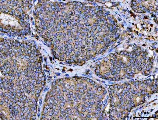
| Sample | Paraffin-embedded human breast cancer tissue; fixative not specified (datasheet A04279-1) |
| Fixation | Image fixative and duration unreported (datasheet A04279-1); verify before use. |
| Sectioning | 4–5 µm sections on charged slides (standard) |
| Deparaffinisation | Xylene, graded ethanol series to water (standard) |
| Antigen retrieval | Heat retrieval: EDTA pH 8.0 (datasheet A04279-1); 20 min, 95–100 °C (standard) |
| Peroxidase block | 3% H2O2, 10 min, room temperature (standard) |
| Blocking | 10% goat serum (datasheet A04279-1) |
| Primary antibody | Rabbit anti-ARL13B, 2-5 μg/ml (datasheet A04279-1) |
| Primary incubation | Overnight at 4 °C (datasheet A04279-1) |
| Detection | HRP-conjugated secondary, DAB chromogen (datasheet A04279-1) |
| Counterstain | Hematoxylin, blue, dehydrate and mount (standard) |
| Expected result | ARL13B-positive staining in glandular cells of adrenal gland (HPA tissue IHC: High). HPA tissue profile: Cytoplasmic expression in most tissues. No signal in the no-primary control. |
ARL13B is associated with the cilium membrane and cytoskeleton and has no annotated transmembrane segment (UniProt Q3SXY8). In paraffin sections, expect cytoplasmic staining across many tissues, with high staining reported in adrenal and gallbladder glandular cells and lung alveolar type I cells (HPA tissue IHC). HPA rates its tissue IHC evidence Approved, with medium consistency between antibody staining and RNA expression (HPA tissue IHC).
| Cytoplasmic stain in adrenal or gallbladder glandular cells, or lung alveolar type I cells. | This matches the cells and high staining levels reported in tissue sections (HPA tissue IHC). A discernible ciliary profile can add support, but its absence in a section does not by itself overturn a matching tissue pattern (UniProt Q3SXY8 localization; general IHC practice). |
| A sharply nuclear pattern dominates while the expected cytoplasmic pattern is absent. | Nuclear localization is outside the supplied ARL13B locations (UniProt Q3SXY8; HPA tissue IHC). Treat this as suspect staining; review the antibody, detection controls and morphology before assigning it to ARL13B (general IHC practice). |
| Strong stain appears in a cell population reported as low, while expected high-staining cells are blank. | For example, respiratory epithelial cells are reported low, whereas lung alveolar type I cells are high (HPA tissue IHC). Check cell identification and controls; cross-reactivity or endogenous detection activity is possible, but this pattern alone cannot distinguish them (general IHC practice). |
| Broad, diffuse colour obscures cell boundaries and the expected cell-to-cell pattern. | The result is difficult to score against the reported cytoplasmic and cell-specific distribution (HPA tissue IHC). Background from detection reagents or insufficient blocking is possible; compare controls before calling the diffuse colour positive (general IHC practice). |
| No stain is visible in an adrenal gland, gallbladder or lung section containing the reported cells. | These are useful positive-reference cells because HPA reports high staining in them (HPA tissue IHC). First confirm that the relevant cells are present, then check the IHC run and antibody conditions; one blank section does not establish biological absence (general IHC practice). |
| Tissue and cell selection | Adrenal and gallbladder glandular cells and lung alveolar type I cells are reported High; endometrial glandular cells and adipocytes are reported Medium (HPA tissue IHC). Compare like cell populations when judging intensity. |
| Compartment and section geometry | ARL13B associates with proximal ciliary membranes and also colocalizes with microtubules (UniProt Q3SXY8). HPA reports cytoplasmic staining in most tissue sections (HPA tissue IHC); a thin cilium may be hard to resolve by chromogenic IHC (general IHC practice). |
| Antibody evidence | The listed antibody HPA048926 has IHC Approved and ICC Supported status (HPA antibodies). Tissue IHC has medium consistency with RNA data (HPA tissue IHC), so use morphology and controls when interpreting an unexpected distribution (general IHC practice). |
| Isoform coverage | Three isoforms are annotated (UniProt Q3SXY8). The supplied record gives no epitope or isoform coverage for the IHC-validated antibody, so a stain cannot be assigned to a particular isoform from these data. |
| Situation | Likely cause | Next action |
|---|---|---|
| The expected high-staining cells are present, but the section is blank. | The result conflicts with HPA's reported High staining in those cell populations (HPA tissue IHC); an IHC run or reagent issue remains possible (general IHC practice). | Check the positive-reference section and detection controls, then review retrieval and antibody dilution under the laboratory's validated IHC conditions (general IHC practice). |
| Colour is mainly nuclear. | This does not match the supplied cytoplasmic tissue pattern or ciliary and cytoskeletal locations (HPA tissue IHC; UniProt Q3SXY8). Background or off-target binding is possible (general IHC practice). | Compare a reagent control and a positive-reference tissue; inspect cell boundaries before scoring the nuclear colour (general IHC practice). |
| Unexpected cells stain more strongly than the reported high-staining cells. | Cell identification, cross-reactivity or endogenous detection activity could account for the mismatch (general IHC practice); HPA's levels apply to named cell populations (HPA tissue IHC). | Recheck morphology and compare appropriate detection controls with the HPA cell-level pattern before attributing the signal to ARL13B (HPA tissue IHC; general IHC practice). |
| Diffuse colour makes cytoplasmic staining hard to distinguish. | Detection background or insufficient blocking may obscure the reported pattern (HPA tissue IHC; general IHC practice). | Inspect a no-primary control and review blocking, washes and chromogen development within the validated IHC workflow (general IHC practice). |
| Ciliary staining is not individually visible in an otherwise plausible section. | Section plane and chromogenic resolution can limit visibility of a small cilium (general IHC practice); HPA describes tissue IHC broadly as cytoplasmic (HPA tissue IHC). | Judge the named cell populations and cytoplasmic distribution first; reserve finer subcellular assessment for an IF/ICC experiment (HPA tissue IHC; general IHC practice). |
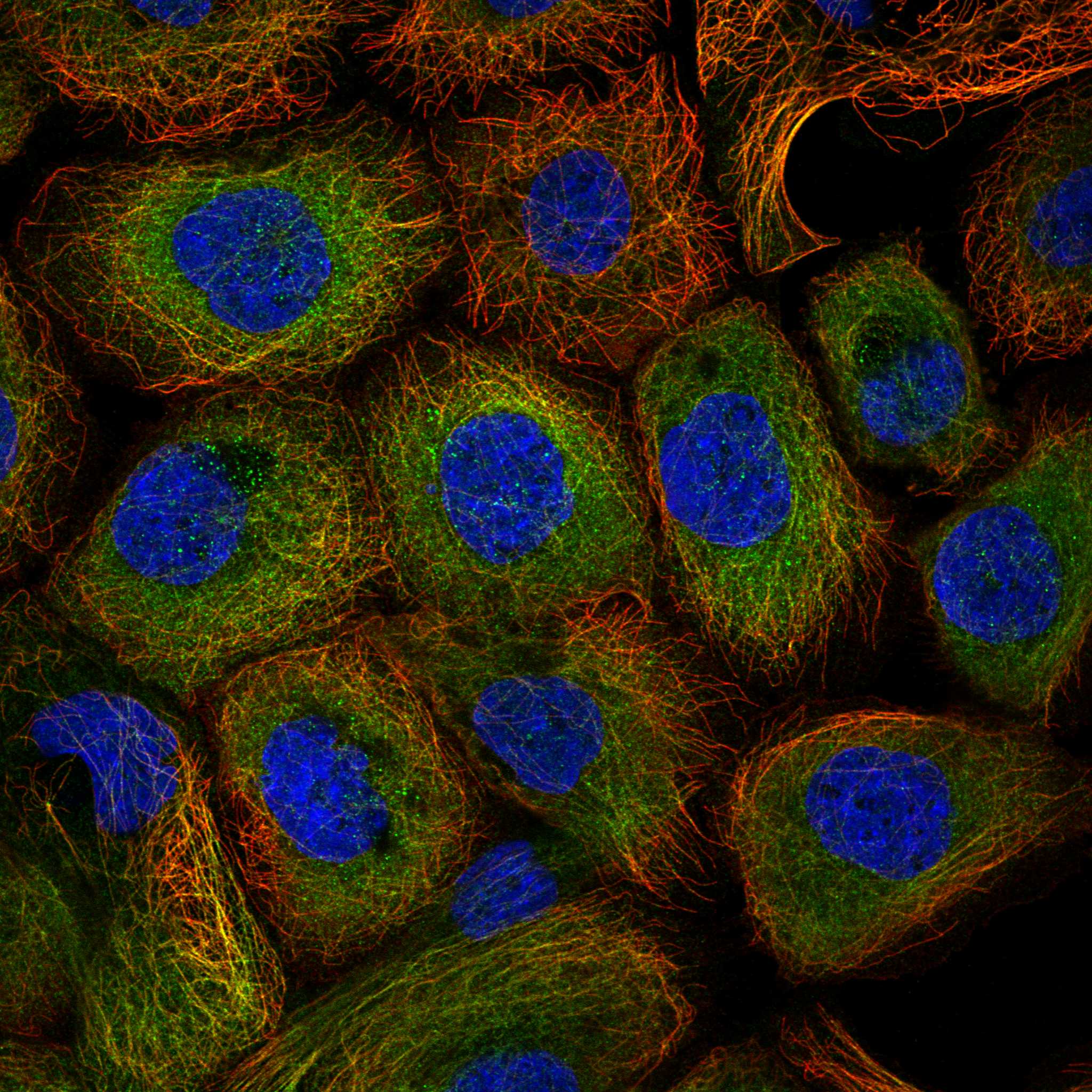
| What should IF/ICC show if finer localization is needed? | HPA reports supported localization at microtubules, primary cilia and the primary cilium transition zone in ICC-IF (HPA subcellular). UniProt describes a proximal ciliary membrane compartment that excludes the transition zone (UniProt Q3SXY8). | Use the separate IF/ICC guide for experiment design; interpret transition-zone assignment with the distinction between the HPA imaging summary and UniProt's more specific membrane description in mind (HPA subcellular; UniProt Q3SXY8). |


Comprehensive Human Protein Atlas IHC scoring per tissue (reliability: Approved — Medium consistency between antibody staining and RNA expression data.). Rows are taken directly from the HPA tissue chart — click any row's HPA link to view the source.
| Tissue | Cell type | Level | Evidence | Source |
|---|---|---|---|---|
| Adrenal gland | Glandular cells | High | Protein (IHC) | HPA → |
| Endometrium | Glandular cells | Medium | Protein (IHC) | HPA → |
| Gallbladder | Glandular cells | High | Protein (IHC) | HPA → |
| Lung | Alveolar cells type I | High | Protein (IHC) | HPA → |
| Adipose tissue | Adipocytes | Medium | Protein (IHC) | HPA → |
| Tissue | Cell type | Level | Evidence | Source |
|---|---|---|---|---|
| None in HPA: ARL13B is detected in all 45 scored tissues. Use a no-primary (secondary-only) and an isotype control instead. | ||||
Troubleshoot ARL13B staining in paraffin sections by checking retrieval, staining compartment, controls and scoring before interpreting diffuse signal (UniProt Q3SXY8; HPA tissue IHC).
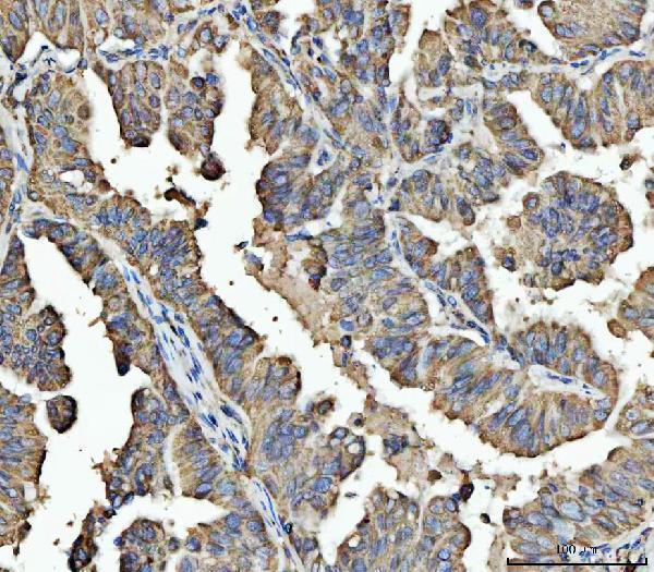
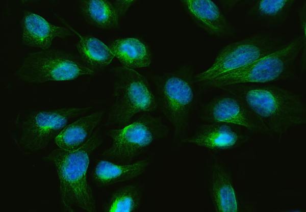
Anti-ARL13B A04279-1 has IHC images from human paraffin-embedded breast, colorectal, gastric, and testicular tumor sections (IHC image captions) and an IF image from U20S cells (IF image caption).
A04279-1 will render with its human breast cancer paraffin-section IHC image; the catalog also shows colorectal, gastric, and testicular cancer paraffin-section IHC images (IHC image captions). Its IF image shows U20S cells (IF image caption), and its listed reactivity is human, mouse, and rat (catalog: reactivity).
Which to pick: Choose A04279-1 for paraffin-section tissue IHC: its human breast cancer image used EDTA retrieval at pH 8.0 and 2 μg/ml primary antibody (IHC image caption); the fixative is unreported (IHC image caption). For IF/ICC, A04279-1 is listed for both applications and has an IF image from U20S cells (catalog: applications; IF image caption). For mouse or rat work, A04279-1 lists those species as reactive, while its supplied IHC images document human tissue only (catalog: reactivity; IHC image captions).