This website uses cookies to ensure you get the best experience on our website.


- Table of Contents
Plan chromogenic ARL6 IHC on paraffin sections using ciliated bronchus or fallopian tube as high-staining controls (HPA tissue IHC). This guide covers fixation, antigen retrieval, antibody dilution of 1:50–1:100 (datasheet), and assessment of cytoplasmic and ciliary staining (HPA tissue IHC).

Expected localisation, validated protocols, controls and antibodies — the at-a-glance facts below, then the full design guide.
| Expected localisation | Cytoplasmic staining with cilia positivity (HPA tissue IHC) | |
| Staining pattern | Mainly cytoplasmic in most tissues, including cilia (HPA tissue IHC) | |
| Antigen retrieval | Citrate pH 6.0 HIER, 95–98 °C, 20 min (rule: cytoplasmic / membrane antigen) | |
| Positive control | Bronchus+4 more · see all | |
| Negative control | Adipose tissue+3 more · see all |
| Fixation | Keep paraffin-section fixation consistent (standard IHC practice; not target-specific); Matched tissue-IHC evidence does not establish the fixation claim. Validate the specimen-specific method before use. (selected-SKU IHC image A05003) | |
| Caveat | Antibody staining and RNA show medium consistency (HPA tissue IHC) | |
| Regulation | Retina-enhanced RNA; staining effect unknown (HPA tissue RNA + IHC) | |
| Isoform / epitope | 2 isoforms; epitope coverage is unspecified (UniProt) |
Compare the catalog antibody’s IHC-P protocol (datasheet) with a published mouse tissue IHC protocol (PMC5154108).
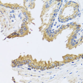
| Sample | Paraffin-embedded human prostate tissue; fixative not specified (datasheet A05003) |
| Fixation | Image fixative and duration unreported (datasheet A05003); verify before use. |
| Sectioning | 4–5 µm sections on charged slides (standard) |
| Deparaffinisation | Xylene, graded ethanol series to water (standard) |
| Antigen retrieval | Heat-induced epitope retrieval in citrate buffer, pH 6.0, 20 min at 95–98 °C (standard rule: cytoplasmic / membrane antigen) |
| Peroxidase block | 3% H2O2, 10 min, room temperature (standard) |
| Blocking | 10% normal serum of the secondary host, 30 min, room temperature (standard) |
| Primary antibody | Rabbit anti-ARL6, 1:50-1:100 (datasheet A05003) |
| Primary incubation | Overnight at 4 °C (standard) |
| Detection | HRP-polymer secondary, DAB chromogen 5–10 min (standard) |
| Counterstain | Hematoxylin, blue, dehydrate and mount (standard) |
| Expected result | ARL6-positive staining in ciliated cells (cilia axoneme) of bronchus (HPA tissue IHC: High). HPA tissue profile: Mainly cytoplasmic expression in most tissues, including cilia positivity. No signal in the no-primary control. |
ARL6 localizes to the cilium membrane, axoneme and basal body, with puncta near the axoneme (UniProt Q9H0F7). In paraffin sections, expect mainly cytoplasmic staining in many tissues and conspicuous cilia staining in ciliated epithelium, including bronchus, fallopian tube and nasopharynx (HPA tissue IHC). ARL6 has no transmembrane segment (UniProt Q9H0F7 topology). HPA rates its tissue IHC evidence Approved, with medium consistency between staining and RNA expression (HPA tissue IHC).
| Cilia and adjacent cytoplasm stain in bronchial, fallopian tube or nasopharyngeal ciliated cells (HPA tissue IHC). | This fits the reported high cilia axoneme signal and mainly cytoplasmic tissue profile (HPA tissue IHC). Fine puncta near an axoneme are also compatible with ARL6 localization (UniProt Q9H0F7). Judge the pattern against recognizable ciliated cells, rather than expecting every cell in the section to look alike (general IHC practice). |
| Strong signal is confined to nuclei while identifiable cilia and cytoplasm lack signal. | That is discordant with the main tissue IHC pattern (HPA tissue IHC) and warrants a specificity check. Nucleoplasm is an uncertain additional location in ICC-IF (HPA subcellular ICC-IF), so isolated nuclear chromogen in IHC should not be accepted as the expected result on that basis. |
| Intense staining appears in adipocytes or in prostate glandular cells, especially without signal in a positive control. | Both cell types are reported as not detected in tissue IHC (HPA tissue IHC). Investigate nonspecific antibody binding or endogenous detection activity with appropriate controls (general IHC practice). A single discordant cell type does not, by itself, identify which source caused the stain. |
| Chromogen covers the section diffusely, obscuring cell borders and cilia. | The reported cell and compartment pattern cannot be scored reliably through diffuse background (HPA tissue IHC; general IHC practice). Examine a no-primary control and review blocking, washes and detection conditions before interpreting weak cellular staining (general IHC practice). |
| No staining appears in an adequately preserved bronchial ciliated epithelium control. | This conflicts with the reported high bronchial cilia signal (HPA tissue IHC). Check antibody and detection controls, section quality, and the retrieval conditions specified for the antibody (general IHC practice). HPA does not establish ARL6-specific fixation sensitivity or a universally effective retrieval condition. |
| Anatomical compartment | UniProt places ARL6 at the cilium membrane, axoneme and basal body, including puncta flanking the axoneme (UniProt Q9H0F7). HPA describes mainly cytoplasmic tissue staining with cilia positivity (HPA tissue IHC). Read ciliary signal in its cellular context. |
| Tissue and cell selection | HPA reports high signal in bronchial, fallopian tube and nasopharyngeal ciliated cells, and in kidney tubular cells (HPA tissue IHC). Adipocytes and prostate glandular cells are reported not detected (HPA tissue IHC). Select controls by cell type as well as tissue. |
| Strength of tissue evidence | The tissue IHC reliability label is Approved, with medium consistency between antibody staining and RNA expression (HPA tissue IHC). This supports a working reference pattern, while discordant staining still requires slide-level controls. |
| Isoforms and antibody coverage | Two ARL6 isoforms are listed (UniProt Q9H0F7). Neither their tissue-specific distribution nor recognition of each isoform by the selected antibody is established in the supplied record; avoid assigning a stained compartment or a negative result to one isoform. |
| Membrane association | ARL6 has no transmembrane segment, although its reported localization includes ciliary membrane and small membrane-associated patches (UniProt Q9H0F7). A ciliary outline or puncta can fit this localization; a transmembrane staining requirement would be unsupported. |
| IF/ICC cross-check | Q: Should IF/ICC reproduce the chromogenic tissue pattern? A: Use its separate guide. HPA supports primary cilium and cilium-tip localization in ICC-IF, but marks microtubules, cytosol and nucleoplasm uncertain (HPA subcellular ICC-IF). These observations do not establish an IHC-P protocol. |
| Situation | Likely cause | Next action |
|---|---|---|
| Cilia are hard to distinguish from surrounding cytoplasmic stain. | ARL6 tissue staining is mainly cytoplasmic and includes cilia positivity (HPA tissue IHC); crowded or poorly resolved structures can complicate compartment scoring (general IHC practice). | Locate intact, recognizable ciliated epithelium and compare cilia with adjacent cytoplasm at suitable magnification (general IHC practice). Record cytoplasmic and ciliary signal separately rather than scoring a uniformly brown edge as cilia. |
| The expected positive tissue has no signal. | Possible assay or specimen failure is a general IHC consideration; HPA reports high staining in bronchial ciliated cells (HPA tissue IHC). No ARL6-specific fixation effect is documented here. | Confirm that ciliated cells are present, then check the antibody's IHC-P instructions, detection reagents and an appropriate positive control (general IHC practice). Review retrieval as an assay variable without assuming an ARL6-specific optimum. |
| A reported not-detected cell type stains strongly. | Adipocytes, prostate glandular cells, cervix glandular cells and vaginal squamous epithelial cells are reported not detected (HPA tissue IHC). Nonspecific binding or endogenous detection activity can produce misleading chromogen (general IHC practice). | Compare a no-primary control, inspect whether staining follows cell anatomy, and review blocking and detection steps (general IHC practice). Treat persistent discordance as unresolved rather than reclassifying the cell type as ARL6-positive. |
| Background is widespread, including spaces without cells. | This pattern does not match the reported cellular and ciliary localization (HPA tissue IHC). In chromogenic IHC, excess detection background or inadequate washing can obscure a specific pattern (general IHC practice). | Use a no-primary control and review wash, blocking and detection conditions (general IHC practice). Reassess ARL6 only after cell borders and cilia can be evaluated. |
| Nuclear staining dominates the section. | Mainly cytoplasmic tissue staining is reported (HPA tissue IHC). Nucleoplasm is listed only as an uncertain additional ICC-IF location (HPA subcellular ICC-IF). | Check controls and compare nuclear signal with ciliary and cytoplasmic staining in a known-positive tissue (general IHC practice; HPA tissue IHC). Do not use the uncertain ICC-IF annotation alone to validate nuclear IHC staining. |


Comprehensive Human Protein Atlas IHC scoring per tissue (reliability: Approved — Medium consistency between antibody staining and RNA expression data.). Rows are taken directly from the HPA tissue chart — click any row's HPA link to view the source.
| Tissue | Cell type | Level | Evidence | Source |
|---|---|---|---|---|
| Bronchus | Ciliated cells (cilia axoneme) | High | Protein (IHC) | HPA → |
| Cerebral cortex | Neuronal cells | High | Protein (IHC) | HPA → |
| Duodenum | Glandular cells | High | Protein (IHC) | HPA → |
| Fallopian tube | Ciliated cells (cilia axoneme) | High | Protein (IHC) | HPA → |
| Kidney | Cells in tubules | High | Protein (IHC) | HPA → |
| Tissue | Cell type | Level | Evidence | Source |
|---|---|---|---|---|
| Adipose tissue | Adipocytes | Not detected | Protein (IHC) | HPA → |
| Cervix | Glandular cells | Not detected | Protein (IHC) | HPA → |
| Prostate | Glandular cells | Not detected | Protein (IHC) | HPA → |
| Vagina | Squamous epithelial cells | Not detected | Protein (IHC) | HPA → |
Troubleshoot ARL6 staining in paraffin section IHC by checking retrieval, compartment, controls and scoring before interpreting chromogenic signal.
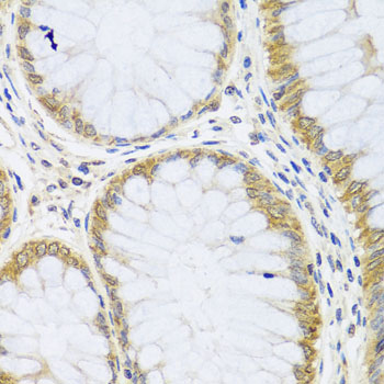
The catalog includes one anti-ARL6 antibody with real IHC images of paraffin-embedded human prostate, colon, and liver damage samples; human and mouse reactivity is listed (A05003 image captions; catalog: reactivity).
A05003 is listed for IHC and has images of paraffin-embedded human prostate, colon, and liver damage samples at 1:100 (catalog: applications; A05003 image captions). Mouse reactivity is listed, but the supplied images show human samples only (catalog: reactivity; A05003 image captions).
Which to pick: Choose A05003 for tissue IHC: its application list includes IHC, and its images document paraffin-embedded human tissue with microwave retrieval in 10 mM PBS, pH 7.2; the fixative is unreported (catalog: applications; A05003 image captions). No IF/ICC application or image is supplied for A05003, so the payload does not support an IF/ICC choice (catalog: applications; catalog: IF images). A05003 lists human and mouse reactivity, but mouse tissue IHC is not shown; its host is rabbit and clonality is unreported (catalog: reactivity and host; A05003 image captions; catalog: clone).