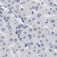
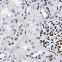
ARNT / Aryl hydrocarbon receptor nuclear translocator · IHC design guide

Design Immunohistochemistry for ARNT

Plan ARNT staining in paraffin sections with nuclear signal as the expected tissue pattern (HPA tissue IHC). Start with the catalog antibody at 1:50 and compare nuclear staining across sections fixed and processed consistently (datasheet M02263; standard IHC practice).

Evidence assembled Oct 2026 · For research use; verify linked source records and product datasheet before use
Immunohistochemistry protocol sheet for ARNT (IHC for ARNT): expected localisation Nuclear staining in most tissues (HPA tissue IHC), antibody M02263, validated IHC image, and IHC protocol steps
Printable ARNT IHC protocol sheet — expected localisation Nuclear staining in most tissues (HPA tissue IHC), antibody M02263, controls and protocol steps. Open the full ARNT IHC guide →

ARNT Immunohistochemistry Experimental Design Guide

Expected localisation, validated protocols, controls and antibodies — the at-a-glance facts below, then the full design guide.

Must know before staining
Expected localisation Nuclear staining in most tissues (HPA tissue IHC)
Staining pattern Nuclear; high in lung type I alveolar and fallopian glandular cells (HPA tissue IHC)
Antigen retrieval EDTA pH 8.0 HIER, heat-mediated (datasheet M02263)
Positive control ⓘ Endometrium+4 more · see all
Negative control ⓘ Cerebellum+1 more · see all
Important caveats
Reasons your staining may differ from the expected pattern.
Fixation Matched tissue-IHC evidence does not establish the fixation claim. Validate the specimen-specific method before use. (selected-SKU IHC image M02263)
Caveat Staining has medium consistency with RNA expression (HPA tissue IHC)
Regulation Expression regulation unannotated (UniProt)
Isoform / epitope 4 isoforms; epitope coverage is unestablished (UniProt)
Section 1

Recommended ARNT IHC & IF Protocols

The catalog antibody’s IHC-P protocol uses heat-mediated EDTA retrieval (datasheet M02263). Four published ARNT chromogenic IHC protocols provide comparison conditions (PMC2938742; PMC7418509; PMC4484474; PMC11133443).

Recommended immunohistochemistry (IHC-P) protocol parameters
SampleParaffin-embedded human renal cancer tissue; fixative not specified (datasheet M02263)
FixationImage fixative and duration unreported (datasheet M02263); verify before use.
Sectioning4–5 µm sections on charged slides (standard)
DeparaffinisationXylene, graded ethanol series to water (standard)
Antigen retrievalHeat retrieval: EDTA pH 8.0 (datasheet M02263); 20 min, 95–100 °C (standard)
Peroxidase block3% H2O2, 10 min, room temperature (standard)
Blocking10% goat serum (datasheet M02263)
Primary antibodyRabbit monoclonal (clone EAD-1) anti-ARNT, 1:50 (datasheet M02263)
Primary incubationOvernight at 4 °C (datasheet M02263)
DetectionHRP-conjugated secondary, DAB chromogen (datasheet M02263)
CounterstainHematoxylin, blue, dehydrate and mount (standard)
Expected resultARNT-positive staining in cells in endometrial stroma of endometrium (HPA tissue IHC: Medium). HPA tissue profile: Nuclear expression in most tissues. No signal in the no-primary control.
💡Decision noteStart with EDTA pH 8.0 for the catalog antibody (datasheet M02263). Citrate pH 6.0 is a published alternative to evaluate during retrieval optimization (PMC2938742; PMC4484474).
Section 2

What Is the Expected ARNT Staining Pattern?

ARNT is a nuclear transcription factor with no transmembrane segment (UniProt P27540: nucleus, topology). In paraffin sections, expect nuclear staining across many tissues, including fallopian tube glandular cells, lung alveolar type I cells and placental decidual cells (HPA: nuclear expression in most tissues; High in these cells). HPA rates the tissue IHC pattern Supported, with medium consistency between staining and RNA expression (HPA: reliability).

What am I looking at on my slide?
Nuclei stain in the expected cell population, with little signal outside nuclei.This fits the reported ARNT location (UniProt P27540: nucleus). Judge intensity by the tissue and cell type: fallopian tube glandular cells are High, whereas endometrial stromal cells are Medium (HPA: tissue IHC). Nuclear staining need not be equally strong throughout a section because HPA reports cell-specific levels (HPA: tissue IHC).
Strong staining is mainly cytoplasmic, membranous or extracellular.That distribution conflicts with ARNT's nuclear location and lack of a transmembrane segment (UniProt P27540: location, topology). Treat it as suspect and review a known-positive nuclear control, tissue morphology and detection controls before scoring it as ARNT (general IHC practice).
Cardiomyocytes or cerebellar granular-layer cells stain strongly.HPA reports ARNT as Not detected in those cells, although its tissue pattern is only Supported (HPA: heart muscle, cerebellum; reliability). Review the staining against adjacent cell types and controls; antibody cross-reactivity or endogenous detection activity is possible, but this observation alone does not establish either cause (general IHC practice).
Brown signal coats many structures without a clear nuclear boundary.This is diffuse background rather than a convincing nuclear pattern (UniProt P27540: nucleus; general IHC practice). Examine a section processed without primary antibody for detection background, and check blocking, washes and chromogen development as general IHC variables (general IHC practice).
A selected known-positive cell population has no nuclear staining.First verify that the section contains the relevant cells: fallopian tube glandular cells, lung alveolar type I cells and placental decidual cells are reported High (HPA: tissue IHC). If morphology confirms their presence, check the IHC workflow and antibody performance before interpreting a blank section biologically (general IHC practice).
💡Expected ARNT appearanceCall ARNT positive when nuclei of the relevant cells stain, potentially strongly in HPA High populations (UniProt P27540: nucleus; HPA: tissue IHC); isolated strong cytoplasmic or membranous color is suspect (UniProt P27540: topology; general IHC practice).
How each factor affects the staining
Tissue and cell selectionUse HPA's cell-level observations when choosing a positive comparator: fallopian tube glandular cells, lung alveolar type I cells and placental decidual cells are High; endometrial stromal cells are Medium (HPA: tissue IHC). HPA's Not detected calls for cardiomyocytes and cerebellar granular-layer cells describe those cells, not whole-tissue absence (HPA: tissue IHC).
Evidence strength and antibodiesThe overall tissue IHC pattern is Supported, with medium consistency against RNA data (HPA: reliability). HPA lists IHC status as Supported for HPA001759 and CAB004318 (HPA: antibodies). These labels support a comparison with the reported pattern; they do not establish that every unexpected signal is specific.
Molecular form and locationARNT has four listed isoforms and no signal peptide, propeptide or transmembrane segment (UniProt P27540: isoforms, processing, topology). Its reported location is nuclear (UniProt P27540: subcellular location). The supplied record gives no antibody epitope, so it cannot establish which isoforms an individual antibody detects.
IF/ICC Q&A: where should ARNT appear?Nucleoplasm and nuclear bodies are the supported locations in HPA ICC-IF images from A-431, U-251MG and U2OS (HPA: subcellular). This is an IF/ICC localisation reference; compare paraffin-section results primarily with HPA tissue IHC observations (HPA: tissue IHC).
Why is my staining missing, weak or wrong?
SituationLikely causeNext action
A High comparator has no visible nuclear signal.The relevant cells may be absent from the viewed field, or a general IHC workflow step may have failed (HPA: High cell populations; general IHC practice).Confirm the expected cell type on the counterstained section, then review the IHC-validated antibody's prescribed procedure, detection reagents and a parallel positive control (general IHC practice). Do not infer a target-specific fixation effect; none is supplied.
Only faint nuclear signal appears in a chosen tissue.The sampled cells may have a lower reported level: adipocytes and endometrial stromal cells are Medium, while several listed cell populations are Low (HPA: tissue IHC).Identify the stained cell type before changing conditions. Compare it with an HPA High cell population processed in parallel, and interpret any intensity difference within the limits of HPA's Supported rating (HPA: tissue IHC; reliability).
Strong color appears outside nuclei.The location disagrees with ARNT's nuclear annotation; nonspecific staining or detection background may explain it (UniProt P27540: location; general IHC practice).Compare with a primary-omission control and examine whether a known-positive population retains a clear nuclear pattern. Review blocking and chromogen development as general IHC checks (general IHC practice).
A reported Not detected cell population appears positive.Cross-reactivity, endogenous detection activity or misidentification of nearby cells may contribute (HPA: cardiomyocytes and cerebellar granular-layer cells Not detected; general IHC practice).Recheck cell identity and nuclear boundaries, then compare primary-omission and known-positive controls. Treat the HPA call as a reference observation rather than proof that every positive cell is an artefact (HPA: tissue IHC; reliability).
The whole section has diffuse brown background.Blocking, washing or chromogen development may be contributing to nonspecific signal (general IHC practice).Inspect a primary-omission section and review those general IHC steps. Score only cell-associated nuclear staining that can be distinguished from background (UniProt P27540: nucleus; general IHC practice).
IF/ICC images show nuclear bodies, but IHC nuclei look more uniform.HPA reports nucleoplasm and nuclear bodies in ICC-IF, while its tissue IHC summary reports nuclear expression without requiring resolvable nuclear bodies (HPA: subcellular; tissue IHC).For this paraffin-section guide, assess nuclear localisation and the relevant tissue cell pattern (HPA: tissue IHC). Use the ICC-IF observation as a localisation comparison, without requiring nuclear-body detail in chromogenic IHC.

Sample controls for ARNT IHC & IF

🧪Run fallopian tube first: glandular cells should stain for ARNT (HPA: High in fallopian tube glandular cells). Use heart muscle as the negative tissue (HPA: Not detected in cardiomyocytes); on the positive slide, score neighboring cells with counterstain-only nuclei and no nuclear DAB as internal negatives, without assuming a particular neighboring cell type is negative.
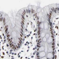
Positive control tissue: Endometrium (Cells in endometrial stroma, HPA Medium)
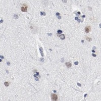
Negative control tissue: Cerebellum (HPA Not detected)
ICC-IF cell lines (HPA subcellular resource): HPA ICC-IF images show ARNT in A-431, U-251MG, U2OS, with annotated localisation: Nucleoplasm (supported), Nuclear bodies (supported) (HPA subcellular).
Technical controls: Include a no-primary, secondary-only slide and a concentration-matched rabbit immunoglobulin isotype control (M02263 caption: rabbit primary antibody). Use identically processed ARNT-knockout material as a biological negative if available; quench endogenous peroxidase and check for nonspecific DAB deposits in fallopian tube glands (M02263 caption: peroxidase detection with DAB).
⚠️Feasibility: A target-specific fixation window or fixation effect is unreported, and the selected M02263 paraffin-section caption does not state a fixative (M02263 caption). Heat retrieval with EDTA at pH 8.0 was used in the caption, but its necessity for fallopian tube is unreported (M02263 caption). The evidence does not establish whether frozen sections or IF/ICC are easier; for fallopian tube IHC, distinguish nuclear glandular staining from any luminal DAB deposit (HPA: High in fallopian tube glandular cells; UniProt P27540: nucleus).

HPA tissue IHC evidence for ARNT

Comprehensive Human Protein Atlas IHC scoring per tissue (reliability: Supported — Medium consistency between antibody staining and RNA expression data.). Rows are taken directly from the HPA tissue chart — click any row's HPA link to view the source.

Positive expression · recommended positive controls

TissueCell typeLevelEvidenceSource
Endometrium Cells in endometrial stroma Medium Protein (IHC) HPA →
Fallopian tube Glandular cells High Protein (IHC) HPA →
Lung Alveolar cells type I High Protein (IHC) HPA →
Placenta Decidual cells High Protein (IHC) HPA →
Adipose tissue Adipocytes Medium Protein (IHC) HPA →

Undetected expression · recommended negative controls

TissueCell typeLevelEvidenceSource
Cerebellum Cells in granular layer Not detected Protein (IHC) HPA →
Heart muscle Cardiomyocytes Not detected Protein (IHC) HPA →
Section 3

Advanced ARNT IHC Tips

Use nuclear localisation, matched controls, and the catalog antibody’s documented paraffin-section conditions to troubleshoot ARNT chromogenic IHC (UniProt P27540; datasheet M02263).

How should I adjust retrieval when ARNT nuclear staining is weak?
Start with heat-mediated retrieval in EDTA pH 8.0 for paraffin sections (datasheet M02263). The documented image then used a 1:50 primary dilution overnight at 4°C, so hold those conditions constant while checking retrieval (datasheet M02263). Compare adjacent sections with the same heating and cooling schedule, recording both nuclear signal and tissue damage (standard IHC practice). If staining remains weak, test an alternative retrieval buffer only as a controlled fallback, because its performance for this antibody is unreported (datasheet M02263). Score nuclei in comparable, viable regions; ARNT is reported as nuclear (UniProt P27540).
Could fixation explain variable ARNT staining between paraffin blocks?
The catalog image identifies paraffin-embedded human renal cancer tissue but does not state its fixative, so ARNT-specific fixation sensitivity is unknown (datasheet M02263). Record the fixative and processing history for each block, then compare sections cut and stained together (standard IHC practice). If fixation varies, use matched material to assess nuclear signal and morphology before changing the EDTA pH 8.0 retrieval condition (datasheet M02263; standard IHC practice). Keep the 1:50 primary incubation overnight at 4°C during that comparison (datasheet M02263). Neither ARNT’s nuclear location nor its annotated modifications establish a particular fixation effect (UniProt P27540).
What should I check when ARNT staining appears mainly cytoplasmic?
Examine the chromogenic deposit against a nuclear counterstain: ARNT is annotated in the nucleus, and tissue profiling describes nuclear expression in most tissues (UniProt P27540; HPA tissue IHC). Supported IF localisation includes nucleoplasm and nuclear bodies, which gives a useful compartment check without proving the catalog antibody’s specificity (HPA subcellular). If cytoplasmic colour dominates, review section thickness, counterstain, focus, and diffuse background on an adjacent control section (standard IHC practice). Compare the same cell population across slides; HPA reports high staining in fallopian tube glandular cells and no detection in cerebellar granular-layer cells (HPA tissue IHC).
Can isoforms or epitope masking explain inconsistent nuclear signal?
ARNT has 4 annotated isoforms, but the supplied catalog image does not identify the antibody’s epitope or establish isoform coverage (UniProt P27540; datasheet M02263). Its annotated bHLH domain spans residues 89–142, and PAS domains span 161–235 and 349–419; these locations alone cannot assign the recognized site (UniProt P27540). Annotated modifications include N-acetylalanine at residue 2 and phosphoserine at 77, without evidence here that either changes staining (UniProt P27540). Check the antibody’s mapped immunogen before interpreting a negative nucleus as loss of every isoform (standard IHC practice). Compare matched sections under the documented EDTA pH 8.0 retrieval condition (datasheet M02263).
How can I investigate ARNT by IF alongside this IHC result?
Use the paraffin-section IHC result as a localisation reference, while treating IF conditions as a separate optimisation (datasheet M02263; standard IF practice). Multiplex ARNT with a validated marker for the expected cell population, such as the glandular cells reported positive in fallopian tube, and include single-stain controls (HPA tissue IHC; standard IF practice). Choose a fluorophore away from prominent tissue autofluorescence and check spectral overlap before judging nuclear signal (standard IF practice). Because ARNT has no annotated transmembrane segment and localises to the nucleus, permeabilise sufficiently to expose nuclear epitopes, then titrate to preserve morphology (UniProt P27540; standard IF practice). HPA reports supported nucleoplasm and nuclear-body localisation in ICC/IF images (HPA subcellular).
How do I reduce diffuse brown staining without losing nuclear ARNT?
First inspect a no-primary control for endogenous peroxidase activity and nonspecific detection, then apply an appropriate peroxidase block before chromogen development (standard IHC practice). The documented section received 10% goat serum blocking, 1:50 rabbit primary overnight at 4°C, and peroxidase-conjugated secondary for 30 minutes at 37°C (datasheet M02263). Reduce background by checking washes and titrating the primary around that documented condition on matched sections (standard IHC practice; datasheet M02263). Judge the resulting DAB signal within nuclei, since ARNT is annotated as nuclear, and compare it with the no-primary control (UniProt P27540; standard IHC practice).
What is a defensible way to score ARNT IHC across specimens? ⚠ ANSWER MARKED FOR VERIFICATION
Define the target cell population and score only interpretable nuclei on consistently developed, viable sections (UniProt P27540; standard IHC practice). Report the percentage of positive nuclei and staining intensity, or calculate a nuclear H-score from 0–300 using percentages at each intensity level (standard IHC practice). For spatial comparisons, report positive nuclei per mm² of viable tissue and normalise to the total counted nuclei or viable area as appropriate (standard IHC practice). Keep retrieval at EDTA pH 8.0 and acquisition or scoring thresholds consistent across batches (datasheet M02263; standard IHC practice). Record cell type, since HPA staining levels vary across cell populations (HPA tissue IHC).
How can I distinguish genuine ARNT positivity from staining artefacts?
Prioritise well-preserved nuclear staining in the intended cell population: ARNT is nuclear, and HPA describes nuclear expression in most tissues (UniProt P27540; HPA tissue IHC). Check suspicious edge-only colour, necrotic areas, and deposits outside nuclei against neighbouring viable tissue and a no-primary control (standard IHC practice). Endogenous peroxidase can create DAB colour, so review the peroxidase-block control before attributing brown signal to ARNT (standard IHC practice). HPA rates its tissue staining Supported with medium agreement to RNA data; use its reported cell patterns as context, not proof for an individual specimen (HPA tissue IHC). The catalog image documents staining in paraffin-embedded human renal cancer tissue without reporting the fixative (datasheet M02263).
Boster reagents

Best ARNT / Aryl hydrocarbon receptor nuclear translocator IHC Antibodies

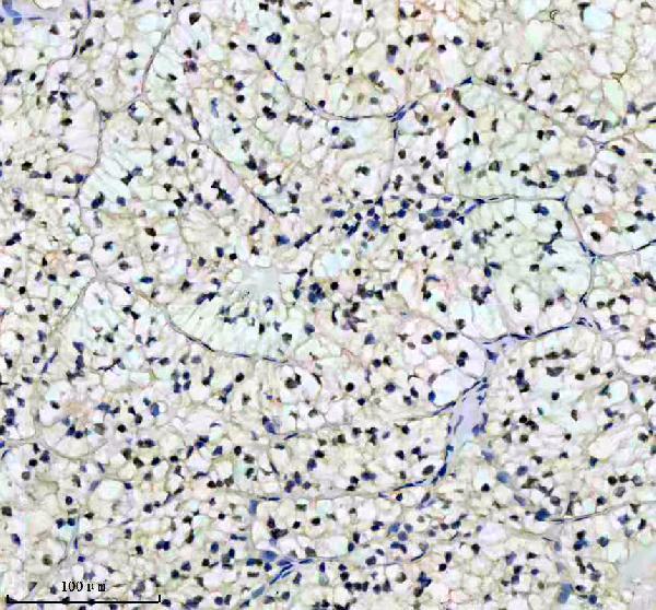
M02263 has chromogenic IHC data from a paraffin-embedded human renal cancer section; no IF image is supplied (M02263 IHC image caption; catalog: if_image_alts).

Real IHC data IHC analysis of ARNT using anti-ARNT antibody (M02263). ARNT was detected in a paraffin-embedded section of human renal cancer tissue. Heat mediated antigen retrieval was performed in EDTA buffer (pH 8.0, epitope retrieval solution). The tissue section was blocked with 10% goat serum. The tissue section was then incubated with 1:50 rabbit anti-ARNT Antibody (M02263) overnight at 4°C. Peroxidase Conjugated Goat Anti-rabbit IgG was used as secondary antibody and incubated for 30 minutes at 37°C. The tissue section was developed using HRP Conjugated Rabbit IgG Super Vision Assay Kit (Catalog # SV0002) with DAB as the chromogen.
Anti-HIF-1 beta ARNT Rabbit Monoclonal Antibody
Cat # M02263

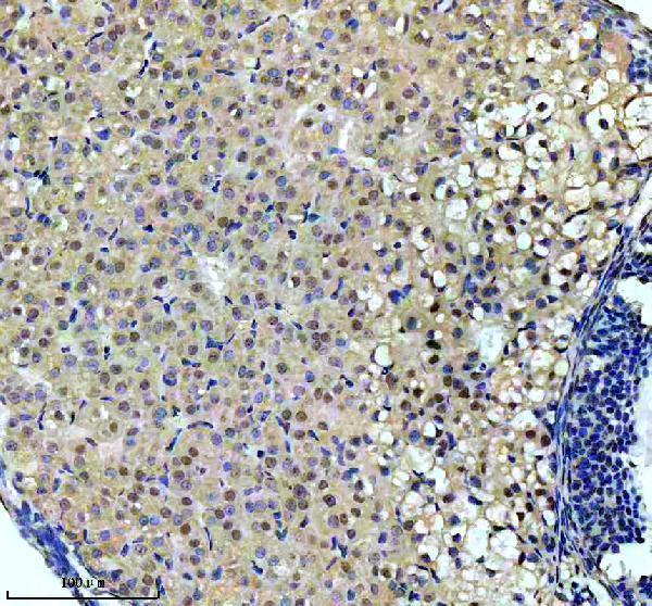
M02263 is listed for human IHC and has an image caption for a paraffin-embedded human renal cancer section (catalog: applications/reactivity; M02263 IHC image caption). A second M02263 caption describes a paraffin-embedded mouse ovary section, while the catalog lists Human reactivity only (M02263 IHC image caption; catalog: reactivity).

Which to pick: Choose M02263 for human paraffin-section IHC: it is a rabbit monoclonal, clone EAD-1, with an IHC example using 1:50 primary antibody (catalog: host/clone/applications; M02263 IHC image caption). The payload does not support an IF/ICC recommendation for M02263 because IF is absent from its application list and no IF dilution or image is supplied (catalog: applications/if_dilution/if_image_alts). Its mouse ovary caption provides a cross-species IHC example, but listed reactivity is Human only; the captions describe paraffin sections without reporting the fixative (M02263 IHC image captions; catalog: reactivity).

Each figure is that product's own IHC / IF validation image from its datasheet.

References

  1. UniProt Consortium. UniProt entry P27540 (ARNT_HUMAN, Aryl hydrocarbon receptor nuclear translocator).
  2. Human Protein Atlas. ARNT tissue IHC expression (reliability: Supported).
  3. Human Protein Atlas. ARNT subcellular location (ICC-IF): Localized to the nucleoplasm and nuclear bodies..
  4. Human Protein Atlas. ARNT antibody validation summary (2 antibodies).
  5. Aryl hydrocarbon nuclear translocator (hypoxia inducible factor 1beta) activity is required more during early than late tumor growth. Molecular carcinogenesis 2010 — PMC2938742.
  6. Aryl hydrocarbon receptor nuclear translocator promotes the proliferation and invasion of clear cell renal cell carcinoma cells potentially by affecting the glycolytic pathway. Oncology letters 2020 — PMC7418509.
  7. Down-regulation of ARNT promotes cancer metastasis by activating the fibronectin/integrin β1/FAK axis. Oncotarget 2015 — PMC4484474.
  8. Targeting ARNT attenuates chemoresistance through destabilizing p38α-MAPK signaling in glioblastoma. Cell death & disease 2024 — PMC11133443.
  9. PubMed PMID:1852076 — UniProt-cited evidence.
  10. PubMed PMID:14702039 — UniProt-cited evidence.
  11. PubMed PMID:11230166 — UniProt-cited evidence.