This website uses cookies to ensure you get the best experience on our website.


- Table of Contents
Plan chromogenic IHC for ARPP21 in paraffin sections using its reported cytoplasmic tissue pattern (HPA tissue IHC). Compare neuronal and glial staining in brain with skeletal muscle myocytes, and interpret intensity alongside the reported medium consistency between staining and RNA (HPA tissue IHC).

Expected localisation, validated protocols, controls and antibodies — the at-a-glance facts below, then the full design guide.
| Expected localisation | Cytoplasmic in brain and skeletal muscle (HPA tissue IHC) | |
| Staining pattern | Cytoplasmic signal in brain cells and skeletal myocytes (HPA tissue IHC) | |
| Antigen retrieval | Citrate pH 6.0 HIER, 95–98 °C, 20 min (rule: cytoplasmic / membrane antigen) | |
| Positive control | Appendix+2 more · see all | |
| Negative control | Adipose tissue+4 more · see all |
| Fixation | Keep fixation conditions consistent across paraffin sections. (standard IHC practice; not target-specific) | |
| Caveat | Staining and RNA show only medium consistency (HPA tissue IHC) | |
| Regulation | Brain-enriched RNA expression (HPA tissue RNA) | |
| Isoform / epitope | Six isoforms; epitope coverage is unspecified (UniProt; datasheets) |
Compare the catalog antibody’s IHC-P protocol with the published ARPP21 dual-staining example in patient spinal cord (PMC13545126).
| Sample | Paraffin-embedded human heart tissue; fixative not specified (datasheet A03525-1) |
| Fixation | Image fixative and duration unreported (datasheet A03525-1); verify before use. |
| Sectioning | 4–5 µm sections on charged slides (standard) |
| Deparaffinisation | Xylene, graded ethanol series to water (standard) |
| Antigen retrieval | Heat-induced epitope retrieval in citrate buffer, pH 6.0, 20 min at 95–98 °C (standard rule: cytoplasmic / membrane antigen) |
| Peroxidase block | 3% H2O2, 10 min, room temperature (standard) |
| Blocking | 10% normal serum of the secondary host, 30 min, room temperature (standard) |
| Primary antibody | Rabbit anti-ARPP21, 1:100-1:300 (datasheet A03525-1) |
| Primary incubation | Overnight at 4 °C (standard) |
| Detection | HRP-polymer secondary, DAB chromogen 5–10 min (standard) |
| Counterstain | Hematoxylin, blue, dehydrate and mount (standard) |
| Expected result | ARPP21-positive staining in goblet cells of appendix (HPA tissue IHC: Medium). HPA tissue profile: Cytoplasmic expression in the brain and skeletal muscles. No signal in the no-primary control. |
ARPP21 is a cytoplasmic protein with no transmembrane segment (UniProt Q9UBL0 topology). In paraffin-section IHC, expect cytoplasmic staining in cerebral-cortex neurons, caudate glial cells and appendix goblet cells, each reported at medium intensity (HPA tissue IHC). HPA also describes cytoplasmic expression in brain and skeletal muscle, but rates tissue IHC reliability as Enhanced with medium consistency between antibody staining and RNA expression (HPA tissue IHC).
| Medium cytoplasmic staining in cerebral-cortex neurons or caudate glial cells, with tissue detail preserved. | This matches the reported cell types, intensity and tissue-IHC compartment (HPA tissue IHC). Score the named cells separately from neighboring cells; a positive field alone does not establish that every cell in it expresses ARPP21 (general IHC interpretation). |
| Predominantly nuclear or nucleolar staining in paraffin-section IHC, without the expected cytoplasmic pattern. | Treat this as a compartment mismatch against the tissue-IHC profile and UniProt cytoplasmic annotation (HPA tissue IHC; UniProt Q9UBL0). Check morphology and controls before calling it ARPP21; HPA's approved nucleolar observation comes from ICC-IF, a different application (HPA ICC-IF). |
| Strong staining in adipocytes or adrenal glandular cells while the expected positive cells are unstained. | Those cell types are listed as not detected (HPA tissue IHC). Suspect cross-reactivity or endogenous chromogenic activity, then compare a no-primary control and a known-positive section processed in the same run (general IHC practice). |
| Diffuse color across cells, stroma or section edges, obscuring cell boundaries. | This is difficult to score as cytoplasmic ARPP21 (HPA tissue IHC). Review blocking, washes, primary-antibody concentration and detection controls; diffuse deposition can arise from general IHC workflow rather than the reported cell-specific pattern (general IHC practice). |
| No staining in cerebral-cortex neuronal cells or caudate glial cells. | Both are reported at medium intensity, so absence calls for a run-level check before a biological negative call (HPA tissue IHC). Verify that the tissue and target cells are present, then review retrieval, antibody incubation and chromogen performance (general IHC practice). |
| Tissue and cell selection | Cortex neurons, caudate glial cells and appendix goblet cells are medium; skeletal-muscle myocytes are low, so a weak muscle result alone is a less decisive positive control (HPA tissue IHC). |
| Antibody validation | HPA017303 has Enhanced IHC validation (HPA antibodies). The tissue-IHC profile still has medium antibody/RNA consistency, so interpret unexpected cells and intensity with controls (HPA tissue IHC). |
| Isoform context | Isoform 2 is expressed in brain; isoform 1 is present in immature thymocytes at protein level (UniProt Q9UBL0). The supplied IHC record does not assign individual tissue-staining observations to an isoform (HPA tissue IHC). |
| Topology and processing | ARPP21 has no transmembrane segment or signal peptide; its listed chain spans residues 2–812 (UniProt Q9UBL0). Interpret a crisp cell-surface-only pattern cautiously against the cytoplasmic tissue-IHC profile (HPA tissue IHC). |
| IF/ICC Q: Should nucleoli stain? | A: HPA ICC-IF lists approved nucleoli as the main location and approved cytosol as an additional location (HPA ICC-IF). That application-specific result does not replace the cytoplasmic paraffin-section IHC expectation (HPA tissue IHC). |
| Situation | Likely cause | Next action |
|---|---|---|
| The known-positive cortex or caudate section has no visible signal. | The target cells may be absent from the field, or an IHC step may have failed (general IHC practice); HPA reports medium staining in these cells (HPA tissue IHC). | Confirm cell identity and section integrity, then check the run's retrieval, primary incubation and detection controls before scoring the sample negative (general IHC practice). |
| Only nuclei or nucleoli are colored in an IHC section. | The pattern conflicts with cytoplasmic tissue IHC and UniProt localization (HPA tissue IHC; UniProt Q9UBL0); ICC-IF has a separate approved nucleolar observation (HPA ICC-IF). | Inspect cell boundaries and a no-primary control, then repeat or confirm with the IHC-validated antibody and an appropriate positive section (general IHC practice; HPA antibodies). |
| Adipocytes or adrenal glandular cells stain strongly. | Both are reported as not detected in those cells (HPA tissue IHC); nonspecific antibody binding or endogenous detection activity is possible (general IHC practice). | Compare no-primary and known-positive controls; review blocking and detection chemistry before assigning ARPP21 positivity (general IHC practice). |
| Background color obscures the cytoplasm in most cells. | Excess primary-antibody signal, incomplete washing or endogenous chromogenic activity may obscure localization (general IHC practice). | Check the no-primary control and reagent instructions; optimize blocking, washes and antibody concentration while retaining a positive-control section (general IHC practice). |
| Skeletal muscle looks faint beside a clearly positive brain section. | Myocytes are listed as low, while cerebral-cortex neuronal cells and caudate glial cells are medium (HPA tissue IHC). | Score each cell population against its reported level and morphology; use the medium brain staining to judge whether the run detected the target (HPA tissue IHC; general IHC interpretation). |
| A stained field is reported positive although the named target cells are unstained. | A field-level impression can conceal staining in another cell type (general IHC interpretation); HPA observations specify cells within each tissue (HPA tissue IHC). | Record tissue, cell identity, compartment and intensity separately, and compare the result with the corresponding HPA cell-level observation (HPA tissue IHC; general IHC interpretation). |
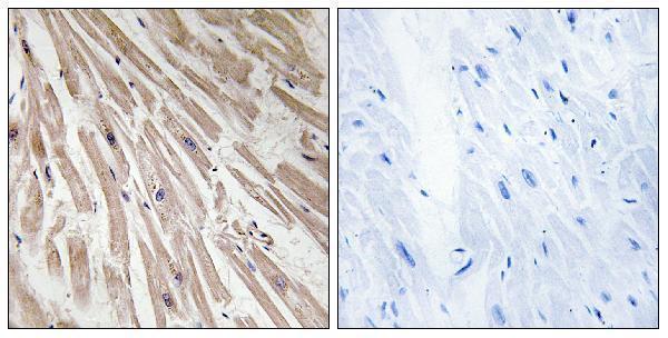
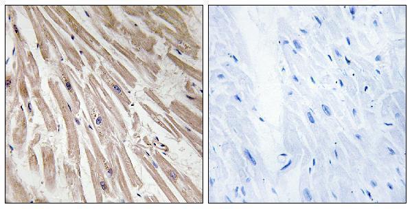


Comprehensive Human Protein Atlas IHC scoring per tissue (reliability: Enhanced — Medium consistency between antibody staining and RNA expression data.). Rows are taken directly from the HPA tissue chart — click any row's HPA link to view the source.
| Tissue | Cell type | Level | Evidence | Source |
|---|---|---|---|---|
| Appendix | Goblet cells | Medium | Protein (IHC) | HPA → |
| Caudate | Glial cells | Medium | Protein (IHC) | HPA → |
| Cerebral cortex | Neuronal cells | Medium | Protein (IHC) | HPA → |
| Tissue | Cell type | Level | Evidence | Source |
|---|---|---|---|---|
| Adipose tissue | Adipocytes | Not detected | Protein (IHC) | HPA → |
| Adrenal gland | Glandular cells | Not detected | Protein (IHC) | HPA → |
| Bone marrow | Hematopoietic cells | Not detected | Protein (IHC) | HPA → |
| Breast | Adipocytes | Not detected | Protein (IHC) | HPA → |
| Bronchus | Respiratory epithelial cells | Not detected | Protein (IHC) | HPA → |
Troubleshoot ARPP21 staining in paraffin sections by checking retrieval, compartment, cell type and controls before comparing signal intensity across samples.
Two anti-ARPP21 antibodies have paraffin-section IHC images from human heart (each SKU’s IHC image caption); both list IF and human/mouse reactivity (catalog applications/reactivity).
A03525-1 lists IHC and IF, with an IHC image of paraffin-embedded human heart tissue (A03525-1 catalog applications; IHC image caption). A30584 lists IHC, IF and ICC, with its own IHC image of paraffin-embedded human heart tissue (A30584 catalog applications; IHC image caption).
Which to pick: For tissue IHC, either A03525-1 or A30584 has an image supporting paraffin sections; the fixative is unreported in both captions (each SKU’s IHC image caption). For IF/ICC, choose A30584 if ICC is required because it lists both applications; A03525-1 lists IF but not ICC (catalog applications). Both are rabbit polyclonals listed as reactive with human and mouse, though their IHC images show human tissue only (catalog host/dilution data and reactivity; each SKU’s IHC image caption).