This website uses cookies to ensure you get the best experience on our website.


- Table of Contents
Plan ARSD staining in paraffin sections with the catalog antibody at 1:100–1:300 (datasheet). Compare granular cytoplasmic staining in bronchial respiratory epithelial cells with adipocytes, where staining was not detected (HPA tissue IHC).

Expected localisation, validated protocols, controls and antibodies — the at-a-glance facts below, then the full design guide.
| Expected localisation | Granular cytoplasm (HPA tissue IHC); lysosome expected (UniProt) | |
| Staining pattern | Granular cytoplasm in glandular and respiratory epithelial cells (HPA tissue IHC) | |
| Antigen retrieval | Citrate pH 6.0 HIER, 95–98 °C, 20 min (rule: cytoplasmic / membrane antigen) | |
| Positive control | Adrenal gland+4 more · see all | |
| Negative control | Adipose tissue+2 more · see all |
| Fixation | Keep fixation consistent across sections. (standard IHC practice; not target-specific) | |
| Caveat | Staining and RNA show medium consistency (HPA tissue IHC) | |
| Regulation | Expression regulation not annotated (UniProt) | |
| Isoform / epitope | 3 isoforms; epitope coverage is unspecified (UniProt; datasheet) |
Compare the catalog antibody's IHC-P protocol with published ARSD staining in breast cancer and glioma specimens (PMC8560752; PMC10521731; PMC13547030).
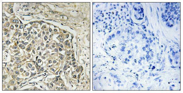
| Sample | Paraffin-embedded human lung carcinoma tissue; fixative not specified (datasheet A08673-1) |
| Fixation | Image fixative and duration unreported (datasheet A08673-1); verify before use. |
| Sectioning | 4–5 µm sections on charged slides (standard) |
| Deparaffinisation | Xylene, graded ethanol series to water (standard) |
| Antigen retrieval | Heat-induced epitope retrieval in citrate buffer, pH 6.0, 20 min at 95–98 °C (standard rule: cytoplasmic / membrane antigen) |
| Peroxidase block | 3% H2O2, 10 min, room temperature (standard) |
| Blocking | 10% normal serum of the secondary host, 30 min, room temperature (standard) |
| Primary antibody | Rabbit anti-ARSD, 1:100-1:300 (datasheet A08673-1) |
| Primary incubation | Overnight at 4 °C (standard) |
| Detection | HRP-polymer secondary, DAB chromogen 5–10 min (standard) |
| Counterstain | Hematoxylin, blue, dehydrate and mount (standard) |
| Expected result | ARSD-positive staining in glandular cells of adrenal gland (HPA tissue IHC: High). HPA tissue profile: General cytoplasmic expression with a granular pattern. No signal in the no-primary control. |
ARSD is lysosomal and has no annotated transmembrane segment (UniProt P51689). In paraffin IHC, expect granular cytoplasmic staining in positive cells, including glandular, respiratory epithelial, neuronal and glial cells (HPA tissue IHC). HPA rates its tissue staining Approved, with medium consistency between staining and RNA expression; interpret individual fields with that qualification (HPA tissue IHC).
| Granular cytoplasmic staining is strong in adrenal or appendix glandular cells. | This matches HPA's general cytoplasmic, granular profile and its High staining calls for these cells (HPA tissue IHC). Score the identified cells and their cytoplasm, rather than treating every stained structure in the section as ARSD positive (standard IHC practice). |
| The dominant signal is nuclear or outlines the cell surface, with little cytoplasmic granularity. | That distribution conflicts with the reported IHC pattern and lysosomal assignment (HPA tissue IHC; UniProt P51689). Check morphology and the antibody control before assigning specificity; compartment mismatch alone does not identify the artefact's cause (standard IHC practice). |
| Adipocytes, ovarian stroma cells or soft tissue fibroblasts stain as strongly as known-positive cells. | HPA reports ARSD as Not detected in those cell populations (HPA tissue IHC). Confirm cell identity and compare controls; unexpected staining can reflect cross-reactivity or endogenous detection activity, but the image alone cannot distinguish them (standard IHC practice). |
| Brown signal is diffuse across tissue, with no cell-defined granular pattern. | This is difficult to reconcile with HPA's granular cytoplasmic profile (HPA tissue IHC). Review the detection control, background distribution and counterstain before scoring it as ARSD; diffuse color alone is insufficient evidence of target localization (standard IHC practice). |
| A well-preserved adrenal or appendix glandular field has no detectable cytoplasmic signal. | HPA calls those glandular cells High, so an absent result warrants investigation (HPA tissue IHC). Check that the expected cells are present and assess the staining run with suitable controls; a single blank field does not establish biological absence (standard IHC practice). |
| Compartment and topology | UniProt places ARSD in lysosomes and annotates no transmembrane segment (UniProt P51689). HPA describes granular cytoplasmic tissue staining (HPA tissue IHC). These records support a cytoplasmic pattern; they do not prove that each chromogenic granule is a lysosome. |
| Tissue and cell selection | HPA reports High staining in bronchial respiratory epithelial cells, caudate neurons, cerebellar granular-layer cells and cortical glia, as well as several glandular populations (HPA tissue IHC). Its RNA classification is low tissue specificity, so assess named cell populations rather than expecting a tissue-exclusive signal (HPA tissue IHC). |
| Antibody evidence | The tissue IHC profile is Approved, with medium consistency between antibody staining and RNA expression (HPA tissue IHC). HPA004694 is listed as IHC Approved; HPA063835 is listed as ICC Approved, with no IHC status supplied for it (HPA antibodies). These statuses do not validate another reagent. |
| Processing, glycosylation and isoforms | UniProt annotates signal residues 1–33, a 34–593 chain, glycosylation at 61, 128 and 347, and three isoforms (UniProt P51689). The supplied records give no antibody epitope or isoform-specific staining evidence, so do not attribute a weak field to cleavage, glycosylation or one isoform. |
| What should ICC-IF show? | HPA reports vesicles and lipid droplets as Approved ICC-IF locations, with images from A-549, MCF-7 and U2OS (HPA subcellular). Use that finding to interpret ICC-IF on its own guide page; the tissue IHC record establishes granular cytoplasm, without resolving those structures in paraffin sections (HPA tissue IHC). |
| Situation | Likely cause | Next action |
|---|---|---|
| No signal in an HPA High glandular population | The expected cells may be absent from the field, or the IHC run may have failed; HPA's High call alone cannot identify the cause (HPA tissue IHC; standard IHC practice). | Confirm glandular morphology, then compare a suitable positive control and the run's staining controls. Review the validated IHC conditions for the antibody used before calling the sample negative (standard IHC practice). |
| Mostly nuclear or continuous surface staining | The dominant distribution disagrees with ARSD's lysosomal assignment and HPA's granular cytoplasmic profile (UniProt P51689; HPA tissue IHC). | Recheck the compartment at higher magnification and compare antibody and detection controls. Record the mismatch rather than scoring that signal as expected ARSD (standard IHC practice). |
| Strong staining in a reported negative cell population | Adipocytes, ovarian stroma cells and soft tissue fibroblasts are Not detected in the supplied HPA tissue profile; misidentified cells or nonspecific detection are possibilities (HPA tissue IHC; standard IHC practice). | Identify the stained cells with morphology and compare nearby positive cells. Use an appropriate detection control to investigate endogenous activity before assigning cross-reactivity (standard IHC practice). |
| Uniform color or haze obscures granules | Diffuse background obscures the cell-defined granular pattern reported for ARSD, but does not establish a target-specific failure (HPA tissue IHC; standard IHC practice). | Inspect the detection control and background outside expected cells; review blocking, washes and chromogen development as general IHC variables (standard IHC practice). |
| Positive cells are weak while background is low | A weak result differs from HPA's High calls in selected cells, yet HPA rates tissue staining only medium-consistent with RNA expression (HPA tissue IHC). No ARSD-specific fixation or retrieval sensitivity is supplied. | Confirm the cell population and control performance. Review the antibody's validated IHC conditions and any retrieval used as general workflow checks; avoid claiming an ARSD-specific fixation effect (standard IHC practice). |
| A granular field contains both plausible positive and negative cells | HPA's calls differ by cell population, including High glandular or neural populations and Not detected adipocytes or fibroblasts (HPA tissue IHC). Mixed morphology can make a field-wide score misleading (standard IHC practice). | Score cytoplasmic granularity and intensity within identified cell types, using the HPA calls as comparators. Keep uncertain cells separate until morphology and controls support their identity (HPA tissue IHC; standard IHC practice). |


Comprehensive Human Protein Atlas IHC scoring per tissue (reliability: Approved — Medium consistency between antibody staining and RNA expression data.). Rows are taken directly from the HPA tissue chart — click any row's HPA link to view the source.
| Tissue | Cell type | Level | Evidence | Source |
|---|---|---|---|---|
| Adrenal gland | Glandular cells | High | Protein (IHC) | HPA → |
| Appendix | Glandular cells | High | Protein (IHC) | HPA → |
| Breast | Glandular cells | High | Protein (IHC) | HPA → |
| Bronchus | Respiratory epithelial cells | High | Protein (IHC) | HPA → |
| Caudate | Neuronal cells | High | Protein (IHC) | HPA → |
Troubleshoot ARSD staining in paraffin sections by checking retrieval, cellular pattern, controls and scoring before interpreting chromogenic signal.
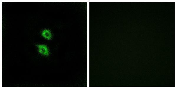
A08673-1 has IHC data from paraffin-embedded human lung carcinoma tissue and IF data from MCF7 cells; listed reactivity is human (IHC and IF image captions; catalog reactivity).
A08673-1 shows IHC staining of paraffin-embedded human lung carcinoma tissue alongside a synthesized-peptide-blocked image (IHC image caption). The same SKU shows IF staining of MCF7 cells alongside a synthesized-peptide-blocked image (IF image caption).
Which to pick: Choose A08673-1 for paraffin-section tissue IHC based on its own IHC image; the fixative is unreported (IHC image caption). For cellular IF, A08673-1 lists IF and shows MCF7 cells; ICC is not separately listed (catalog applications; IF image caption). No cross-species option is documented: A08673-1 lists human reactivity, has a rabbit host, and has no specified clone (catalog reactivity; host; clone).