ASGR1 / Asialoglycoprotein receptor 1 · IHC design guide

Design Immunohistochemistry for ASGR1

Plan chromogenic ASGR1 IHC in paraffin sections with the catalog antibody (datasheet A05376-1). Use liver hepatocytes as a positive reference and assess their distinct cytoplasmic staining (HPA tissue IHC).

Evidence assembled Oct 2026 · For research use; verify linked source records and product datasheet before use
Immunohistochemistry protocol sheet for ASGR1 (IHC for ASGR1): expected localisation Hepatocyte cytoplasm (HPA tissue IHC); cell membrane (UniProt), antibody A05376-1, validated IHC image, and IHC protocol steps
Printable ASGR1 IHC protocol sheet — expected localisation Hepatocyte cytoplasm (HPA tissue IHC); cell membrane (UniProt), antibody A05376-1, controls and protocol steps. Open the full ASGR1 IHC guide →

ASGR1 Immunohistochemistry Experimental Design Guide

Expected localisation, validated protocols, controls and antibodies — the at-a-glance facts below, then the full design guide.

Must know before staining
Expected localisation Hepatocyte cytoplasm (HPA tissue IHC); cell membrane (UniProt)
Staining pattern Distinct cytoplasmic staining in hepatocytes (HPA tissue IHC)
Antigen retrieval EDTA pH 8.0 HIER, heat-mediated (datasheet A05376-1)
Positive control ⓘ Liver
Negative control ⓘ Adipose tissue+4 more · see all
Important caveats
Reasons your staining may differ from the expected pattern.
Fixation Keep fixation consistent across sections (standard IHC practice; not target-specific)
Caveat A secreted variant may complicate tissue staining (HPA tissue IHC)
Regulation No specific expression regulator reported (UniProt)
Isoform / epitope 2 isoforms; map the epitope to extracellular or cytoplasmic regions (UniProt)
Section 1

Recommended ASGR1 IHC & IF Protocols

The catalog antibody has an EDTA pH 8.0 IHC protocol (datasheet: A05376-1). Published paraffin-section examples cover mouse liver and humanized liver (PMC13491154; PMC10104468).

Recommended immunohistochemistry (IHC-P) protocol parameters
SampleParaffin-embedded human liver cancer tissue; fixative not specified (datasheet A05376-1)
FixationImage fixative and duration unreported (datasheet A05376-1); verify before use.
Sectioning4–5 µm sections on charged slides (standard)
DeparaffinisationXylene, graded ethanol series to water (standard)
Antigen retrievalHeat retrieval: EDTA pH 8.0 (datasheet A05376-1); 20 min, 95–100 °C (standard)
Peroxidase block3% H2O2, 10 min, room temperature (standard)
Blocking10% goat serum (datasheet A05376-1)
Primary antibodyRabbit anti-ASGR1, 1:50 recommended; image 1:100 (datasheet A05376-1)
Primary incubationOvernight at 4 °C (datasheet A05376-1)
DetectionHRP-conjugated secondary, DAB chromogen (datasheet A05376-1)
CounterstainHematoxylin, blue, dehydrate and mount (standard)
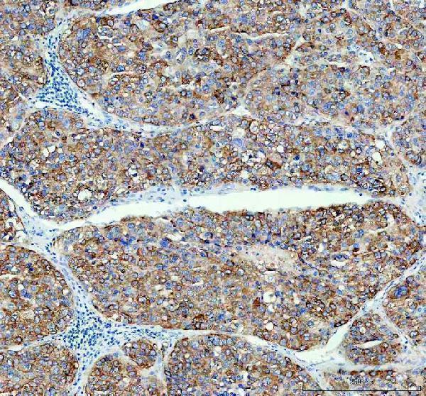
Expected resultASGR1-positive staining in hepatocytes of liver (HPA tissue IHC: High). HPA tissue profile: Distinct cytoplasmic expression in hepatocytes. No signal in the no-primary control.
💡Decision noteStart with heat retrieval in EDTA pH 8.0 for the catalog antibody (datasheet: A05376-1). The published mouse-liver workflow uses Tris-EDTA pH 9.0 (PMC13491154).
Section 2

What Is the Expected ASGR1 Staining Pattern?

In paraffin-section IHC, expect strong cytoplasmic staining in hepatocytes (HPA: High in liver hepatocytes; distinct cytoplasmic expression). ASGR1 is a membrane receptor with a cytoplasmic segment at residues 1–40, a transmembrane segment at 41–61, and an extracellular segment at 62–291 (UniProt P07306 topology). HPA rates the tissue IHC pattern Enhanced, while noting only medium consistency between staining and RNA expression (HPA: reliability description).

What am I looking at on my slide?
Strong, distinct cytoplasmic chromogen in liver hepatocytes, with little staining outside them.This matches the reported positive tissue and cell type (HPA: liver hepatocytes High; distinct cytoplasmic expression). A visible cell border may fit receptor topology, but border staining is not required for a positive paraffin-section result (UniProt P07306 topology; HPA: tissue IHC profile).
Nuclei dominate the signal while hepatocyte cytoplasm is weak or clear.Predominantly nuclear staining conflicts with the reported cytoplasmic tissue pattern and membrane-receptor topology (HPA: tissue IHC profile; UniProt P07306 topology). Review the nuclear counterstain and detection controls before scoring the nuclear color as ASGR1 (standard IHC practice).
Strong staining appears in non-hepatocyte cells, including cells in a tissue reported as negative.That distribution warrants a specificity check rather than automatic scoring as ASGR1 (UniProt P07306: hepatic parenchymal-cell specificity; HPA: tissue IHC profile). Cross-reactivity or endogenous detection activity are possible IHC explanations; neither is established by this slide alone (standard IHC practice).
Pale, diffuse color covers cells and surrounding tissue without a distinct hepatocyte pattern.Diffuse color is harder to interpret than the reported distinct hepatocyte staining (HPA: tissue IHC profile). Residual detection reagent, inadequate blocking, or insufficient washing can create background in chromogenic IHC; compare a no-primary control (standard IHC practice).
No convincing hepatocyte signal appears in the liver positive control.An absent signal conflicts with the reported High hepatocyte staining (HPA: liver hepatocytes High). First check that the section contains preserved, assessable hepatocytes and that the detection system worked; the supplied sources do not establish ASGR1-specific fixation sensitivity (standard IHC practice; HPA: source scope).
💡Expected ASGR1 appearanceCall the result positive when hepatocytes show High, distinct cytoplasmic staining (HPA: liver hepatocytes High; tissue IHC profile); dominant nuclear color, widespread staining in unrelated cells, or diffuse background is a warning to investigate (standard IHC practice).
How each factor affects the staining
Compartment and epitope locationThe receptor spans the membrane, but HPA describes a cytoplasmic IHC pattern (UniProt P07306 topology; HPA: tissue IHC profile). Without the antibody’s epitope information, topology cannot predict whether its stain will trace the cell surface, intracellular receptor, or both (UniProt P07306 topology; standard IHC interpretation).
Tissue selection and validationLiver hepatocytes provide the documented positive pattern (HPA: High). HPA reports adipose-tissue adipocytes as Not detected, while stomach and gallbladder glandular cells are Low; the latter are less clear negative comparators (HPA: tissue IHC). Two listed antibodies, HPA011954 and HPA012852, have Enhanced IHC validation (HPA: antibodies).
Protein variants and secreted annotationUniProt lists 2 isoforms, H1a and H1b, and includes a Secreted annotation (UniProt P07306). HPA notes that at least one protein variant is secreted, complicating agreement between tissue RNA and protein location; this does not establish a separate diagnostic staining pattern (HPA: reliability description).
Antigen retrievalRetrieval is a general paraffin-section IHC variable to optimize with tissue controls (standard IHC practice). The supplied HPA and UniProt records specify neither an ASGR1 retrieval condition nor a target-specific fixation effect; do not infer either from the extracellular domain or High liver staining (UniProt P07306 topology; HPA: tissue IHC).
IF/ICC Q&A: where is ASGR1 seen?HPA reports mainly vesicles (supported), with additional cell-junction localization (approved), in its ICC-IF assessment (HPA: subcellular). These IF observations complement the cytoplasmic liver IHC pattern; they do not set a chromogenic paraffin-section scoring requirement (HPA: subcellular; HPA: tissue IHC).
Why is my staining missing, weak or wrong?
SituationLikely causeNext action
Liver positive control is blank.A failed staining or detection step is possible; blank liver conflicts with the reported High hepatocyte signal (standard IHC practice; HPA: liver hepatocytes High).Check section integrity and reagent sequence, then review retrieval and detection with appropriate run controls (standard IHC practice). No ASGR1-specific retrieval condition or fixation sensitivity is supplied (HPA: source scope).
Nuclear color is mistaken for target staining.Counterstain or nonspecific nuclear color can obscure the expected cytoplasmic pattern (standard IHC practice; HPA: tissue IHC profile).Compare the no-primary control and distinguish chromogen from counterstain before scoring; require a distinct hepatocyte cytoplasmic component (standard IHC practice; HPA: tissue IHC profile).
Non-hepatocyte or reported-negative tissue stains strongly.Cross-reactivity or endogenous detection activity may account for an unexpected distribution (standard IHC practice; UniProt P07306: hepatic parenchymal-cell specificity).Compare liver with a documented Not detected cell type and inspect a no-primary control; investigate any persistent unexpected signal before assigning ASGR1 positivity (HPA: tissue IHC; standard IHC practice).
Chromogen is widespread and obscures cell boundaries.Background from insufficient blocking or washing is possible when color lacks the reported distinct hepatocyte pattern (standard IHC practice; HPA: tissue IHC profile).Check no-primary background, blocking, washing, and detection development; score only interpretable cellular staining (standard IHC practice).
Only weak staining appears in stomach or gallbladder.Low staining in glandular cells is reported for both tissues (HPA: tissue IHC).Treat these as low-expression comparators rather than definitive negative controls; use liver hepatocytes to judge whether the run detects the documented strong pattern (HPA: tissue IHC).
ICC-IF shows puncta or junctional signal that differs from the IHC slide.HPA reports vesicles as the main ICC-IF location and cell junctions as an additional location, while liver IHC is described as cytoplasmic (HPA: subcellular; HPA: tissue IHC profile).Interpret each preparation against its own reported pattern; assess the paraffin-section result by hepatocyte distribution and cytoplasmic chromogen (HPA: tissue IHC profile; standard IHC practice).

Sample controls for ASGR1 IHC & IF

🧪Run liver first: hepatocytes should stain strongly (HPA: High in hepatocytes). Use adipose tissue as a negative, where adipocytes are not detected (HPA: Not detected in adipocytes); nonparenchymal cells on the liver slide should lack comparable hepatocyte staining (UniProt P07306: expression exclusively in hepatic parenchymal cells).
Positive control tissue: Liver (Hepatocytes, HPA High)
Negative control tissue: Adipose tissue (HPA Not detected)
ICC-IF cell lines (HPA subcellular resource): HPA ICC-IF images show ASGR1 in Hep-G2, CACO-2, U2OS, with annotated localisation: Vesicles (supported) (HPA subcellular).
Technical controls: Include a no-primary, secondary-only control; a concentration-matched rabbit IgG isotype control for the rabbit primary (selected-SKU caption: rabbit anti-ASGR1); and ASGR1 knockout material, if available, as a biological negative (standard IHC control practice). Block endogenous peroxidase before HRP/DAB detection in liver sections (standard IHC practice; selected-SKU caption: HRP/DAB detection).
⚠️Feasibility: A target-specific fixation window or fixation effect is unreported, and the fixative in the A05376-1 tissue-IHC caption is unreported (selected-SKU caption). The caption reports heat retrieval in EDTA at pH 8.0, but does not establish that retrieval is required under other conditions (selected-SKU caption); the supplied evidence does not establish whether frozen sections or IF are easier. Liver pigment can resemble DAB signal, so assess staining against the no-primary control (standard IHC practice).

HPA tissue IHC evidence for ASGR1

Comprehensive Human Protein Atlas IHC scoring per tissue (reliability: Enhanced — Medium consistency between antibody staining and RNA expression data. At least one protein variant secreted, tissue location of RNA and protein might differ and correlation is complex.). Rows are taken directly from the HPA tissue chart — click any row's HPA link to view the source.

Positive expression · recommended positive controls

TissueCell typeLevelEvidenceSource
Liver Hepatocytes High Protein (IHC) HPA →

Undetected expression · recommended negative controls

TissueCell typeLevelEvidenceSource
Adipose tissue Adipocytes Not detected Protein (IHC) HPA →
Adrenal gland Glandular cells Not detected Protein (IHC) HPA →
Appendix Glandular cells Not detected Protein (IHC) HPA →
Bone marrow Hematopoietic cells Not detected Protein (IHC) HPA →
Breast Adipocytes Not detected Protein (IHC) HPA →
Section 3

Advanced ASGR1 IHC Tips

Use hepatocyte localisation and the catalog antibody’s paraffin-section conditions to troubleshoot ASGR1 chromogenic IHC; evaluate IF/ICC separately (HPA tissue IHC; datasheet A05376-1).

What retrieval should I start with for weak ASGR1 staining in paraffin sections?
Start with heat-mediated EDTA pH 8.0 antigen retrieval for paraffin-section ASGR1 IHC (datasheet A05376-1). The selected image then used 10% goat serum blocking and antibody at 1:100 overnight at 4°C, so those conditions provide a comparison point (datasheet A05376-1). If staining remains weak, compare shorter and longer heating on adjacent sections while holding antibody dilution and detection constant (standard IHC practice). Check tissue morphology alongside signal, because excessive heating can damage sections and make apparent staining difficult to interpret (standard IHC practice). The image caption does not report a fixative, so it cannot establish retrieval performance for a particular fixation method (datasheet A05376-1).
How should I troubleshoot fixation when ASGR1 staining varies between specimens?
Record each specimen’s fixative, fixation duration, processing history, and section age before changing the staining method (standard IHC practice). The selected paraffin-section image does not state its fixative, and target-specific ASGR1 fixation sensitivity is therefore unknown from that evidence (datasheet A05376-1). Compare specimens processed similarly and include a consistently stained liver control in each run, since hepatocytes provide an expected positive population (HPA tissue IHC; standard IHC practice). If a new fixation condition gives weak signal, compare retrieval and antibody titration on matched sections while monitoring morphology (standard IHC practice). Do not treat a fixation-associated difference as biological loss of ASGR1 without controls for tissue processing (standard IHC practice).
Should ASGR1 appear only at hepatocyte membranes in chromogenic IHC?
No: assess hepatocyte cytoplasm as well as membrane-associated staining, because tissue IHC reports distinct cytoplasmic expression in hepatocytes (HPA tissue IHC). ASGR1 spans the membrane at residues 41–61, with a cytoplasmic region at 1–40 and an extracellular region at 62–291 (UniProt P07306 topology). Its receptor–ligand complex is internalised and the receptor recycled, providing a biological reason to examine intracellular staining patterns (UniProt P07306 function). Separate subcellular imaging places ASGR1 mainly in vesicles, with additional cell-junction localisation (HPA subcellular). In chromogenic sections, judge whether staining follows intact hepatocyte structures rather than demanding a continuous membrane outline (HPA tissue IHC; standard IHC practice).
Can the antibody distinguish ASGR1 isoforms or glycosylation states in tissue?
Do not assign an isoform from chromogenic staining alone: the record lists 2 isoforms, H1a and H1b, without establishing this antibody’s isoform coverage (UniProt P07306 isoforms; datasheet A05376-1). The protein has an extracellular region at residues 62–291 and a C-type lectin domain at 161–278, but the selected caption does not identify the antibody epitope (UniProt P07306 topology and domains; datasheet A05376-1). Glycosylation is annotated at residues 79 and 147; staining differences alone cannot identify either modification state (UniProt P07306 glycosylation; standard IHC practice). Request epitope or immunogen information before inferring which variants the antibody detects (standard antibody-validation practice). If variant specificity matters, validate it with independently characterised material (standard antibody-validation practice).
How can I assess ASGR1 by IF alongside a hepatocyte marker?
For the separate IF/ICC workflow, multiplex ASGR1 with a validated hepatocyte marker and assess whether signals occur in the expected cells (HPA tissue IHC; standard IF practice). Include an unstained tissue image and choose spectrally separated fluorophores, favouring a longer-wavelength channel for a weak signal if tissue autofluorescence interferes (standard IF practice). Establish whether the antibody recognises the extracellular residues 62–291 or cytoplasmic residues 1–40 before choosing permeabilisation conditions (UniProt P07306 topology; standard IF practice). Compare permeabilised and nonpermeabilised preparations when epitope accessibility is uncertain, using matched acquisition settings (standard IF practice). Vesicular signal can be plausible, but the ICC/IF localisation record does not validate this catalog antibody’s IF performance (HPA subcellular; datasheet A05376-1).
What should I check when ASGR1 DAB staining looks diffuse?
First inspect a section processed without primary antibody to assess staining contributed by the detection reagents (standard IHC practice). The selected tissue image used 10% goat serum blocking, a peroxidase-conjugated secondary antibody, and DAB detection (datasheet A05376-1). Include an endogenous peroxidase block and assess whether DAB signal persists in the no-primary control; these are general chromogenic IHC checks, not ASGR1-specific validation (standard IHC practice). If background remains high, titrate the primary around the reported 1:100 condition and review washing and detection exposure on matched sections (datasheet A05376-1; standard IHC practice). Compare background with intact hepatocytes before calling diffuse colour ASGR1 expression (HPA tissue IHC; standard IHC practice).
How should I score ASGR1 IHC across liver specimens? ⚠ ANSWER MARKED FOR VERIFICATION
Define the cell population and scoring rule before comparing sections, and score viable hepatocytes separately from other cells (HPA tissue IHC; standard IHC practice). An H-score combines the percentage of cells at intensity levels 0–3 into a 0–300 score; alternatively, report percentage positive under a fixed threshold (standard IHC practice). Keep retrieval, antibody dilution, DAB development, imaging, and counterstain consistent across specimens (standard IHC practice). Normalise positive-cell counts to the number of evaluable hepatocytes, or report stained area per measured area when using density, and document exclusions for necrosis and section edges (standard IHC practice). Preserve compartment-specific scores if cytoplasmic and membrane-associated signals are both evaluated (HPA tissue IHC; UniProt P07306 topology; standard IHC practice).
How do I distinguish true ASGR1 staining from artefact?
Look for reproducible staining in intact hepatocytes, where tissue IHC reports high ASGR1 expression and distinct cytoplasmic signal (HPA tissue IHC). Membrane-associated and intracellular patterns can both fit the receptor’s topology and trafficking, but staining confined to an implausible compartment warrants review (UniProt P07306 topology and function; HPA subcellular). Be cautious with strong signal limited to section edges or necrotic areas, since those patterns can reflect processing or detection artefacts (standard IHC practice). Check a no-primary control and endogenous peroxidase blocking when DAB appears outside the expected cell population (standard IHC practice). Interpret staining in unexpected cells against controls and morphology before claiming expression, especially because the tissue profile is enriched in liver hepatocytes (HPA tissue IHC; standard IHC practice).
Boster reagents

Best ASGR1 / Asialoglycoprotein receptor 1 IHC Antibodies

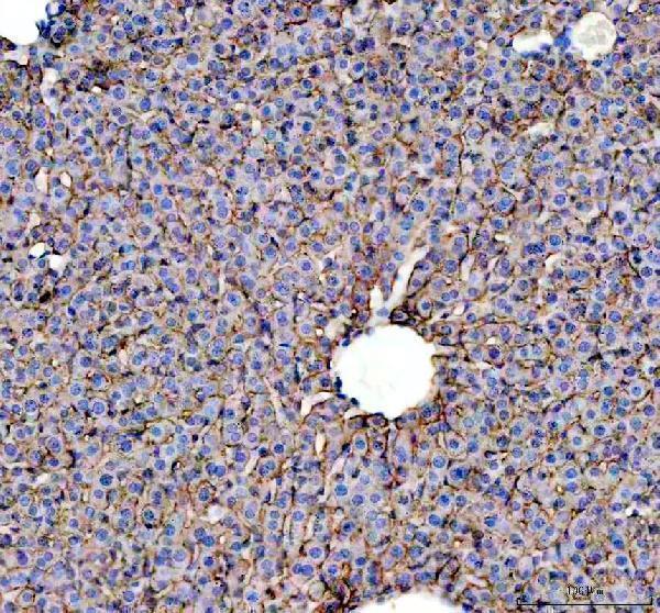
The IHC-validated catalog antibody has paraffin-section images from human liver cancer and mouse and rat liver (A05376-1 IHC image captions). IF is listed, but no IF image is supplied (A05376-1 catalog).

Real IHC data IHC analysis of ASGR1 using anti-ASGR1 antibody (A05376-1). ASGR1 was detected in a paraffin-embedded section of human liver cancer tissue. Heat mediated antigen retrieval was performed in EDTA buffer (pH 8.0, epitope retrieval solution). The tissue section was blocked with 10% goat serum. The tissue section was then incubated with 1:100 rabbit anti-ASGR1 Antibody (A05376-1) overnight at 4°C. Peroxidase Conjugated Goat Anti-rabbit IgG was used as secondary antibody and incubated for 30 minutes at 37°C. The tissue section was developed using HRP Conjugated Rabbit IgG Super Vision Assay Kit (Catalog # SV0002) with DAB as the chromogen.
Anti-ASGR1 Antibody
Cat # A05376-1

A05376-1 has IHC images from paraffin sections of human liver cancer and mouse and rat liver; its applications also include IF and ICC (A05376-1 IHC image captions; catalog applications). A05376 lists IHC and human, mouse, and rat reactivity, with no IHC image supplied (A05376 catalog applications, reactivity, and image alts).

Which to pick: Choose A05376-1 for paraffin-section IHC because its own captions document staining in human liver cancer and mouse and rat liver (A05376-1 IHC image captions); the fixative is unreported (A05376-1 IHC image captions). Choose A05376-1 for IF/ICC because both applications are listed, although no IF image is supplied (A05376-1 catalog applications and image alts). For cross-species IHC, A05376-1 has image evidence across all three listed species, while A05376 lists the same reactivity without an IHC image (A05376-1 IHC image captions; A05376 catalog reactivity and image alts).

Each figure is that product's own IHC / IF validation image from its datasheet.

References

  1. UniProt Consortium. UniProt entry P07306 (ASGR1_HUMAN, Asialoglycoprotein receptor 1).
  2. Human Protein Atlas. ASGR1 tissue IHC expression (reliability: Enhanced).
  3. Human Protein Atlas. ASGR1 subcellular location (ICC-IF): Mainly localized to vesicles. In addition localized to the cell junctions..
  4. Human Protein Atlas. ASGR1 antibody validation summary (2 antibodies).
  5. ASGR1 Deficiency Promotes Liver Fibrosis by Enhancing Pro-Inflammatory Arachidonic Acid Metabolism and ALP Secretion. Immunity, inflammation and disease 2026 — PMC13491154.
  6. Asialoglycoprotein receptor 1 is a novel PCSK9-independent ligand of liver LDLR cleaved by furin. The Journal of biological chemistry 2021 — PMC8479480.
  7. IL-6-GP130 signaling protects human hepatocytes against lipid droplet accumulation in humanized liver models. Science advances 2023 — PMC10104468.
  8. Asialoglycoprotein receptor 1 promotes SARS-CoV-2 infection of human normal hepatocytes. Signal transduction and targeted therapy 2024 — PMC10866945.
  9. PubMed PMID:2982798 — UniProt-cited evidence.
  10. PubMed PMID:3753585 — UniProt-cited evidence.
  11. PubMed PMID:20886072 — UniProt-cited evidence.