This website uses cookies to ensure you get the best experience on our website.


- Table of Contents
Plan ASH2L chromogenic IHC on paraffin sections using the mainly nuclear tissue pattern (HPA tissue IHC). This guide covers the catalog antibody’s documented IHC workflow (datasheet A06129-1) and the staining-to-RNA discordance to consider when interpreting results (HPA tissue IHC).

Expected localisation, validated protocols, controls and antibodies — the at-a-glance facts below, then the full design guide.
| Expected localisation | Mainly nuclear in several tissues (HPA tissue IHC) | |
| Staining pattern | Nuclear staining in several tissue cell types (HPA tissue IHC) | |
| Antigen retrieval | EDTA pH 8.0 HIER, heat-mediated (datasheet A06129-1) | |
| Positive control | Appendix+4 more · see all | |
| Negative control | Caudate+4 more · see all |
| Fixation | Keep paraffin-section fixation consistent (standard IHC practice; not target-specific); Matched tissue-IHC evidence does not establish the fixation claim. Validate the specimen-specific method before use. (selected-SKU IHC image A06129-1) | |
| Caveat | Staining has low concordance with RNA expression (HPA tissue IHC) | |
| Regulation | Higher expression in adult heart and testis (UniProt) | |
| Isoform / epitope | 3 isoforms; epitope differences unspecified (UniProt) |
The catalog antibody has an IHC-P protocol (datasheet A06129-1); one published ASH2L protocol reports staining of Pelodiscus sinensis testis (PMC10215499).
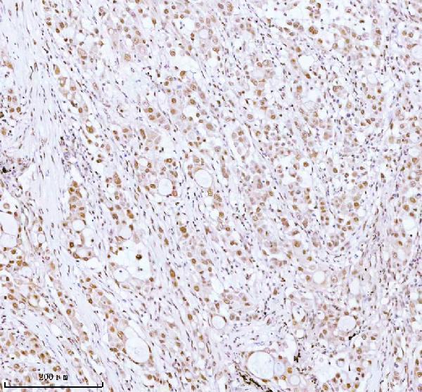
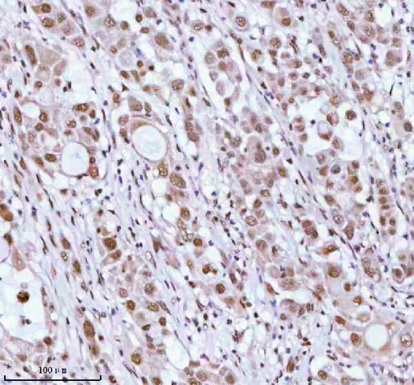
| Sample | Paraffin-embedded human lung adenocarcinoma tissue; fixative not specified (datasheet A06129-1) |
| Fixation | Image fixative and duration unreported (datasheet A06129-1); verify before use. |
| Sectioning | 4–5 µm sections on charged slides (standard) |
| Deparaffinisation | Xylene, graded ethanol series to water (standard) |
| Antigen retrieval | Heat retrieval: EDTA pH 8.0 (datasheet A06129-1); 20 min, 95–100 °C (standard) |
| Peroxidase block | 3% H2O2, 10 min, room temperature (standard) |
| Blocking | 10% goat serum (datasheet A06129-1) |
| Primary antibody | Rabbit anti-ASH2L, 2-5 μg/ml (datasheet A06129-1) |
| Primary incubation | Overnight at 4 °C (datasheet A06129-1) |
| Detection | HRP-conjugated secondary, DAB chromogen (datasheet A06129-1) |
| Counterstain | Hematoxylin, blue, dehydrate and mount (standard) |
| Expected result | ASH2L-positive staining in glandular cells of appendix (HPA tissue IHC: Medium). HPA tissue profile: Mainly nuclear expression in several different tissue types. No signal in the no-primary control. |
ASH2L is a nuclear protein with no transmembrane segment (UniProt Q9UBL3). In paraffin tissue sections, expect mainly nuclear staining in several cell types, including appendix and colon glandular cells and kidney tubule cells with medium reported staining (HPA tissue IHC). HPA rates its IHC staining Approved but reports low consistency with RNA expression data, so use cell-specific tissue patterns as guides rather than universal positive controls (HPA tissue IHC).
| Medium nuclear staining in appendix or colon glandular cells, or kidney tubule cells (HPA tissue IHC). | This matches the reported cell-specific IHC pattern and the nuclear localisation of ASH2L (HPA tissue IHC; UniProt Q9UBL3). Compare nuclei with nearby background before scoring, and record the stained cell type as well as intensity (general IHC practice). |
| Predominantly cytoplasmic staining, with little nuclear signal, in an otherwise positive field. | This does not match the mainly nuclear tissue pattern (HPA tissue IHC). Check morphology, chromogen deposits and control sections before interpreting it as ASH2L; ICC-IF also reports a supported plasma membrane location, but that observation alone does not establish a matching paraffin IHC pattern (HPA subcellular; general IHC practice). |
| Staining in a cell population reported as undetected, such as lung alveolar cells (HPA tissue IHC). | Treat this as unexpected for that specific population, not proof that the entire lung must be negative (HPA tissue IHC). Cross-reactivity or endogenous detection activity is possible; review an antibody-omission control and cell identity before scoring (general IHC practice). |
| Diffuse colour across nuclei, cytoplasm and acellular areas. | A diffuse field cannot establish nuclear localisation (UniProt Q9UBL3; general IHC practice). Background from detection reagents, incomplete blocking or inadequate washing is possible; compare an antibody-omission control and inspect whether the signal follows tissue structures (general IHC practice). |
| No nuclear signal in appendix or colon glandular cells (HPA tissue IHC). | These cells were reported at medium staining, so an absent signal warrants a run-level check (HPA tissue IHC). The HPA Approved rating has low staining-to-RNA consistency; a single negative section cannot establish absence of ASH2L protein (HPA tissue IHC). |
| Compartment and topology (UniProt Q9UBL3). | ASH2L is nuclear and has no transmembrane segment, making nuclear staining the main IHC readout (UniProt Q9UBL3; HPA tissue IHC). Do not infer that every membrane-like signal is false: supported plasma membrane localisation was observed by ICC-IF (HPA subcellular). |
| Cell-specific reference patterns and validation (HPA tissue IHC). | Several epithelial and other populations show medium staining, while named populations such as lung alveolar cells and liver cholangiocytes were not detected (HPA tissue IHC). The Approved IHC assessment also notes low consistency with RNA data; interpret controls by cell type (HPA tissue IHC). |
| Three reported isoforms (UniProt Q9UBL3). | Isoforms 1, 2 and 3 are listed, but the supplied record gives no antibody epitope or isoform-specific tissue pattern (UniProt Q9UBL3). A positive or negative IHC result cannot be assigned to one isoform from these data alone (UniProt Q9UBL3). |
| Unreported target-specific preparation response. | Target-specific fixation effects are not established by the supplied assay evidence. Verify with a matched IHC source before attributing a result to fixation. |
| Situation | Likely cause | Next action |
|---|---|---|
| No signal appears in expected appendix or colon glandular cells (HPA tissue IHC). | Possible run failure, inadequate detection, or a section-dependent result; HPA reports medium staining in these cells but low consistency with RNA data (HPA tissue IHC; general IHC practice). | Confirm the expected cell type and morphology, inspect a known-working positive section processed in the same run, then review reagent sequence and detection steps (HPA tissue IHC; general IHC practice). |
| Signal is mainly cytoplasmic instead of nuclear. | The compartment differs from the predominant tissue IHC pattern and UniProt localisation (HPA tissue IHC; UniProt Q9UBL3). Nonspecific staining or deposited chromogen may account for it (general IHC practice). | Compare antibody-omission and positive controls, inspect deposits at higher magnification, and score nuclear signal separately from cytoplasmic colour (general IHC practice). |
| Cells reported as undetected show convincing-looking colour, such as liver cholangiocytes (HPA tissue IHC). | Cross-reactivity, endogenous detection activity, or mistaken cell identification is possible (general IHC practice). The HPA observation refers to cholangiocytes, not every cell in liver (HPA tissue IHC). | Check cell morphology and an antibody-omission control; if colour persists without primary antibody, investigate the detection system before assigning ASH2L positivity (general IHC practice). |
| Background obscures nuclear boundaries throughout the section. | Nonspecific reagent binding, endogenous detection activity, or incomplete washing can reduce contrast (general IHC practice). The diffuse pattern does not resolve the expected nuclear signal (HPA tissue IHC). | Review blocking, washing and detection controls using standard IHC practice; score only cells whose nuclei can be distinguished reliably (general IHC practice). |
| Intensity differs across tissues or between nearby cell populations. | HPA reports medium staining in selected populations, low staining in others, and no detection in some named populations; its antibody assessment notes low consistency with RNA data (HPA tissue IHC). | Record tissue, cell type, compartment and intensity separately. Compare each population with its own HPA observation instead of applying a whole-tissue positive or negative label (HPA tissue IHC; general IHC practice). |
| Does an ICC-IF membrane signal change the expected paraffin IHC result? | HPA ICC-IF lists nucleoplasm as the main location and plasma membrane as an additional supported location (HPA subcellular). Tissue IHC is described as mainly nuclear (HPA tissue IHC). | Use predominantly nuclear staining for the IHC decision; assess ICC-IF localisation on its own guide page, and do not infer a paraffin IHC membrane pattern from ICC-IF alone (HPA tissue IHC; HPA subcellular). |
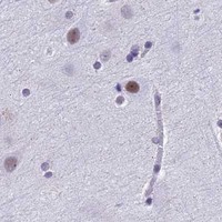


Comprehensive Human Protein Atlas IHC scoring per tissue (reliability: Approved — Low consistency between antibody staining and RNA expression data.). Rows are taken directly from the HPA tissue chart — click any row's HPA link to view the source.
| Tissue | Cell type | Level | Evidence | Source |
|---|---|---|---|---|
| Appendix | Glandular cells | Medium | Protein (IHC) | HPA → |
| Colon | Glandular cells | Medium | Protein (IHC) | HPA → |
| Esophagus | Squamous epithelial cells | Medium | Protein (IHC) | HPA → |
| Fallopian tube | Glandular cells | Medium | Protein (IHC) | HPA → |
| Gallbladder | Glandular cells | Medium | Protein (IHC) | HPA → |
| Tissue | Cell type | Level | Evidence | Source |
|---|---|---|---|---|
| Caudate | Glial cells | Not detected | Protein (IHC) | HPA → |
| Cerebellum | Cells in granular layer | Not detected | Protein (IHC) | HPA → |
| Hippocampus | Glial cells | Not detected | Protein (IHC) | HPA → |
| Liver | Cholangiocytes | Not detected | Protein (IHC) | HPA → |
| Lung | Alveolar cells | Not detected | Protein (IHC) | HPA → |
Troubleshoot ASH2L staining in paraffin sections by checking retrieval, nuclear localisation, controls, and cell specific scoring before interpreting differences between tissues.
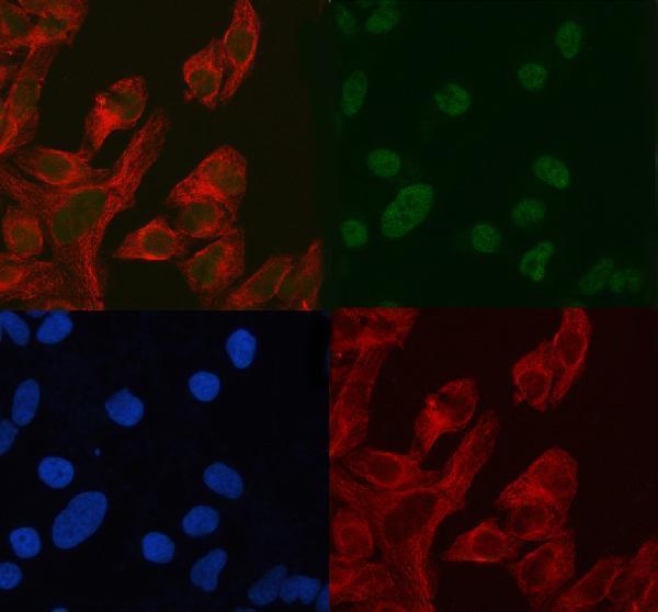
A06129-1 and M06129 list human, mouse and rat reactivity. Both have human paraffin-section IHC images; A06129-1 also has a HELA cell IF/ICC image (catalog reactivity and image captions).
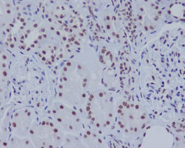
A06129-1 has IHC images from human lung adenocarcinoma and placenta paraffin sections, plus an IF/ICC image from HELA cells (catalog image captions). M06129 has an IHC image from a human kidney paraffin section and lists IF/ICC as an application (catalog image caption; catalog applications).
Which to pick: For tissue IHC, choose A06129-1 if its captioned EDTA pH 8.0 retrieval and 2 μg/ml primary condition are useful starting points; choose M06129 if a rabbit monoclonal with a human kidney paraffin-section image is preferred (A06129-1 IHC caption; M06129 catalog host/clone and IHC caption). For IF/ICC, A06129-1 has a HELA cell image at 5 μg/ml, while M06129 lists IF/ICC at 1:50 without an IF image (catalog IF images and dilutions). Both list human, mouse and rat reactivity, although the supplied IHC images show human tissue only; neither IHC caption reports the fixative (catalog reactivity and IHC captions).