ASNS / Asparagine synthetase [glutamine-hydrolyzing] · IHC design guide

Design Immunohistochemistry for ASNS

Plan ASNS staining in paraffin sections using pancreatic exocrine glandular cells as a high-staining reference (HPA tissue IHC). Start the IHC-validated antibody at 2–5 μg/mL (datasheet A03302-2), and score cytoplasmic staining (HPA tissue IHC).

Evidence assembled Oct 2026 · For research use; verify linked source records and product datasheet before use
Immunohistochemistry protocol sheet for ASNS (IHC for ASNS): expected localisation Cytoplasmic staining in tissue sections (HPA tissue IHC), antibody A03302-2, validated IHC image, and IHC protocol steps
Printable ASNS IHC protocol sheet — expected localisation Cytoplasmic staining in tissue sections (HPA tissue IHC), antibody A03302-2, controls and protocol steps. Open the full ASNS IHC guide →

ASNS Immunohistochemistry Experimental Design Guide

Expected localisation, validated protocols, controls and antibodies — the at-a-glance facts below, then the full design guide.

Must know before staining
Expected localisation Cytoplasmic staining in tissue sections (HPA tissue IHC)
Staining pattern Cytoplasmic staining in pancreas, brain and stomach (HPA tissue IHC)
Antigen retrieval EDTA pH 8.0 HIER, heat-mediated (datasheet A03302-2)
Positive control ⓘ Cerebellum+4 more · see all
Negative control ⓘ Adrenal gland+4 more · see all
Important caveats
Reasons your staining may differ from the expected pattern.
Fixation Keep fixation consistent across sections (standard IHC practice; not target-specific)
Caveat Staining and RNA show medium consistency; verify locally (HPA tissue IHC)
Regulation Expression regulation not annotated (UniProt)
Isoform / epitope 3 isoforms; epitope effects are unknown (UniProt)
Section 1

Recommended ASNS IHC & IF Protocols

The catalog antibody’s IHC-P protocol (datasheet: A03302-2) is accompanied by three published ASNS IHC protocols (PMC9424662; PMC10393983; PMC8440958).

Recommended immunohistochemistry (IHC-P) protocol parameters
SampleParaffin-embedded human gastric carcinoma tissue; fixative not specified (datasheet A03302-2)
FixationImage fixative and duration unreported (datasheet A03302-2); verify before use.
Sectioning4–5 µm sections on charged slides (standard)
DeparaffinisationXylene, graded ethanol series to water (standard)
Antigen retrievalHeat retrieval: EDTA pH 8.0 (datasheet A03302-2); 20 min, 95–100 °C (standard)
Peroxidase block3% H2O2, 10 min, room temperature (standard)
Blocking10% goat serum (datasheet A03302-2)
Primary antibodyRabbit anti-ASNS, 2-5 μg/ml (datasheet A03302-2)
Primary incubationOvernight at 4 °C (datasheet A03302-2)
DetectionHRP-conjugated secondary, DAB chromogen (datasheet A03302-2)
CounterstainHematoxylin, blue, dehydrate and mount (standard)
Expected resultASNS-positive staining in purkinje cells of cerebellum (HPA tissue IHC: High). HPA tissue profile: Cytoplasmic expression in several different tissue types, including pancreas, brain and stomach. No signal in the no-primary control.
💡Decision noteStart with heat-mediated EDTA pH 8.0 retrieval (datasheet: A03302-2); the published excerpts do not specify a retrieval buffer (PMC9424662; PMC10393983; PMC8440958).
Section 2

What Is the Expected ASNS Staining Pattern?

ASNS staining should be predominantly cytoplasmic, consistent with HPA tissue IHC and supported cytosolic ICC-IF localization (HPA: tissue IHC; HPA: cytosol supported). Strong examples include pancreatic exocrine glandular cells, cerebellar Purkinje cells, stomach glandular cells and placental trophoblastic cells (HPA: High in each). HPA rates the tissue IHC pattern Approved, with medium consistency against RNA data and external verification pending (HPA: tissue IHC). UniProt reports no transmembrane segment (UniProt P08243 topology).

What am I looking at on my slide?
Cytoplasmic staining in pancreatic exocrine glandular cells or cerebellar Purkinje cells.This matches two High IHC examples (HPA: High in pancreatic exocrine glandular cells; HPA: High in Purkinje cells). Compare cells within the section before judging overall intensity: an appropriate positive pattern is cell specific and cytoplasmic (HPA: tissue IHC profile).
Predominantly nuclear, membranous or sharply extracellular staining.The compartment conflicts with supported cytosolic ICC-IF localization and the cytoplasmic tissue IHC profile (HPA: cytosol supported; HPA: tissue IHC). Treat it as suspect and review morphology, controls and detection background before interpreting it as ASNS; UniProt provides no subcellular annotation (UniProt P08243).
Strong staining in adrenal glandular cells or lung alveolar cells.These particular cell populations were Not detected in HPA tissue IHC (HPA: adrenal glandular cells Not detected; HPA: lung alveolar cells Not detected). Check cell identity and controls; unexpected color can reflect antibody cross-reactivity or endogenous detection activity. An HPA negative example does not establish that every cell in that organ is negative.
Diffuse color across cells, stroma and blank areas.A broad haze does not resemble HPA's cytoplasmic, cell-specific tissue pattern (HPA: tissue IHC profile). General IHC causes include insufficient washing, nonspecific antibody binding or endogenous enzyme activity. Assess a no-primary control and review blocking, washing and detection settings before scoring cells.
No convincing cytoplasmic signal in pancreatic exocrine glandular cells.That conflicts with a High HPA example (HPA: High in pancreatic exocrine glandular cells), but a single section cannot establish ASNS absence. Confirm that the expected cells are present, then review the antibody's IHC-P conditions, antigen retrieval and detection controls; HPA does not report ASNS-specific fixation sensitivity.
💡Expected ASNS appearanceA credible positive is predominantly cytoplasmic staining in the relevant cells, including strong pancreatic exocrine glandular or Purkinje-cell staining (HPA: tissue IHC profile; HPA: High in both), while dominant nuclear or diffuse stromal color is suspect against the supported cytosolic pattern (HPA: cytosol supported).
How each factor affects the staining
Tissue and cell selectionHPA records High staining in pancreatic exocrine glandular, Purkinje, placental trophoblastic, stomach glandular and tonsillar germinal center cells (HPA: tissue IHC). It also records Low neuronal staining in hippocampus and Not detected staining in adrenal glandular cells (HPA: tissue IHC). Select and score the named cell population, rather than assigning one result to an entire organ.
Strength of the tissue evidenceThe HPA tissue profile is Approved, with medium consistency between staining and RNA expression and external verification pending (HPA: tissue IHC reliability). HPA lists IHC as Approved for HPA029318 and HPA064737 (HPA: antibodies); those labels support cautious comparison with the reported pattern, not a universal result for every antibody or specimen.
Topology and isoformsUniProt lists no transmembrane segment and three ASNS isoforms (UniProt P08243 topology; UniProt P08243 isoforms). The supplied record gives no epitope map for the catalog antibody, so it cannot establish whether all isoforms stain equally or explain an unexpected compartment. Interpret staining location against the observed HPA cytosolic pattern (HPA: cytosol supported).
Processing and modificationsUniProt lists no signal peptide or propeptide and a chain spanning residues 2–561; it also records modifications including acetyllysine 385 and phosphorylation at 545 and 557 (UniProt P08243 processing; UniProt P08243 modified residues). No supplied source links these features to IHC staining strength, retrieval response or epitope accessibility.
IF/ICC Q: Where should ASNS fluorescence appear?A: Predominantly in the cytosol (HPA: cytosol supported). HPA lists ICC as Enhanced for HPA004924 and HPA064737 (HPA: antibodies). Compare that localization with the IHC pattern, but use a dedicated IF/ICC guide for the separate workflow; the supplied record does not support an IF protocol here.
Why is my staining missing, weak or wrong?
SituationLikely causeNext action
No signal in a pancreatic positive control.The result conflicts with High staining reported for exocrine glandular cells (HPA: pancreas IHC); the relevant cells may be absent, or a general IHC workflow step may have failed.Locate exocrine glandular cells, confirm control-slide performance, and check the chosen antibody's IHC-P instructions for retrieval, dilution and detection. Do not infer ASNS-specific fixation sensitivity from HPA.
Strong nuclei but little cytoplasm.Nuclear dominance conflicts with HPA cytoplasmic tissue staining and supported cytosolic ICC-IF localization (HPA: tissue IHC; HPA: cytosol supported).Review morphology and the no-primary control, then reassess antibody specificity and detection conditions before scoring nuclear color as ASNS.
Color in stromal or cell-free regions.That distribution differs from the cell-associated cytoplasmic profile (HPA: tissue IHC); general IHC background or endogenous detection activity is possible.Check a no-primary control and review blocking, washing and detection steps. Score only identifiable cells with a plausible compartment pattern.
Strong signal in an HPA negative cell population.HPA reports Not detected staining for cells such as adrenal glandular cells and lung alveolar cells (HPA: tissue IHC). Misidentified cells, cross-reactivity or detection background may explain a mismatch.Verify cell identity and include negative and no-primary controls; investigate reproducible signal before calling that population ASNS positive.
Unexpectedly weak staining in a selected tissue.HPA levels differ by cell population: pancreatic exocrine cells are High, while hippocampal neuronal cells are Low (HPA: tissue IHC). A weak result may therefore fit the selected population.Compare the named cells and their HPA level, then check a High positive control before changing general IHC conditions or calling the run a failure.
IHC and ICC-IF appear to disagree.HPA reports cytoplasmic tissue IHC and supported cytosolic ICC-IF localization, but uses application-specific antibody validation labels (HPA: tissue IHC; HPA: subcellular; HPA: antibodies).Confirm that the same cell population and compartment are being compared. Review each antibody's validation for its application; an ICC Enhanced label alone does not establish its IHC performance (HPA: antibodies).

Sample controls for ASNS IHC & IF

🧪Run cerebellum first and confirm staining in Purkinje cells (HPA: High in Purkinje cells); use adrenal glandular cells as the negative tissue (HPA: Not detected in adrenal glandular cells). On the cerebellum slide, assess nearby cells without specific staining as internal negative comparators, but verify them visually because HPA identifies Purkinje cells as High without specifying an internal negative cell type (HPA: High in Purkinje cells).
Positive control tissue: Cerebellum (Purkinje cells, HPA High)
Negative control tissue: Adrenal gland (HPA Not detected)
ICC-IF cell lines (HPA subcellular resource): HPA ICC-IF images show ASNS in A-431, U-251MG, U2OS, MCF-7, SK-MEL-30, with annotated localisation: Cytosol (supported) (HPA subcellular).
Technical controls: Include a no-primary, secondary-only control, a matched rabbit IgG isotype control, and ASNS-knockout material or peptide competition if the immunizing peptide is available (selected-SKU IHC caption: rabbit primary antibody). For chromogenic cerebellar IHC, block endogenous peroxidase and check for pigment that could mimic DAB staining (selected-SKU IHC caption: HRP/DAB detection).
⚠️Feasibility: A target-specific fixation window or fixation effect is unreported, and the selected-SKU paraffin-section caption does not state the fixative (selected-SKU IHC caption: fixative not stated). The demonstrated paraffin-section procedure uses heat retrieval in EDTA at pH 8.0, but retrieval dependence has not been established (selected-SKU IHC caption: EDTA retrieval); the supplied evidence does not establish that frozen sections or IF are easier. Cerebellar pigment may complicate chromogenic scoring, so compare signal with the no-primary control (selected-SKU IHC caption: HRP/DAB detection).

HPA tissue IHC evidence for ASNS

Comprehensive Human Protein Atlas IHC scoring per tissue (reliability: Approved — Medium consistency between antibody staining and RNA expression data. Pending external verification.). Rows are taken directly from the HPA tissue chart — click any row's HPA link to view the source.

Positive expression · recommended positive controls

TissueCell typeLevelEvidenceSource
Cerebellum Purkinje cells High Protein (IHC) HPA →
Pancreas Exocrine glandular cells High Protein (IHC) HPA →
Placenta Trophoblastic cells High Protein (IHC) HPA →
Stomach Glandular cells High Protein (IHC) HPA →
Tonsil Germinal center cells High Protein (IHC) HPA →

Undetected expression · recommended negative controls

TissueCell typeLevelEvidenceSource
Adrenal gland Glandular cells Not detected Protein (IHC) HPA →
Breast Adipocytes Not detected Protein (IHC) HPA →
Bronchus Respiratory epithelial cells Not detected Protein (IHC) HPA →
Endometrium Cells in endometrial stroma Not detected Protein (IHC) HPA →
Fallopian tube Glandular cells Not detected Protein (IHC) HPA →
Section 3

Advanced ASNS IHC Tips

Use the catalog antibody’s paraffin-section result as the IHC starting point, then assess cytoplasmic staining against appropriate tissue and detection controls.

What retrieval should I try when ASNS staining is weak in paraffin sections?
Start with heat-mediated retrieval in EDTA at pH 8.0 (datasheet A03302-2). The selected paraffin-section example used this retrieval before an overnight incubation at 4°C with 2 μg/ml primary antibody (datasheet A03302-2). Keep section thickness, heating, cooling and detection conditions consistent across the comparison, and include a glandular-cell-rich stomach section as a reference with reported high staining (HPA: stomach glandular cells High). If signal remains weak, compare a carefully controlled alternative retrieval condition as a fallback, recording both staining intensity and tissue damage (standard IHC practice). A stronger signal is useful only if the expected cellular pattern and control separation also improve (HPA: cytoplasmic tissue profile; standard IHC practice).
Could fixation explain weak ASNS staining in my IHC sections?
Target-specific sensitivity of ASNS staining to fixation is unknown from the supplied evidence (datasheet A03302-2: fixative not stated). The selected image documents a paraffin-embedded gastric carcinoma section, but does not identify its fixative or fixation duration (datasheet A03302-2). Record the fixative, duration and processing history for each specimen, then compare sections using the same EDTA pH 8.0 retrieval and detection conditions (datasheet A03302-2; standard IHC practice). Include a reference section with reported ASNS-positive cells to distinguish a run-wide failure from a specimen-specific problem (HPA: stomach glandular cells High; standard IHC practice). Avoid assigning a fixation effect to ASNS without a controlled comparison (standard IHC practice).
Should ASNS staining appear in nuclei or along cell membranes?
Assess ASNS primarily as cytoplasmic staining in tissue sections (HPA: cytoplasmic expression in several tissue types). Cytosolic localisation is also supported by subcellular imaging, while UniProt provides no subcellular annotation for this record (HPA: Cytosol supported; UniProt P08243: subcellular location not annotated). ASNS has no annotated transmembrane segment or signal peptide, so a crisp membrane-only pattern calls for additional specificity checks (UniProt P08243 topology and processing). Compare the suspicious pattern with a no-primary control and a section containing reported high-staining cells (standard IHC practice; HPA: stomach glandular cells High). Score the cytoplasmic compartment separately from nuclear or membranous colour so unexpected localisation cannot inflate the result (standard IHC practice).
Can this antibody distinguish ASNS isoforms or modified protein in tissue?
Do not assign an IHC signal to a particular ASNS isoform without epitope information and isoform-specific validation (UniProt P08243: 3 isoforms; standard IHC practice). The record identifies a glutamine amidotransferase region at residues 2–191 and an asparagine synthetase region at 213–536, but the selected caption does not map this antibody’s epitope (UniProt P08243 domains; datasheet A03302-2 caption). Acetyllysine at 385 and phosphorylation at 545 and 557 are annotated modifications, without evidence here that they alter staining (UniProt P08243 modified residues). If discrepant staining matters, compare independently validated antibodies with known epitopes and assess their compartment patterns in matched sections (standard IHC practice).
How should I investigate ASNS by multiplex IF alongside this IHC assay?
Treat multiplex IF as a separate assay requiring its own controls, because the selected product evidence is a chromogenic paraffin-section example (datasheet A03302-2). Pair ASNS with a validated marker for the cell population under study; for a tissue reference, HPA reports high ASNS staining in stomach glandular cells (HPA: stomach glandular cells High; standard IF practice). Check unstained tissue in each imaging channel, then place the weaker signal in a fluorophore channel with less tissue autofluorescence and verify spectral separation (standard IF practice). ASNS is supported in the cytosol and has no annotated transmembrane segment, so optimise permeabilisation for access to an intracellular epitope, once its location is established (HPA: Cytosol supported; UniProt P08243 topology; standard IF practice).
What should I check when DAB appears throughout the ASNS section?
Run a no-primary section to assess detection-system staining, including endogenous peroxidase, before attributing diffuse DAB to ASNS (standard IHC practice). The selected workflow used 10% goat serum, 2 μg/ml rabbit primary antibody overnight at 4°C, and a peroxidase-linked secondary for 30 minutes at 37°C (datasheet A03302-2). Apply an appropriate peroxidase block and check whether secondary-only staining or excessive DAB development explains the background (standard chromogenic IHC practice). If controls are clean, compare primary concentration and washing conditions while preserving the documented EDTA pH 8.0 retrieval as the starting condition (datasheet A03302-2; standard IHC practice). Judge improvement by clearer cytoplasmic cell boundaries, not simply paler slides (HPA: cytoplasmic tissue profile; standard IHC practice).
How can I compare ASNS staining across sections without confusing cellularity with expression? ⚠ ANSWER MARKED FOR VERIFICATION
Define the cell population and cytoplasmic compartment before scoring, because the reported tissue pattern is cytoplasmic and varies by cell type (HPA: cytoplasmic tissue profile; HPA: cell-specific staining entries). For comparable regions, record the percentage of positive target cells and an intensity-based H-score using the same thresholds across sections (standard IHC quantification practice). If measuring positive-cell density per mm², also report the number of eligible cells or tissue area assessed so differences in cellularity remain visible (standard IHC quantification practice). Normalise each comparison to its prespecified target-cell population and exclude folds, section edges and necrotic areas consistently (standard IHC quantification practice). Keep exposure, colour separation and scoring rules fixed for the entire set (standard IHC quantification practice).
How do I separate credible ASNS positivity from staining artefact?
A credible result has cytoplasmic staining in identifiable cells and separates from the no-primary control (HPA: cytoplasmic tissue profile; standard IHC practice). Reported high-staining references include exocrine glandular cells in pancreas and glandular cells in stomach, whereas adrenal glandular cells were reported as not detected (HPA: pancreas exocrine glandular cells High; HPA: stomach glandular cells High; HPA: adrenal glandular cells Not detected). Check membrane-only or nuclear-only colour against the supported cytosolic location and lack of a transmembrane segment (HPA: Cytosol supported; UniProt P08243 topology). Treat staining concentrated at section edges, in necrosis or in a no-primary control as a reason to investigate processing or endogenous enzyme activity before interpretation (standard IHC practice). HPA rates its tissue IHC evidence Approved with medium staining-to-RNA consistency and pending external verification (HPA: reliability description).
Boster reagents

Best ASNS / Asparagine synthetase [glutamine-hydrolyzing] IHC Antibodies

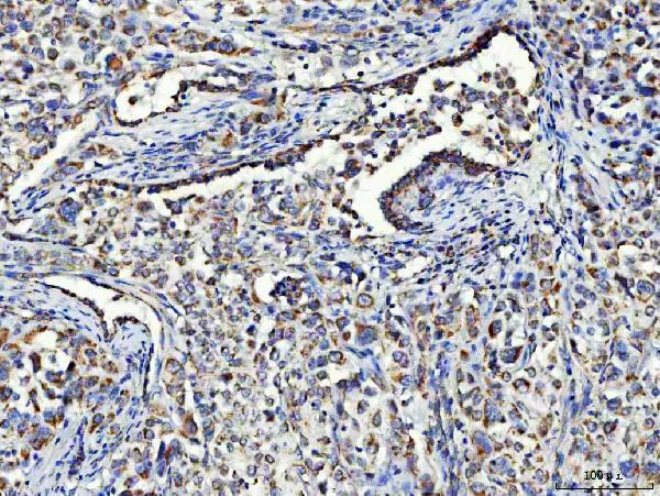
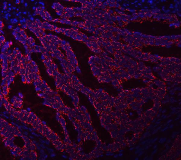
Both anti-ASNS antibodies have human paraffin-section IHC images; one also has a human paraffin-section IF image. Both list human, mouse, and rat reactivity (catalog image captions; catalog reactivity).

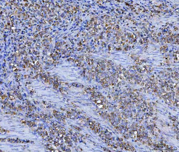
Real IHC data IHC analysis of Asparagine synthetase/ASNS using anti-Asparagine synthetase/ASNS antibody (A03302-2). Asparagine synthetase/ASNS was detected in a paraffin-embedded section of human gastric carcinoma tissue. Heat mediated antigen retrieval was performed in EDTA buffer (pH 8.0, epitope retrieval solution). The tissue section was blocked with 10% goat serum. The tissue section was then incubated with 2 μg/ml rabbit anti-Asparagine synthetase/ASNS Antibody (A03302-2) overnight at 4°C. Peroxidase Conjugated Goat Anti-rabbit IgG was used as secondary antibody and incubated for 30 minutes at 37°C. The tissue section was developed using HRP Conjugated Rabbit IgG Super Vision Assay Kit (Catalog # SV0002) with DAB as the chromogen.
Anti-Asparagine synthetase/ASNS Antibody ®
Cat # A03302-2
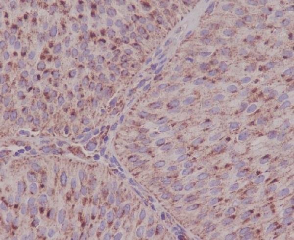
Real IHC data Immunohistochemical analysis of paraffin-embedded human bladder, using ASNS Antibody.
Anti-ASNS/Asparagine Synthetase Rabbit Monoclonal Antibody
Cat # M03302

A03302-2 has IHC images from paraffin-embedded human gastric carcinoma, lung cancer, and testicular germ cell tumors, plus an IF image from paraffin-embedded human intestinal cancer (A03302-2 IHC/IF captions). M03302 lists IHC and ICC/IF applications and has an IHC image from paraffin-embedded human bladder (M03302 applications; M03302 IHC caption).

Which to pick: For tissue IHC, choose A03302-2 when its documented human tumor examples match your sample; M03302 provides a paraffin-embedded human bladder example (A03302-2 IHC captions; M03302 IHC caption). For IF, A03302-2 has a human tissue image; M03302 lists ICC/IF but has no supplied IF image (A03302-2 IF caption; M03302 applications and image alts). Both list human, mouse, and rat reactivity, although the supplied tissue images are human only; M03302 is a rabbit monoclonal, A03302-2 has no clone specified, and neither IHC caption reports a fixative (catalog reactivity and clone fields; IHC captions).

Each figure is that product's own IHC / IF validation image from its datasheet.

References

  1. UniProt Consortium. UniProt entry P08243 (ASNS_HUMAN, Asparagine synthetase [glutamine-hydrolyzing]).
  2. Human Protein Atlas. ASNS tissue IHC expression (reliability: Approved).
  3. Human Protein Atlas. ASNS subcellular location (ICC-IF): Localized to the cytosol..
  4. Human Protein Atlas. ASNS antibody validation summary (3 antibodies).
  5. ASNS can predict the poor prognosis of clear cell renal cell carcinoma. Frontiers in oncology 2022 — PMC9424662.
  6. From ALL to Myeloid and NK Malignancies: Operationalizing "ASNS-Low" for L-Asparaginase Repurposing and Combination Therapy. Biomolecules 2026 — PMC13296854.
  7. NUCKS1, a LINC00629-upregulated gene, facilitated osteosarcoma progression and metastasis by elevating asparagine synthesis. Cell death & disease 2023 — PMC10393983.
  8. Proteomics Analysis Identified ASNS as a Novel Biomarker for Predicting Recurrence of Skull Base Chordoma. Frontiers in oncology 2021 — PMC8440958.
  9. PubMed PMID:2886907 — UniProt-cited evidence.
  10. PubMed PMID:2565875 — UniProt-cited evidence.
  11. PubMed PMID:14702039 — UniProt-cited evidence.