This website uses cookies to ensure you get the best experience on our website.


- Table of Contents
Plan chromogenic ASRGL1 IHC in paraffin sections using cytoplasmic staining in seminiferous ducts and female genital glands as a reference (HPA tissue IHC). Start the catalog antibody at 1:50 (datasheet A08869-1), and assess staining by cell type and compartment.

Expected localisation, validated protocols, controls and antibodies — the at-a-glance facts below, then the full design guide.
| Expected localisation | Cytoplasmic staining in tissue (HPA tissue IHC) | |
| Staining pattern | Cytoplasmic in seminiferous ducts and genital glands (HPA tissue IHC) | |
| Antigen retrieval | EDTA pH 8.0 HIER, heat-mediated (datasheet A08869-1) | |
| Positive control | Cerebellum+4 more · see all | |
| Negative control | Adipose tissue+4 more · see all |
| Fixation | Keep fixation consistent across sections (standard IHC practice; not target-specific); Matched tissue-IHC evidence does not establish the fixation claim. Validate the specimen-specific method before use. (selected-SKU IHC image A08869-1) | |
| Caveat | RNA may poorly predict staining: medium consistency (HPA tissue IHC) | |
| Regulation | Overexpressed in uterine and ovarian carcinomas (UniProt) | |
| Isoform / epitope | 2 isoforms; cleavage yields alpha and beta chains (UniProt) |
The catalog antibody’s IHC-P protocol is paired with published chromogenic ASRGL1 protocols for human brain and cervical tissue (PMC11099023; PMC6059725).
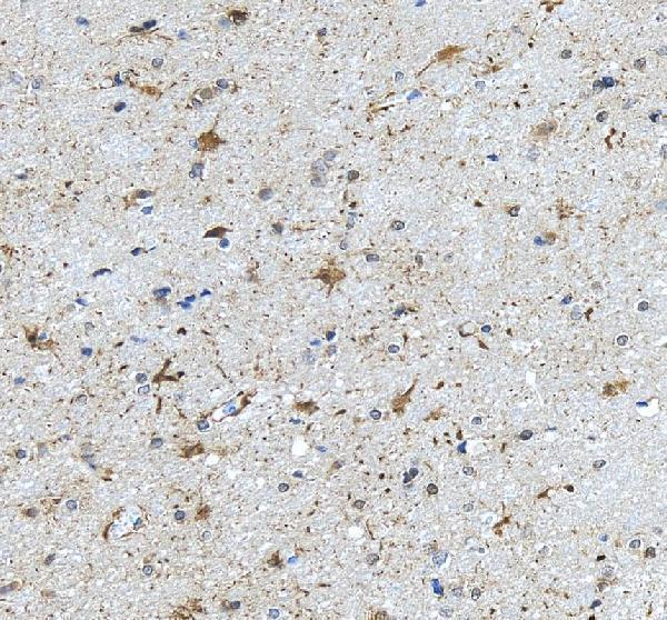
| Sample | Paraffin-embedded human brain tissue; fixative not specified (datasheet A08869-1) |
| Fixation | Image fixative and duration unreported (datasheet A08869-1); verify before use. |
| Sectioning | 4–5 µm sections on charged slides (standard) |
| Deparaffinisation | Xylene, graded ethanol series to water (standard) |
| Antigen retrieval | Heat retrieval: EDTA pH 8.0 (datasheet A08869-1); 20 min, 95–100 °C (standard) |
| Peroxidase block | 3% H2O2, 10 min, room temperature (standard) |
| Blocking | 10% goat serum (datasheet A08869-1) |
| Primary antibody | Rabbit anti-ASRGL1, 1:50 recommended; image 2 μg/ml (datasheet A08869-1) |
| Primary incubation | Overnight at 4 °C (datasheet A08869-1) |
| Detection | HRP-conjugated secondary, DAB chromogen (datasheet A08869-1) |
| Counterstain | Hematoxylin, blue, dehydrate and mount (standard) |
| Expected result | ASRGL1-positive staining in granular cells - cytoplasm/membrane of cerebellum (HPA tissue IHC: High). HPA tissue profile: Cytoplasmic expression most abundant in seminiferous ducts and glandular cells of female genital tract. No signal in the no-primary control. |
ASRGL1 is a cytoplasmic protein with no transmembrane segment (UniProt Q7L266). In paraffin section IHC, expect prominent staining in seminiferous ducts and female genital tract glandular cells (HPA: tissue IHC profile). HPA reports high staining in cervix and endometrium glandular cells, epididymis glandular cells, and cerebellar granular cells; its tissue IHC reliability is Enhanced, with medium consistency between staining and RNA data (HPA: tissue IHC).
| Distinct staining in cervix or endometrium glandular cells, or epididymis glandular cells. | This fits the reported High staining in those cell populations (HPA: tissue IHC). Judge the labelled cells and their cytoplasmic pattern, rather than calling an entire section positive from isolated deposits (HPA: tissue IHC profile; general IHC practice). |
| Predominantly nuclear staining in tissue IHC, with little cytoplasmic staining. | That differs from the reported tissue cytoplasmic profile (HPA: tissue IHC profile; UniProt Q7L266: cytoplasm). Check the counterstain and control sections before interpreting it as ASRGL1; nuclear localisation in ICC-IF does not establish a nuclear tissue IHC pattern (HPA: subcellular ICC-IF). |
| Strong signal in adipocytes or alveolar cells while the expected positive cells stain weakly. | HPA reports ASRGL1 as not detected in adipose tissue adipocytes and lung alveolar cells (HPA: tissue IHC). Treat this cell-specific mismatch as possible nonspecific binding or detection background; it does not establish that every cell in those tissues is negative (general IHC practice; HPA: tissue IHC). |
| Uniform colour over cells and surrounding section, without a readable cellular boundary. | Diffuse deposition cannot establish the cell-specific cytoplasmic pattern reported by HPA (HPA: tissue IHC profile). Review background controls and detection conditions before scoring, since nonspecific antibody binding or chromogen background can obscure localisation (general IHC practice). |
| No staining in glandular cells of a known-positive cervix or endometrium control. | Both populations have High reported staining (HPA: tissue IHC). First check whether the control section and detection run worked; absence in one section alone does not establish ASRGL1 absence in the specimen (general IHC practice). |
| Cell-specific comparison tissue | Cervix and endometrium glandular cells, epididymis glandular cells, and cerebellar granular cells are reported High; bronchus basal cells and selected glial cells are Medium (HPA: tissue IHC). Compare like cell types when judging intensity. |
| Antibody validation | HPA lists IHC as Enhanced for HPA029725, HPA055572 and CAB070192 (HPA: antibodies). Its tissue profile still notes medium consistency with RNA data (HPA: tissue IHC); validation supports comparison with the reported pattern, not an assumption that every specimen stains. |
| Topology and compartment | ASRGL1 is cytoplasmic and has no transmembrane segment (UniProt Q7L266). Interpret tissue staining against the HPA cytoplasmic profile; cerebellar granular cells are specifically annotated as cytoplasm/membrane, so a membrane-associated edge there is not automatically discordant (HPA: tissue IHC). |
| Processing and isoforms | ASRGL1 autocleaves into alpha residues 1–167 and beta residues 168–308, and has two annotated isoforms (UniProt Q7L266). The supplied sources do not identify the antibodies’ epitopes, so they cannot establish which chain or isoform a stain detects. |
| IF/ICC Q&A: should its pattern match tissue IHC? | HPA reports mainly microtubules, with additional nucleoplasm and cytokinetic bridge localisation in ICC-IF (HPA: subcellular ICC-IF). Tissue IHC instead has a cytoplasmic profile (HPA: tissue IHC); interpret each application against its own reported pattern. |
| Situation | Likely cause | Next action |
|---|---|---|
| The positive control has no visible signal. | A failed staining or detection step is possible; high staining is reported in cervix and endometrium glandular cells (HPA: tissue IHC; general IHC practice). | Verify control tissue identity, antibody addition, retrieval, and detection reagents on the same run before scoring test sections (general IHC practice). |
| Only nuclei appear strongly labelled. | This conflicts with the reported tissue cytoplasmic profile, although HPA separately notes nucleoplasm in ICC-IF (HPA: tissue IHC; HPA: subcellular ICC-IF). | Compare with counterstain and negative controls, then review primary antibody concentration and detection background (general IHC practice). |
| Unexpected cells stain more strongly than the expected population. | Cell-specific cross-reactivity or detection background is possible when staining departs from the HPA cell map (HPA: tissue IHC; general IHC practice). | Identify the stained cell type, compare it with HPA’s cell-level entries, and inspect an appropriate negative control (HPA: tissue IHC; general IHC practice). |
| Colour is diffuse across tissue or outside cells. | Nonspecific binding, inadequate blocking, or excess chromogen development can obscure cellular staining (general IHC practice). | Review blocking, washes, antibody concentration, and development time; score only a resolved cellular pattern (general IHC practice). |
| Colour appears in a control without primary antibody. | Endogenous detection activity or detection reagent background may produce chromogenic signal independently of primary antibody (general IHC practice). | Check the detection controls and apply the appropriate endogenous activity block for the chosen detection system (general IHC practice). |
| A low-signal population appears negative. | HPA reports Low staining in several populations, including colon glandular cells and urinary bladder urothelial cells (HPA: tissue IHC). | Check a reported high-staining control in the same run before interpreting weak or absent staining in that population (HPA: tissue IHC; general IHC practice). |


Comprehensive Human Protein Atlas IHC scoring per tissue (reliability: Enhanced — Medium consistency between antibody staining and RNA expression data.). Rows are taken directly from the HPA tissue chart — click any row's HPA link to view the source.
| Tissue | Cell type | Level | Evidence | Source |
|---|---|---|---|---|
| Cerebellum | Granular cells - cytoplasm/membrane | High | Protein (IHC) | HPA → |
| Cervix | Glandular cells | High | Protein (IHC) | HPA → |
| Endometrium | Glandular cells | High | Protein (IHC) | HPA → |
| Epididymis | Glandular cells | High | Protein (IHC) | HPA → |
| Bronchus | Basal cells | Medium | Protein (IHC) | HPA → |
| Tissue | Cell type | Level | Evidence | Source |
|---|---|---|---|---|
| Adipose tissue | Adipocytes | Not detected | Protein (IHC) | HPA → |
| Adrenal gland | Glandular cells | Not detected | Protein (IHC) | HPA → |
| Bone marrow | Hematopoietic cells | Not detected | Protein (IHC) | HPA → |
| Breast | Adipocytes | Not detected | Protein (IHC) | HPA → |
| Esophagus | Squamous epithelial cells | Not detected | Protein (IHC) | HPA → |
Troubleshoot ASRGL1 staining in paraffin sections by checking retrieval, cellular pattern, controls, and scoring before interpreting signal intensity.
A08869-1 has an IHC image from a paraffin-embedded human brain section (A08869-1 IHC caption); human, mouse and rat reactivity is listed (catalog: reactivity).
A08869-1 is listed for IHC and for human, mouse and rat reactivity (catalog: applications/reactivity). Its own image documents chromogenic IHC on paraffin-embedded human brain with EDTA retrieval at pH 8.0 and DAB detection; the fixative is unreported (A08869-1 IHC caption).
Which to pick: For tissue IHC, choose A08869-1: this rabbit polyclonal antibody has an IHC application listing and an image from a paraffin-embedded human brain section (catalog: host/clonality/applications; A08869-1 IHC caption). For IF/ICC, A08869-1 has a listed 1:50 dilution, but no IF/ICC application listing or image; validate its use in your sample (catalog: if_dilution/applications/if_image_alts). For mouse or rat tissue, A08869-1 lists reactivity with both species, while its IHC image documents human tissue only (catalog: reactivity; A08869-1 IHC caption).