This website uses cookies to ensure you get the best experience on our website.


- Table of Contents
Use the catalog antibody at 1:100–1:200 for paraffin-section IHC (datasheet: A06057-1), with granular cytoplasmic staining as the tissue reference (HPA tissue IHC). Compare high-staining glandular cells with low-staining smooth muscle cells (HPA tissue IHC), and assess specificity because staining may reflect protein from more than one gene (HPA tissue IHC).

Expected localisation, validated protocols, controls and antibodies — the at-a-glance facts below, then the full design guide.
| Expected localisation | Granular cytoplasm (HPA tissue IHC); mitochondrial inner membrane (UniProt) | |
| Staining pattern | Granular cytoplasmic staining across tissues (HPA tissue IHC) | |
| Antigen retrieval | Citrate pH 6.0 HIER, 95–98 °C, 20 min (rule: cytoplasmic / membrane antigen) | |
| Positive control | Adrenal gland+4 more · see all | |
| Negative control | None in HPA (detected in all 45 tissues); use no-primary + isotype controls |
| Fixation | Keep fixation consistent across paraffin sections. (standard IHC practice; not target-specific) | |
| Caveat | Staining may reflect protein from more than one gene (HPA tissue IHC) | |
| Regulation | Overexpressed in lung adenocarcinoma (UniProt) | |
| Isoform / epitope | 3 isoforms; epitope coverage is unknown (UniProt) |
The catalog antibody protocol (datasheet) appears alongside published ATAD3A IHC protocols for bladder and lung tissues (PMC10757082; PMC8358594; PMC10808738).
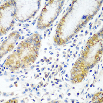
| Sample | Paraffin-embedded human stomach tissue; fixative not specified (datasheet A06057-1) |
| Fixation | Image fixative and duration unreported (datasheet A06057-1); verify before use. |
| Sectioning | 4–5 µm sections on charged slides (standard) |
| Deparaffinisation | Xylene, graded ethanol series to water (standard) |
| Antigen retrieval | Heat-induced epitope retrieval in citrate buffer, pH 6.0, 20 min at 95–98 °C (standard rule: cytoplasmic / membrane antigen) |
| Peroxidase block | 3% H2O2, 10 min, room temperature (standard) |
| Blocking | 10% normal serum of the secondary host, 30 min, room temperature (standard) |
| Primary antibody | Rabbit anti-ATAD3A, 1:100-1:200 (datasheet A06057-1) |
| Primary incubation | Overnight at 4 °C (standard) |
| Detection | HRP-polymer secondary, DAB chromogen 5–10 min (standard) |
| Counterstain | Hematoxylin, blue, dehydrate and mount (standard) |
| Expected result | ATAD3A-positive staining in glandular cells of adrenal gland (HPA tissue IHC: High). HPA tissue profile: Granular cytoplasmic expression in all tissues. No signal in the no-primary control. |
ATAD3A is associated with the mitochondrial inner membrane and matrix nucleoids; its single transmembrane segment spans residues 247–264 (UniProt Q9NVI7 topology). In paraffin section IHC, expect granular cytoplasmic staining across tissues, including strong staining in several glandular and epithelial cell populations (HPA: tissue IHC). Treat tissue intensity as a guide: HPA rates the tissue pattern Approved, with medium consistency against RNA data and a warning that the antibody targets proteins from more than one gene (HPA: tissue reliability).
| Granular cytoplasmic staining is prominent in adrenal, appendix, breast, cervical, colonic or duodenal glandular cells, or bronchial respiratory epithelium (HPA: High in these cell populations). | This matches the reported tissue pattern (HPA: granular cytoplasmic expression). Mitochondrial localisation offers a compartment explanation (UniProt Q9NVI7: inner membrane and matrix nucleoids), but chromogenic IHC alone does not resolve the inner membrane. |
| A predominantly nuclear, membranous or extracellular signal replaces granular cytoplasmic staining (HPA: tissue IHC pattern). | That distribution conflicts with the reported mitochondrial localisation (UniProt Q9NVI7; HPA: subcellular). Check morphology, counterstain and detection artefacts before interpreting it as ATAD3A; the supplied sources do not establish those compartments as the expected IHC result. |
| The strongest signal falls in smooth muscle cells, chondrocytes or adipocytes while adjacent expected positive cells are weak (HPA: Low in those cell types; High in listed glandular cells). | The relative pattern conflicts with HPA tissue observations. Consider antibody cross-reactivity or endogenous detection activity as possible causes (general IHC practice); HPA specifically cautions that its tissue antibody targets proteins from more than one gene (HPA: tissue reliability). |
| Diffuse colour covers many cells and extracellular spaces, obscuring discrete cytoplasmic granules (HPA: granular cytoplasmic profile). | This prevents a reliable localisation call. Uneven blocking, excess primary antibody or detection background can produce diffuse staining (general IHC practice); compare a no-primary control before assigning the colour to ATAD3A. |
| Little or no signal appears in a run that includes an HPA-reported high-staining tissue, such as colon glandular cells (HPA: High in colon glandular cells). | First assess tissue preservation, retrieval, antibody incubation and detection with appropriate run controls (general IHC practice). An absent signal does not by itself prove ATAD3A absence; HPA reports staining levels, not guaranteed positivity under every laboratory condition. |
| Mitochondrial topology (UniProt Q9NVI7) | A single transmembrane segment at 247–264 separates an intermembrane region from a matrix region (UniProt Q9NVI7 topology). The payload gives no antibody epitope, so topology cannot predict which region the IHC antibody recognises or which retrieval condition works. |
| Tissue reference strength (HPA: Approved IHC) | HPA describes granular cytoplasmic staining across tissues, with medium consistency between staining and RNA expression and a multiple-gene targeting caution (HPA: tissue reliability). Use its high and low cell populations as comparisons, without treating their intensity as proof of antibody specificity. |
| Reported isoforms and processing (UniProt Q9NVI7) | UniProt lists 3 isoforms and an annotated chain spanning residues 2–586, with no signal peptide or propeptide (UniProt Q9NVI7). Without the antibody epitope, these annotations cannot establish isoform coverage or explain a missing IHC signal. |
| Retrieval choice (general IHC practice) | Optimise antigen retrieval against a known staining reference and assess whether tissue detail remains interpretable (general IHC practice). No ATAD3A-specific retrieval condition or fixation sensitivity is supplied; HPA staining levels and UniProt topology do not establish either effect. |
| Chromogenic detection controls (general IHC practice) | A no-primary control helps identify background from detection reagents or endogenous enzyme activity (general IHC practice). Keep that signal separate from the granular cytoplasmic reference pattern (HPA: tissue IHC); the payload does not identify a target-specific endogenous activity. |
| Situation | Likely cause | Next action |
|---|---|---|
| The expected high-staining cell population is blank (HPA: High in listed cell populations). | Run failure, unsuitable retrieval or an ineffective primary antibody are possibilities (general IHC practice); absence alone cannot identify which step failed. | Review an appropriate positive run control, then check retrieval, primary incubation and detection in sequence (general IHC practice). Reassess the same cell population against the HPA tissue reference. |
| Most of the section has diffuse chromogenic colour instead of discrete granules (HPA: granular cytoplasmic profile). | Primary antibody excess, insufficient blocking or detection background are possible explanations (general IHC practice). | Compare a no-primary control and optimise blocking, washes and primary concentration (general IHC practice); score localisation only after the cytoplasmic pattern becomes distinguishable. |
| Colour is strongest in HPA-low cells and weak in HPA-high cells (HPA: tissue IHC levels). | Cross-reactivity or endogenous detection activity may contribute (general IHC practice); HPA also warns of antibody targeting proteins from more than one gene (HPA: tissue reliability). | Check a no-primary control, cell identity and the relative pattern in high and low reference populations (general IHC practice; HPA: tissue IHC). Avoid calling the low-cell signal specific on intensity alone. |
| The apparent positive result is chiefly nuclear or extracellular (HPA: granular cytoplasmic profile). | Compartment misassignment, counterstain confusion or background may explain the mismatch (general IHC practice). | Recheck morphology and detection controls, then require a granular cytoplasmic pattern before comparing with the mitochondrial localisation reference (HPA: tissue IHC; UniProt Q9NVI7). |
| Different sections show inconsistent intensity within the same expected cell population (HPA: tissue IHC levels). | Section handling, retrieval or incubation variation can change IHC staining (general IHC practice); the supplied sources do not show ATAD3A-specific fixation sensitivity. | Compare matched run controls and recorded processing conditions (general IHC practice). Interpret intensity only where morphology and the expected cell population remain comparable (HPA: tissue IHC). |
| IF/ICC question: what localisation should an independent fluorescence check show? | A fluorescence image can address subcellular distribution, while chromogenic tissue IHC provides a coarser localisation readout (general microscopy practice). | Look for a mainly mitochondrial signal (HPA: subcellular ICC-IF; UniProt Q9NVI7), while retaining HPA's multiple-gene antibody caution; consult the separate IF/ICC guide for assay design. |
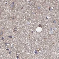
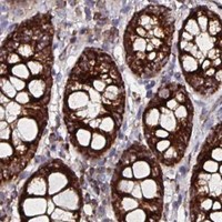
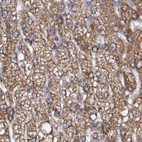


Comprehensive Human Protein Atlas IHC scoring per tissue (reliability: Approved — Medium consistency between antibody staining and RNA expression data. Caution, targets protein from more than one gene.). Rows are taken directly from the HPA tissue chart — click any row's HPA link to view the source.
| Tissue | Cell type | Level | Evidence | Source |
|---|---|---|---|---|
| Adrenal gland | Glandular cells | High | Protein (IHC) | HPA → |
| Appendix | Glandular cells | High | Protein (IHC) | HPA → |
| Breast | Glandular cells | High | Protein (IHC) | HPA → |
| Bronchus | Respiratory epithelial cells | High | Protein (IHC) | HPA → |
| Cerebellum | Purkinje cells | High | Protein (IHC) | HPA → |
| Tissue | Cell type | Level | Evidence | Source |
|---|---|---|---|---|
| None in HPA: ATAD3A is detected in all 45 scored tissues. Use a no-primary (secondary-only) and an isotype control instead. | ||||
Troubleshoot ATAD3A staining in paraffin section chromogenic IHC by checking retrieval, compartmental pattern, controls and scoring against the available evidence.
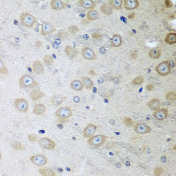
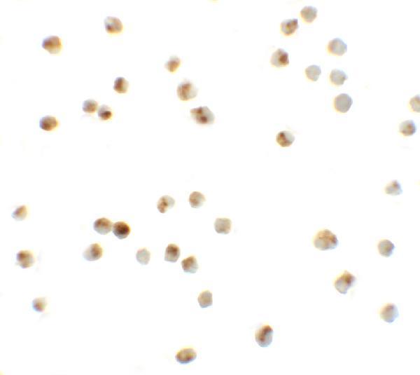
Anti-ATAD3A antibodies have IHC images from paraffin-embedded human stomach and mouse brain (A06057-1 image captions), plus IF/ICC images from Daudi cells (A06057 image captions).
A06057-1 is listed for IHC and has paraffin-section images from human stomach and mouse brain at 1:100 (catalog applications; A06057-1 image captions). A06057 is listed for IF/ICC and has Daudi-cell images at 20 μg/mL (catalog applications; A06057 image captions).
Which to pick: Choose A06057-1 for tissue IHC: its images show paraffin sections with microwave retrieval in 10 mM PBS, pH 7.2; the fixative is unreported (A06057-1 image captions). Choose A06057 for IF/ICC; its images show Daudi cells, while its catalog lists human, mouse and rat reactivity, including rat for cross-species planning (A06057 image captions; catalog reactivity). A06057-1 lists human and mouse reactivity, and clonality is unreported for both antibodies (catalog reactivity; catalog clone fields).