This website uses cookies to ensure you get the best experience on our website.


- Table of Contents
Plan ATP4A paraffin IHC around cytoplasmic staining in gastric parietal cells (HPA tissue IHC) and the expected apical canalicular membrane location (UniProt). The catalog antibody has a human stomach example using 2 µg/mL primary antibody and HRP/DAB detection (datasheet A08198-1).

Expected localisation, validated protocols, controls and antibodies — the at-a-glance facts below, then the full design guide.
| Expected localisation | Observed parietal-cell cytoplasm (HPA tissue IHC); expected apical canalicular membrane (UniProt) | |
| Staining pattern | Cytoplasmic staining in gastric parietal cells (HPA tissue IHC) | |
| Antigen retrieval | EDTA pH 8.0 HIER, heat-mediated (datasheet A08198-1) | |
| Positive control | Stomach+1 more · see all | |
| Negative control | Adipose tissue+4 more · see all |
| Fixation | Keep fixation consistent across sections (standard IHC practice; not target-specific) | |
| Caveat | High staining also reported in late spermatids (HPA tissue IHC) | |
| Regulation | No specific regulator reported (UniProt) | |
| Isoform / epitope | No isoforms annotated; cytoplasmic vs lumenal epitope side matters (UniProt) |
The catalog antibody protocol uses heat-mediated EDTA retrieval at pH 8.0 (datasheet A08198-1). One published ATP4A paraffin-section IHC protocol uses high-temperature retrieval at pH 9 (PMC8874512).
| Sample | Paraffin-embedded rat stomach tissue; fixative not specified (datasheet A08198-1) |
| Fixation | Image fixative and duration unreported (datasheet A08198-1); verify before use. |
| Sectioning | 4–5 µm sections on charged slides (standard) |
| Deparaffinisation | Xylene, graded ethanol series to water (standard) |
| Antigen retrieval | Heat retrieval: EDTA pH 8.0 (datasheet A08198-1); 20 min, 95–100 °C (standard) |
| Peroxidase block | 3% H2O2, 10 min, room temperature (standard) |
| Blocking | 10% goat serum (datasheet A08198-1) |
| Primary antibody | Rabbit anti-ATP4A, 2 μg/ml (datasheet A08198-1) |
| Primary incubation | Overnight at 4 °C (datasheet A08198-1) |
| Detection | HRP-conjugated secondary, DAB chromogen (datasheet A08198-1) |
| Counterstain | Hematoxylin, blue, dehydrate and mount (standard) |
| Expected result | ATP4A-positive staining in glandular cells of stomach (HPA tissue IHC: High). HPA tissue profile: Cytoplasmic expression in gastric parietal cells. No signal in the no-primary control. |
ATP4A should stain gastric parietal cells: UniProt places this 10-transmembrane pump at their apical canalicular membrane (UniProt P20648 topology/localization). HPA describes cytoplasmic expression in gastric parietal cells and rates its tissue IHC profile Enhanced, reflecting high consistency between staining and RNA expression (HPA tissue IHC: Enhanced). Interpret the slide at both cell and compartment level.
| Strong staining in a subset of stomach glandular cells, with apical canalicular or cytoplasmic signal. | This fits ATP4A expression in gastric parietal cells (UniProt P20648 tissue specificity; HPA tissue IHC: stomach glandular cells High). HPA calls the tissue pattern cytoplasmic; UniProt specifies the apical canalicular membrane. Section geometry and assay resolution may make membrane-associated signal appear cytoplasmic (general IHC interpretation). |
| Predominantly nuclear staining, without a plausible parietal-cell cytoplasmic or membrane pattern. | A nuclear-only pattern conflicts with the reported HPA cytoplasmic profile and UniProt apical membrane localization (HPA tissue IHC; UniProt P20648 localization). Review controls and morphology before scoring it as ATP4A; a wrong-compartment signal can reflect nonspecific detection (general IHC practice). |
| Broad staining of cells outside the expected gastric parietal-cell population. | Consider cross-reactivity or endogenous chromogenic activity if the pattern does not match tissue morphology (general IHC practice). Cell identity matters: HPA also reports High staining in testis elongated or late spermatids, so that defined population is a documented exception to a stomach-only reading (HPA tissue IHC). |
| Uniform haze across glands, stroma, and empty spaces obscures cell boundaries. | Diffuse background does not establish ATP4A localization. Compare a no-primary control and assess blocking, wash stringency, and chromogen development (general IHC practice). Score specific signal only where the cell pattern remains distinguishable from background (general IHC practice). |
| No convincing signal in an adequately represented stomach section. | This conflicts with the reported High stomach glandular-cell staining (HPA tissue IHC). Check whether parietal cells are present, then review the assay's positive control, antibody dilution, retrieval conditions, and detection run (general IHC practice). The supplied sources do not establish ATP4A-specific fixation sensitivity. |
| Compartment and section view | ATP4A has 10 transmembrane segments and resides at the parietal-cell apical canalicular membrane (UniProt P20648 topology/localization). HPA describes cytoplasmic IHC staining; use cell morphology and distribution when reconciling these views (HPA tissue IHC; general IHC interpretation). |
| Positive and comparison tissues | Stomach glandular cells stain High; HPA also reports High staining in testis elongated or late spermatids (HPA tissue IHC). The listed adipose, adrenal, appendix, bone marrow, breast, bronchus, caudate, and cerebellum cell populations are Not detected (HPA tissue IHC); those calls apply to the specified cells. |
| Antibody evidence | HPA lists two rabbit polyclonal antibodies, HPA039154 and HPA076684, as IHC Enhanced (HPA antibodies). HPA describes its tissue-profile reliability as Enhanced because staining is highly consistent with RNA expression (HPA tissue IHC). These ratings support pattern assessment; they do not specify a catalog-antibody dilution or retrieval method. |
| Epitope and preparation | The protein spans the membrane repeatedly, with both cytoplasmic and lumenal regions (UniProt P20648 topology). The payload gives no antibody epitope, so topology alone cannot select a retrieval or permeabilisation condition (UniProt P20648 topology; general IHC practice). ATP4A-specific fixation effects are unreported in these sources. |
| IF/ICC: what signal is supported? | HPA's subcellular summary says Membrane, but provides no main-location call or ICC-IF cell-line images; the listed antibodies have no ICC validation entry (HPA subcellular; HPA antibodies). For IF/ICC, treat membrane localization as a hypothesis from UniProt and the HPA summary, and validate it with assay controls (UniProt P20648 localization; general IF practice). |
| Situation | Likely cause | Next action |
|---|---|---|
| Stomach positive control is blank. | Parietal cells may be absent from the viewed area, or an IHC workflow step may have failed (general IHC practice). | Confirm gland morphology and parietal-cell representation; check a known-positive run, primary-antibody setup, retrieval, and detection reagents (general IHC practice). HPA reports High stomach glandular-cell staining (HPA tissue IHC). |
| Signal appears nuclear rather than cytoplasmic or canalicular. | The compartment conflicts with reported ATP4A localization; nonspecific staining is possible (HPA tissue IHC; UniProt P20648 localization; general IHC practice). | Compare a no-primary control, inspect cellular boundaries at higher magnification, and score nuclear-only color separately from plausible ATP4A signal (general IHC practice). |
| Most of the section has weak brown haze. | Background from detection chemistry, insufficient blocking or washing, or excessive chromogen development may obscure the pattern (general chromogenic IHC practice). | Inspect the no-primary control; review blocking, washes, and development time. Require a distinguishable parietal-cell pattern before scoring positivity (general IHC practice). |
| Many cell types stain in the stomach section. | A broad distribution may reflect nonspecific binding or endogenous detection activity rather than the reported parietal-cell profile (HPA tissue IHC; general IHC practice). | Map signal to cell morphology; run a no-primary control and, for peroxidase-based detection, check endogenous peroxidase blocking (general IHC practice). |
| Testis staining seems to contradict stomach enrichment. | HPA reports High protein staining in elongated or late spermatids despite stomach-enriched RNA and the reported parietal-cell expression (HPA tissue IHC; UniProt P20648 tissue specificity). | Identify the stained testis cell population before calling the result off-target. Interpret staining outside that HPA-listed population cautiously and compare appropriate controls (HPA tissue IHC; general IHC practice). |
| A change in retrieval gives a different staining pattern. | Retrieval conditions can alter an IHC assay, but the supplied HPA and UniProt records provide no ATP4A-specific retrieval or fixation-effect result (general IHC practice; HPA tissue IHC; UniProt P20648). | Compare conditions using the same positive tissue and controls, recording cell type, compartment, and background. Retain the condition that yields an interpretable pattern; do not infer an ATP4A-specific fixation mechanism from this comparison (general IHC practice). |
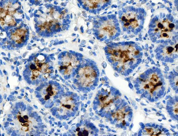
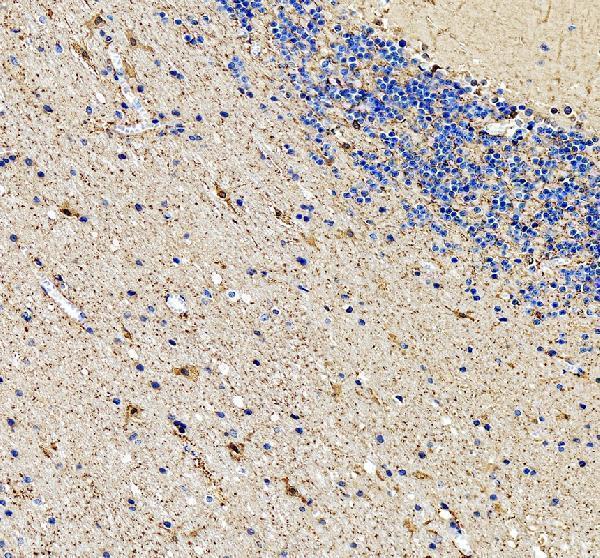


Comprehensive Human Protein Atlas IHC scoring per tissue (reliability: Enhanced — High consistency between antibody staining and RNA expression data.). Rows are taken directly from the HPA tissue chart — click any row's HPA link to view the source.
| Tissue | Cell type | Level | Evidence | Source |
|---|---|---|---|---|
| Stomach | Glandular cells | High | Protein (IHC) | HPA → |
| Testis | Elongated or late spermatids | High | Protein (IHC) | HPA → |
| Tissue | Cell type | Level | Evidence | Source |
|---|---|---|---|---|
| Adipose tissue | Adipocytes | Not detected | Protein (IHC) | HPA → |
| Adrenal gland | Glandular cells | Not detected | Protein (IHC) | HPA → |
| Appendix | Glandular cells | Not detected | Protein (IHC) | HPA → |
| Bone marrow | Hematopoietic cells | Not detected | Protein (IHC) | HPA → |
| Breast | Adipocytes | Not detected | Protein (IHC) | HPA → |
Troubleshoot ATP4A staining in paraffin section IHC by checking retrieval, parietal cell localisation and control tissue before interpreting signal intensity.
The catalog antibody has real paraffin-section IHC images from rat stomach, human stomach, and human stomach cancer; no IF image is supplied (A08198-1 image captions; catalog IF images: none).
A08198-1 will render with a rat stomach paraffin-section IHC figure (A08198-1 card caption). Its catalog also documents human stomach and human stomach cancer IHC images and lists Human, Mouse, and Rat reactivity (A08198-1 image captions; catalog reactivity).
Which to pick: Choose A08198-1 for paraffin-section tissue IHC: its own captions show rat and human stomach sections, with fixative unreported (A08198-1 image captions). For cross-species work, the same SKU lists Human, Mouse, and Rat reactivity, although the supplied IHC images cover Human and Rat only (catalog reactivity; A08198-1 image captions). No IF/ICC application or image is supplied, so there is no supported IF/ICC pick; clonality is unreported.