This website uses cookies to ensure you get the best experience on our website.


- Table of Contents
Plan ATP4B IHC in paraffin sections using stomach as a positive tissue control (HPA tissue IHC). Compare selective cytoplasmic staining in parietal cells with the apical canalicular membrane location annotated for the protein (HPA tissue IHC; UniProt).

Expected localisation, validated protocols, controls and antibodies — the at-a-glance facts below, then the full design guide.
| Expected localisation | Parietal cytoplasm observed; apical membrane expected (HPA tissue IHC; UniProt) | |
| Staining pattern | Selective cytoplasmic staining in gastric parietal cells (HPA tissue IHC) | |
| Antigen retrieval | EDTA pH 8.0 HIER, heat-mediated (datasheet A08719-2) | |
| Positive control | Stomach | |
| Negative control | Adipose tissue+4 more · see all |
| Fixation | Keep fixation consistent across sections (standard IHC practice; not target-specific). Selected-image fixative and duration unreported (datasheet A08719-2); verify before use. | |
| Caveat | Sections lacking parietal cells may appear negative (HPA tissue IHC) | |
| Regulation | Expression regulation not annotated (UniProt) | |
| Isoform / epitope | No isoforms annotated; cytoplasmic versus extracellular epitope matters (UniProt) |
The catalog antibody’s paraffin-section IHC protocol (datasheet A08719-2) is accompanied by four published ATP4B IHC protocols (PMC12697849; PMC8128498; PMC13395671; PMC11134753).
| Sample | Paraffin-embedded mouse stomach tissue; fixative not specified (datasheet A08719-2) |
| Fixation | Image fixative and duration unreported (datasheet A08719-2); verify before use. |
| Sectioning | 4–5 µm sections on charged slides (standard) |
| Deparaffinisation | Xylene, graded ethanol series to water (standard) |
| Antigen retrieval | Heat retrieval: EDTA pH 8.0 (datasheet A08719-2); 20 min, 95–100 °C (standard) |
| Peroxidase block | 3% H2O2, 10 min, room temperature (standard) |
| Blocking | 10% goat serum (datasheet A08719-2) |
| Primary antibody | Rabbit anti-ATP4B, 2-5 μg/ml (datasheet A08719-2) |
| Primary incubation | Overnight at 4 °C (datasheet A08719-2) |
| Detection | HRP-conjugated secondary, DAB chromogen (datasheet A08719-2) |
| Counterstain | Hematoxylin, blue, dehydrate and mount (standard) |
| Expected result | ATP4B-positive staining in glandular cells of stomach (HPA tissue IHC: High). HPA tissue profile: Selective cytoplasmic expression in parietal cells. No signal in the no-primary control. |
ATP4B should stain gastric parietal cells: HPA reports selective cytoplasmic staining within stomach glandular cells, with high staining and Enhanced tissue IHC reliability (HPA tissue IHC). UniProt places the protein at the apical canalicular membrane, with one transmembrane segment at residues 37–57 and an extracellular region at 58–291 (UniProt P51164 topology). Interpret the apparent cytoplasmic pattern in paraffin sections alongside that membrane assignment.
| Strong staining in a subset of stomach glandular cells, with an apparent cytoplasmic pattern (HPA tissue IHC). | This matches HPA’s high, selective parietal-cell result (HPA tissue IHC). Assess the pattern within recognizable gastric glands; UniProt assigns ATP4B to the parietal-cell apical canalicular membrane (UniProt P51164 subcellular location). |
| Staining is predominantly nuclear, rather than in the expected parietal-cell region (HPA tissue IHC; UniProt P51164 subcellular location). | Nuclear localization is unsupported by the supplied sources. Treat it as possible nonspecific staining or detection artefact (general IHC practice); check controls and morphology before counting cells as ATP4B positive. |
| Strong staining appears in nonparietal cells or in a listed tissue where HPA reports no detection (HPA tissue IHC). | Suspect cross-reactivity or endogenous detection activity (general IHC practice). For example, HPA reports no detection in appendix glandular cells and bronchial respiratory epithelial cells; their staining would need independent validation (HPA tissue IHC). |
| Diffuse chromogen covers cells and surrounding tissue without a recognizable selective pattern (HPA tissue IHC). | Background can obscure the expected parietal-cell staining (HPA tissue IHC). Review blocking, antibody concentration, washing and detection controls as general IHC variables (general IHC practice); diffuse color alone is insufficient to score ATP4B. |
| No signal is visible in stomach glandular cells expected to contain parietal cells (HPA tissue IHC). | First verify that the section contains identifiable parietal cells. HPA reports high stomach glandular-cell staining, but a negative slide alone cannot distinguish absent target from assay failure (HPA tissue IHC; general IHC practice). |
| Compartment and epitope orientation (UniProt P51164 topology). | ATP4B has a short cytoplasmic segment at residues 1–36, one membrane span at 37–57, and an extracellular region at 58–291 (UniProt P51164 topology). Antibody epitope location is not supplied, so these features do not predict which retrieval condition will work. |
| Glycosylation (UniProt P51164 PTMs). | UniProt annotates seven glycosylation sites at residues 99, 103, 130, 146, 161, 193 and 222 (UniProt P51164 PTMs). Without the antibody’s epitope or comparative staining data, their effect on this IHC result is unknown. |
| Tissue evidence and validation (HPA tissue IHC; HPA antibodies). | HPA rates the tissue pattern Enhanced because antibody staining is highly consistent with RNA expression; stomach RNA is tissue enriched (HPA tissue IHC). Both listed antibodies, HPA045400 and HPA052649, have Enhanced IHC status (HPA antibodies). |
| Fixation and antigen retrieval (general IHC practice). | Retrieval conditions can be optimized using matched sections and controls (general IHC practice). The supplied HPA and UniProt records give no ATP4B-specific fixation sensitivity or retrieval comparison; no target-specific direction of effect can be assigned. |
| IF/ICC interpretation (HPA subcellular; HPA antibodies). | Q: Should IF/ICC show the same pattern as tissue IHC? A: HPA reports an approved plasma-membrane location in ICC-IF and Approved ICC status for HPA045400 (HPA subcellular; HPA antibodies). Interpret that evidence in its cell-imaging context, not as an IHC-P protocol. |
| Situation | Likely cause | Next action |
|---|---|---|
| Stomach section has no clear parietal-cell signal (HPA tissue IHC). | The section may lack identifiable parietal cells, or the IHC run may have failed (general IHC practice). The supplied sources do not identify an ATP4B-specific fixation cause. | Confirm gland morphology and include a stomach positive control; then review retrieval, antibody concentration and detection using the applicable validated IHC procedure (general IHC practice). |
| Signal appears mainly nuclear (HPA tissue IHC; UniProt P51164 subcellular location). | That compartment disagrees with both the reported tissue pattern and membrane localization; nonspecific detection is possible (HPA tissue IHC; UniProt P51164 subcellular location; general IHC practice). | Check a no-primary control and evaluate whether staining persists independently of the primary antibody; rescore only a selective, morphologically plausible parietal-cell pattern (general IHC practice). |
| Color is widespread across the section (HPA tissue IHC). | Diffuse background may reflect nonspecific binding, incomplete washing or detection background (general IHC practice); it does not match HPA’s selective parietal-cell pattern (HPA tissue IHC). | Review blocking and washes, assess a no-primary control, and adjust antibody concentration within the validated IHC workflow (general IHC practice). |
| Nonparietal cells stain strongly in stomach (HPA tissue IHC). | Cell-type assignment may be mistaken, or the signal may reflect cross-reactivity or endogenous activity (general IHC practice). HPA describes selective parietal-cell expression (HPA tissue IHC). | Recheck gland and cell morphology, compare an appropriate negative tissue, and inspect detection controls before assigning ATP4B positivity (HPA tissue IHC; general IHC practice). |
| A listed negative tissue stains, such as appendix glandular cells (HPA tissue IHC). | HPA reports ATP4B as not detected there; staining could arise from cross-reactivity or the detection system (HPA tissue IHC; general IHC practice). | Repeat alongside a stomach positive control and a no-primary control. Treat persistent unexpected staining as unvalidated until its specificity is independently checked (HPA tissue IHC; general IHC practice). |
| An IF/ICC plasma-membrane image seems inconsistent with cytoplasmic-looking tissue IHC (HPA subcellular; HPA tissue IHC). | HPA describes plasma-membrane localization in ICC-IF and selective cytoplasmic appearance in tissue sections; the records use different imaging contexts (HPA subcellular; HPA tissue IHC). | Score the paraffin IHC slide against its tissue and cell-type pattern, while retaining UniProt’s apical canalicular membrane assignment; use the separate IF/ICC guide for that application (HPA tissue IHC; UniProt P51164 subcellular location). |
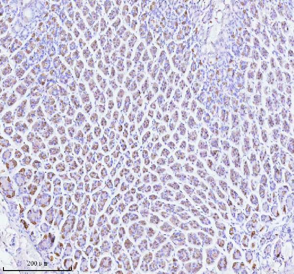
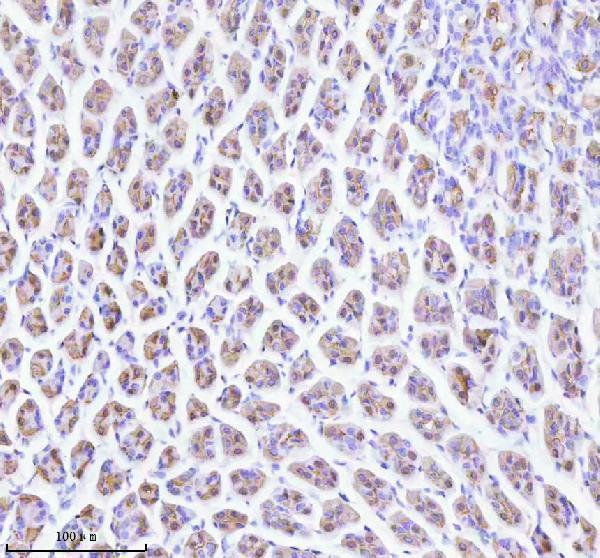
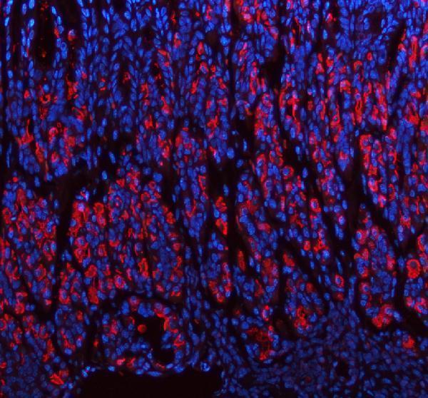
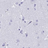


Comprehensive Human Protein Atlas IHC scoring per tissue (reliability: Enhanced — High consistency between antibody staining and RNA expression data.). Rows are taken directly from the HPA tissue chart — click any row's HPA link to view the source.
| Tissue | Cell type | Level | Evidence | Source |
|---|---|---|---|---|
| Stomach | Glandular cells | High | Protein (IHC) | HPA → |
| Tissue | Cell type | Level | Evidence | Source |
|---|---|---|---|---|
| Adipose tissue | Adipocytes | Not detected | Protein (IHC) | HPA → |
| Adrenal gland | Glandular cells | Not detected | Protein (IHC) | HPA → |
| Appendix | Glandular cells | Not detected | Protein (IHC) | HPA → |
| Bone marrow | Hematopoietic cells | Not detected | Protein (IHC) | HPA → |
| Breast | Adipocytes | Not detected | Protein (IHC) | HPA → |
Troubleshoot ATP4B staining in paraffin sections by checking retrieval, cell identity, membrane localisation and the controls used to interpret chromogenic signal.
A08719-2 has IHC images from mouse and rat paraffin stomach sections and IF images from mouse and rat paraffin stomach sections (catalog image captions); human reactivity is listed (catalog applications/reactivity).
A08719-2 is listed for IHC and IF in human, mouse and rat (catalog applications/reactivity). Its images document IHC and IF on mouse and rat paraffin stomach sections (catalog image captions).
Which to pick: For tissue IHC, choose A08719-2: its IHC captions show mouse and rat paraffin stomach sections with EDTA pH 8.0 retrieval and 2 μg/ml primary antibody (A08719-2 IHC image captions); the fixative is unreported (A08719-2 IHC image captions). For IF, A08719-2 has mouse and rat paraffin stomach images at 5 μg/ml (A08719-2 IF image captions); ICC validation is unreported (catalog applications and image captions). For cross-species work, A08719-2 lists human, mouse and rat reactivity, while the supplied IHC and IF images document mouse and rat samples (catalog reactivity; A08719-2 image captions).