This website uses cookies to ensure you get the best experience on our website.


- Table of Contents
Plan chromogenic ATP5F1B IHC in paraffin sections using the granular cytoplasmic pattern and kidney tubules as a high-staining control (HPA tissue IHC). The catalog antibody A32270-3 was demonstrated at 2 μg/mL with DAB detection (datasheet A32270-3).

Expected localisation, validated protocols, controls and antibodies — the at-a-glance facts below, then the full design guide.
| Expected localisation | Granular cytoplasm (HPA tissue IHC); mitochondrial inner membrane (UniProt) | |
| Staining pattern | Granular cytoplasm across tissue cell types (HPA tissue IHC) | |
| Antigen retrieval | EDTA pH 8.0 HIER, heat-mediated (datasheet A32270-3) | |
| Positive control | Duodenum+4 more · see all | |
| Negative control | None in HPA (detected in all 45 tissues); use no-primary + isotype controls |
| Fixation | Keep fixation consistent across paraffin sections (standard IHC practice; not target-specific) | |
| Caveat | Skeletal myocytes stain low despite enhanced tissue RNA (HPA tissue IHC) | |
| Regulation | Regulation not annotated (UniProt) | |
| Isoform / epitope | No isoforms; mature chain spans aa 48–529; map the epitope there (UniProt) |
Compare the catalog antibody’s IHC-P protocol (datasheet A32270-3) with the published ATP5F1B xenograft IHC protocol (PMC9868667).
| Sample | Paraffin-embedded human endometrial adenocarcinoma tissue; fixative not specified (datasheet A32270-3) |
| Fixation | Image fixative and duration unreported (datasheet A32270-3); verify before use. |
| Sectioning | 4–5 µm sections on charged slides (standard) |
| Deparaffinisation | Xylene, graded ethanol series to water (standard) |
| Antigen retrieval | Heat retrieval: EDTA pH 8.0 (datasheet A32270-3); 20 min, 95–100 °C (standard) |
| Peroxidase block | 3% H2O2, 10 min, room temperature (standard) |
| Blocking | 10% goat serum (datasheet A32270-3) |
| Primary antibody | Rabbit anti-ATP5F1B, 2-5 μg/ml (datasheet A32270-3) |
| Primary incubation | Overnight at 4 °C (datasheet A32270-3) |
| Detection | HRP-conjugated secondary, DAB chromogen (datasheet A32270-3) |
| Counterstain | Hematoxylin, blue, dehydrate and mount (standard) |
| Expected result | ATP5F1B-positive staining in glandular cells of duodenum (HPA tissue IHC: High). HPA tissue profile: General granular cytoplasmic expression. No signal in the no-primary control. |
ATP5F1B should produce granular cytoplasmic IHC staining consistent with mitochondria: its F1 catalytic head is associated with the mitochondrial inner membrane and has no transmembrane segment (UniProt P06576 topology; HPA: general granular cytoplasmic expression). High staining is reported in several glandular cell populations, cardiomyocytes, kidney tubule cells, lung macrophages and pancreatic exocrine glandular cells (HPA: tissue IHC). HPA rates the tissue profile Enhanced, with medium consistency between staining and RNA expression (HPA: reliability description).
| Granular cytoplasmic staining is strong in kidney tubule cells, cardiomyocytes or pancreatic exocrine glandular cells, with a clean surrounding field. | This matches the reported compartment and high-staining cell populations (HPA: general granular cytoplasmic expression; HPA: High in kidney tubule cells, cardiomyocytes and pancreatic exocrine glandular cells). Judge the expected pattern in identified cells, rather than treating every cell in the section as an equivalent positive control (general IHC practice). |
| The strongest signal is nuclear, extracellular or confined to section edges rather than granular cytoplasm. | That distribution conflicts with mitochondrial localization and the tissue IHC profile (UniProt P06576: mitochondrial inner membrane; HPA: general granular cytoplasmic expression). Review morphology, counterstain and detection artifacts before assigning it to ATP5F1B (general IHC practice). The localization mismatch alone does not identify its cause. |
| Strong color appears mainly in cells other than the reported high-staining population in a selected tissue. | For example, the reported high signal in lung is in macrophages, while the reported high signal in liver is in cholangiocytes (HPA: tissue IHC). Confirm cell identity and compare an antibody-omission control; cross-reactivity or endogenous detection activity is possible, but cannot be diagnosed from color alone (general IHC practice). |
| Weak, diffuse color covers cells and blank spaces, obscuring individual cytoplasmic granules. | A broad haze is less interpretable than HPA's granular cytoplasmic profile (HPA: tissue IHC). Uneven washing, excess detection reagent or nonspecific antibody binding can produce background (general IHC practice). Evaluate the omission control and reagent handling before calling faint color a true low-level positive. |
| A known high-staining tissue shows no specific signal, while morphology remains readable. | Absence in kidney tubule cells, heart cardiomyocytes or duodenal glandular cells disagrees with their reported high staining (HPA: tissue IHC). Check antibody and detection controls, retrieval conditions and tissue preservation as general IHC troubleshooting steps; these data do not establish ATP5F1B-specific fixation sensitivity (general IHC practice; HPA: tissue IHC). |
| Compartment and topology | ATP5F1B belongs to the soluble F1 catalytic head of mitochondrial ATP synthase, associated with the inner membrane; the beta subunit has no transmembrane segment (UniProt P06576: subunit and topology). In a chromogenic section, assess cytoplasmic granularity rather than expecting the inner membrane itself to be resolved (HPA: general granular cytoplasmic expression; general IHC practice). |
| Cell population and tissue choice | HPA reports High staining in duodenal, fallopian tube and gallbladder glandular cells; cardiomyocytes; kidney tubule cells; liver cholangiocytes; lung macrophages; and pancreatic exocrine glandular cells (HPA: tissue IHC). These named populations offer interpretable positive references, while an unlabeled whole-tissue average can hide cell-specific signal (general IHC practice). |
| Low staining and RNA discordance | HPA records Low protein staining in thyroid glandular cells, skeletal muscle myocytes, chondrocytes, adipocytes and splenic red-pulp cells (HPA: tissue IHC). Its RNA profile is tissue enhanced in skeletal muscle and tongue, yet skeletal muscle myocytes are Low by IHC (HPA: RNA specificity; HPA: Low in skeletal muscle myocytes). Do not substitute RNA enrichment for an expected IHC intensity. |
| Evidence strength across antibodies | The tissue IHC profile is Enhanced, although agreement between antibody staining and RNA is described as medium (HPA: reliability and reliability description). HPA lists IHC Enhanced for HPA001520 and HPA001528, and IHC Supported for CAB017527 (HPA: antibody validation). Record which antibody produced an unusual pattern before comparing it with the atlas profile (general IHC practice). |
| IF/ICC Q: what localization should be seen? | A: Mitochondrial staining is the expected IF/ICC localization (HPA: subcellular location, enhanced); HPA lists images from A-431, U-251MG, U2OS and sperm (HPA: ICC-IF image list). That cell-based observation supports the compartment interpretation, while tissue-section IHC intensity should be judged from the tissue IHC data (HPA: tissue IHC). |
| Situation | Likely cause | Next action |
|---|---|---|
| No color in a selected high-staining population | The expected cell population may be absent from the section, or an IHC workflow step may have failed (HPA: tissue IHC; general IHC practice). | Confirm the cells by morphology, then check a known-positive section and antibody, detection and retrieval steps (general IHC practice). Interpret retrieval changes as workflow checks, not an established ATP5F1B fixation effect. |
| Brown color persists in the antibody-omission control | Endogenous detection activity or detection-reagent background can create antibody-independent color (general chromogenic IHC practice). | Address the detection system's endogenous-activity blocking and inspect reagent background before scoring cells as ATP5F1B positive (general chromogenic IHC practice). |
| Nuclei dominate the signal | A nuclear pattern conflicts with ATP5F1B's reported mitochondrial location (UniProt P06576: mitochondrial inner membrane; HPA: subcellular location). | Compare counterstain and omission controls, inspect whether precipitate overlies nuclei, and reassess antibody specificity before interpreting the signal (general IHC practice). |
| Diffuse haze hides cytoplasmic granules | Nonspecific binding or excess detection background may obscure the reported granular pattern (HPA: tissue IHC; general IHC practice). | Review blocking, antibody dilution, washes and detection exposure using the antibody's IHC instructions; require a discernible cell-associated pattern before scoring (general IHC practice). |
| A low-staining population appears as strong as the positive reference | For example, skeletal muscle myocytes are recorded Low despite skeletal-muscle RNA enhancement (HPA: tissue IHC and RNA specificity); background or cell misidentification remains possible (general IHC practice). | Verify cell identity, compare a reported High population, and use an omission control before revising the expected pattern (HPA: tissue IHC; general IHC practice). |
| Different antibodies produce conflicting cell patterns | Validation status differs across the listed antibodies, and the tissue profile has medium staining-to-RNA consistency (HPA: antibody validation and reliability description). | Document the antibody identifier and controls, compare the same cell population across sections, and qualify any conclusion that depends on one discordant stain (general IHC practice). |


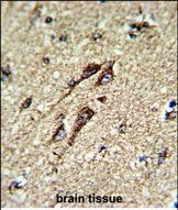
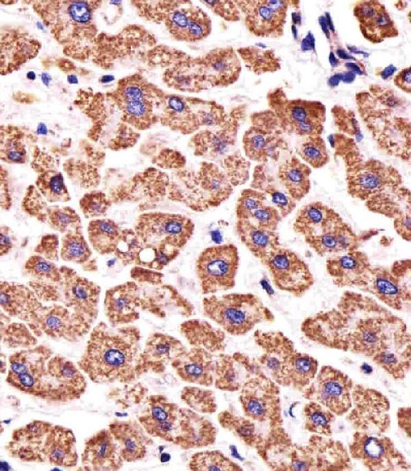

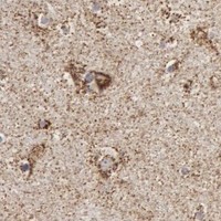


Comprehensive Human Protein Atlas IHC scoring per tissue (reliability: Enhanced — Medium consistency between antibody staining and RNA expression data.). Rows are taken directly from the HPA tissue chart — click any row's HPA link to view the source.
| Tissue | Cell type | Level | Evidence | Source |
|---|---|---|---|---|
| Duodenum | Glandular cells | High | Protein (IHC) | HPA → |
| Fallopian tube | Glandular cells | High | Protein (IHC) | HPA → |
| Gallbladder | Glandular cells | High | Protein (IHC) | HPA → |
| Heart muscle | Cardiomyocytes | High | Protein (IHC) | HPA → |
| Kidney | Cells in tubules | High | Protein (IHC) | HPA → |
| Tissue | Cell type | Level | Evidence | Source |
|---|---|---|---|---|
| None in HPA: ATP5F1B is detected in all 45 scored tissues. Use a no-primary (secondary-only) and an isotype control instead. | ||||
Troubleshoot ATP5F1B staining in paraffin sections using its mitochondrial localisation, the catalog antibody’s tissue IHC conditions, and matched controls.
The catalog antibodies have human tissue IHC images and cell or tissue IF images (catalog image captions); A32270-3 also lists mouse and rat reactivity (catalog reactivity).
A32270-3 has IHC images from human paraffin sections of endometrial adenocarcinoma, ovarian cancer, placenta and rectal cancer, plus IF images from PC-3 cells and human paraffin sections (catalog image captions). A32270-2 has IHC images from formalin-fixed, paraffin-embedded human brain and paraffin sections of human liver and small intestine, plus an IF image from SK-BR-3 cells (catalog image captions).
Which to pick: For tissue IHC, choose A32270-3 for its documented EDTA pH 8.0 retrieval and 2 μg/ml staining on human paraffin sections; their fixative is unreported (A32270-3 IHC captions). Choose A32270-2 when a documented paraffin-section, paraffin-embedded human brain example or its 1:25 IHC-P dilution better matches the study (A32270-2 IHC captions; catalog dilution); its liver and small-intestine captions report paraffin sections without specifying fixative (A32270-2 IHC captions). For IF/ICC, A32270-3 lists both applications and has cell and tissue IF images, while A32270-2 lists IF and has a cell IF image; for mouse or rat tissue, A32270-3 lists reactivity and IHC use in those species, though its supplied IHC images are human (catalog applications and reactivity; catalog image captions). The selected A32270-3 tissue-IHC caption documents paraffin sections, but does not specify the fixative (selected-SKU IHC image A32270-3).