ATP6V1C1 / V-type proton ATPase subunit C 1 · IHC design guide

Design Immunohistochemistry for ATP6V1C1

Plan ATP6V1C1 IHC in paraffin sections around the general cytoplasmic tissue pattern (HPA tissue IHC). Compare high staining in esophageal squamous cells and parathyroid or stomach glandular cells with undetected staining in adipocytes or skeletal myocytes (HPA tissue IHC).

Evidence assembled Oct 2026 · For research use; verify linked source records and product datasheet before use
Immunohistochemistry protocol sheet for ATP6V1C1 (IHC for ATP6V1C1): expected localisation Cytoplasmic staining (HPA tissue IHC); vesicle-associated protein (UniProt), antibody A10380-1, validated IHC image, and IHC protocol steps
Printable ATP6V1C1 IHC protocol sheet — expected localisation Cytoplasmic staining (HPA tissue IHC); vesicle-associated protein (UniProt), antibody A10380-1, controls and protocol steps. Open the full ATP6V1C1 IHC guide →

ATP6V1C1 Immunohistochemistry Experimental Design Guide

Expected localisation, validated protocols, controls and antibodies — the at-a-glance facts below, then the full design guide.

Must know before staining
Expected localisation Cytoplasmic staining (HPA tissue IHC); vesicle-associated protein (UniProt)
Staining pattern General cytoplasmic staining in glandular and squamous cells (HPA tissue IHC)
Antigen retrieval EDTA pH 8.0 HIER, heat-mediated (datasheet A10380-1)
Positive control ⓘ Esophagus+4 more · see all
Negative control ⓘ Adipose tissue+4 more · see all
Important caveats
Reasons your staining may differ from the expected pattern.
Fixation Keep fixation consistent across sections (standard IHC practice; not target-specific); optimize empirically.
Caveat IHC staining has medium concordance with RNA expression (HPA tissue IHC)
Regulation Ubiquitous expression; no induction specified (UniProt)
Isoform / epitope No annotated isoforms; chain 2–382; no transmembrane segment (UniProt)
Section 1

Recommended ATP6V1C1 IHC & IF Protocols

The catalog antibody has an IHC-P protocol (datasheet A10380-1); the published ATP6V1C1 IHC methods below provide additional tissue examples (PMC8806199; PMC3805834; PMC2773039).

Recommended immunohistochemistry (IHC-P) protocol parameters
SampleParaffin-embedded human liver cancer tissue; fixative not specified (datasheet A10380-1)
FixationImage fixative and duration unreported (datasheet A10380-1); verify before use.
Sectioning4–5 µm sections on charged slides (standard)
DeparaffinisationXylene, graded ethanol series to water (standard)
Antigen retrievalHeat retrieval: EDTA pH 8.0 (datasheet A10380-1); 20 min, 95–100 °C (standard)
Peroxidase block3% H2O2, 10 min, room temperature (standard)
Blocking10% goat serum (datasheet A10380-1)
Primary antibodyRabbit anti-ATP6V1C1, 1:50 recommended; image 2 μg/ml (datasheet A10380-1)
Primary incubationOvernight at 4 °C (datasheet A10380-1)
DetectionHRP-conjugated secondary, DAB chromogen (datasheet A10380-1)
CounterstainHematoxylin, blue, dehydrate and mount (standard)
Expected resultATP6V1C1-positive staining in squamous epithelial cells of esophagus (HPA tissue IHC: High). HPA tissue profile: General cytoplasmic expression. No signal in the no-primary control.
💡Decision noteStart with heat-mediated EDTA pH 8.0 retrieval for the catalog antibody (datasheet A10380-1). The published excerpts do not specify a retrieval buffer (PMC8806199; PMC3805834; PMC2773039).
Section 2

What Is the Expected ATP6V1C1 Staining Pattern?

ATP6V1C1 is a V-ATPase V1 subunit associated with cytoplasmic vesicle membranes and has no transmembrane segment (UniProt P21283). In tissue IHC, expect mainly cytoplasmic staining, especially in esophageal squamous epithelial cells and parathyroid or stomach glandular cells (HPA: general cytoplasmic expression; High in these cells). HPA rates its tissue staining Approved, with medium consistency between antibody staining and RNA expression (HPA: tissue IHC reliability).

What am I looking at on my slide?
Cytoplasmic staining in esophageal squamous epithelium or parathyroid or stomach glandular cells.This matches the reported IHC pattern and its strongest listed cell populations (HPA: general cytoplasmic expression; High in these cells). Judge the signal within the relevant cells, since a tissue section also contains other cell types (general IHC practice).
Predominantly nuclear staining with little cytoplasmic signal in a tissue IHC section.Treat this as a possible IHC artefact or assay-specific pattern and check controls before calling it positive (HPA: general cytoplasmic tissue IHC). HPA separately reports approved nucleoplasmic staining in ICC-IF, so nuclear fluorescence in cultured cells requires interpretation in that assay (HPA: subcellular ICC-IF).
Strong staining in adipocytes or skeletal myocytes while expected positive cells are weak.Consider cross-reactivity or endogenous chromogenic detection activity (general IHC practice). HPA reports ATP6V1C1 as Not detected in adipocytes in adipose tissue and breast, and in skeletal myocytes; those observations are useful comparisons, not universal proof of absence (HPA: tissue IHC).
Uniform color across cells, stroma, and empty areas, with no clear cellular pattern.Diffuse background can obscure the reported cytoplasmic distribution (HPA: tissue IHC). Assess a matched negative detection control and the distribution of precipitate; inadequate blocking, washing, or endogenous enzyme control can produce background in chromogenic IHC (general IHC practice).
No convincing staining in esophageal squamous epithelium, parathyroid glandular cells, or stomach glandular cells.These are High-staining examples in HPA, so a blank result raises an assay-performance question (HPA: tissue IHC). Check the control section, tissue preservation, antibody incubation, retrieval conditions, and detection reagents before interpreting the sample as negative (general IHC practice).
💡Expected ATP6V1C1 appearanceCall a tissue IHC result positive when cytoplasmic staining is clear in the relevant cells, particularly the High-staining esophageal squamous or parathyroid and stomach glandular cells (HPA: tissue IHC); diffuse section-wide color or isolated nuclear-only chromogenic staining warrants control review (general IHC practice; HPA: cytoplasmic tissue profile).
How each factor affects the staining
Tissue and cell contextHPA reports Low tissue RNA specificity and a general cytoplasmic protein profile, yet individual cell populations range from High to Not detected (HPA: tissue IHC). Compare like cell types within sections; a negative adipocyte result cannot serve as a whole-tissue negative control (HPA: adipocytes Not detected).
Assay-specific localisationTissue IHC is described as generally cytoplasmic, while approved ICC-IF localisation is mainly nucleoplasmic with additional cytosol signal (HPA: tissue IHC; HPA: subcellular ICC-IF). Can ICC-IF show nuclear signal? Yes, in its own assay; use its separate guide for experimental conditions (HPA: subcellular ICC-IF).
Antibody validationHPA023943 is Approved for IHC, whereas HPA057297 is Approved for ICC-IF; the listed approvals apply to their respective assays (HPA: antibody validation). Tissue IHC reliability is Approved with medium staining–RNA consistency, so corroborate unexpected patterns with controls (HPA: tissue IHC reliability).
Protein location and processingATP6V1C1 is a peripheral V1-complex subunit associated with vesicle membranes, without a transmembrane segment or annotated glycosylation sites (UniProt P21283: function, subcellular location, topology, glycosylation). These annotations support cytoplasmic interpretation but do not predict antigen retrieval or fixation sensitivity (UniProt P21283; HPA: tissue IHC).
Chromogenic detectionEndogenous enzyme activity or nonspecific detection can resemble cell staining, especially when color lacks the expected cytoplasmic distribution (general IHC practice; HPA: tissue IHC pattern). Control the detection system and interpret signal against matched control sections (general IHC practice).
Why is my staining missing, weak or wrong?
SituationLikely causeNext action
A High-staining reference tissue is blank.The expected cells may be absent from the section, or an IHC step may have failed (HPA: High in specified cell populations; general IHC practice).Confirm the relevant cells on the counterstain, then review the IHC-validated antibody, retrieval, incubation, and detection controls (HPA: HPA023943 IHC Approved; general IHC practice).
Only nuclei stain in chromogenic tissue IHC.This diverges from the general cytoplasmic tissue profile; nuclear ICC-IF localisation is a separate HPA observation (HPA: tissue IHC; HPA: subcellular ICC-IF).Check negative detection controls and compare a known positive tissue section before assigning the nuclear IHC signal to ATP6V1C1 (general IHC practice; HPA: High-staining tissues).
Adipocytes or skeletal myocytes show prominent color.These cell types were Not detected in the HPA tissue observations; cross-reactivity or endogenous detection activity is possible (HPA: tissue IHC; general IHC practice).Compare a matched negative detection control and the expected positive cell pattern; investigate persistent cell-specific signal with an independent antibody where available (general IHC practice; HPA: High-staining cells).
The entire section has a weak, diffuse haze.Background from insufficient blocking, washing, or detection controls can hide cell boundaries (general IHC practice).Review blocking and washes, check endogenous enzyme controls for the detection system, and score only interpretable cellular staining (general IHC practice).
Positive and negative tissues both appear equally stained.The assay may lack useful contrast, or the comparison may mix cell populations with different reported levels (general IHC practice; HPA: tissue IHC).Compare the specified High cells with the specified Not detected cells under matched staining conditions; inspect negative detection controls before changing the interpretation (HPA: tissue IHC; general IHC practice).
A negative result follows a change in fixation or retrieval.No target-specific fixation sensitivity or retrieval requirement is established by the supplied HPA and UniProt evidence (HPA: tissue IHC; UniProt P21283).Compare matched positive control sections processed with the old and new conditions; treat any difference as an assay finding rather than a known ATP6V1C1 property (general IHC practice).

Sample controls for ATP6V1C1 IHC & IF

🧪Run esophagus first and require staining in its squamous epithelial cells (HPA: High in esophageal squamous epithelial cells). Use adipose tissue as the negative comparison, with adipocytes expected to lack detectable staining (HPA: Not detected in adipocytes); on the esophagus slide, assess any morphologically identifiable unstained cells as internal background, without assigning them a negative cell type.
Positive control tissue: Esophagus (Squamous epithelial cells, HPA High)
Negative control tissue: Adipose tissue (HPA Not detected)
ICC-IF cell lines (HPA subcellular resource): HPA ICC-IF images show ATP6V1C1 in A-431, MCF-7, U2OS, with annotated localisation: Nucleoplasm (approved) (HPA subcellular).
Technical controls: Include a no-primary (secondary-only) slide and a concentration-matched rabbit IgG isotype control, since the selected antibody is rabbit-derived (caption: rabbit anti-ATP6V1C1); use target knockout material, if available, to test specificity (standard IHC practice). Quench endogenous peroxidase before HRP/DAB detection and check for staining associated with blood or inflammatory cells (standard chromogenic IHC practice; caption: HRP/DAB detection).
⚠️Feasibility: A target-specific fixation window or fixation effect is unreported, and the selected A10380-1 paraffin-section caption does not state the fixative (caption: fixative not stated). The caption uses heat retrieval in EDTA at pH 8.0, but does not establish that retrieval is required (caption: EDTA retrieval); neither frozen sections nor IF are shown to be easier by the supplied evidence. For esophagus, review any peroxidase-associated staining outside the expected squamous epithelial pattern before scoring (HPA: High in squamous epithelial cells; standard chromogenic IHC practice).

HPA tissue IHC evidence for ATP6V1C1

Comprehensive Human Protein Atlas IHC scoring per tissue (reliability: Approved — Medium consistency between antibody staining and RNA expression data.). Rows are taken directly from the HPA tissue chart — click any row's HPA link to view the source.

Positive expression · recommended positive controls

TissueCell typeLevelEvidenceSource
Esophagus Squamous epithelial cells High Protein (IHC) HPA →
Parathyroid gland Glandular cells High Protein (IHC) HPA →
Stomach Glandular cells High Protein (IHC) HPA →
Adrenal gland Glandular cells Medium Protein (IHC) HPA →
Appendix Glandular cells Medium Protein (IHC) HPA →

Undetected expression · recommended negative controls

TissueCell typeLevelEvidenceSource
Adipose tissue Adipocytes Not detected Protein (IHC) HPA →
Bone marrow Hematopoietic cells Not detected Protein (IHC) HPA →
Breast Adipocytes Not detected Protein (IHC) HPA →
Cervix Glandular cells Not detected Protein (IHC) HPA →
Liver Cholangiocytes Not detected Protein (IHC) HPA →
Section 3

Advanced ATP6V1C1 IHC Tips

Troubleshoot ATP6V1C1 staining in paraffin-section chromogenic IHC by checking retrieval, cellular pattern, controls and scoring before interpreting differences between samples.

Which retrieval conditions should I try first when ATP6V1C1 staining is weak?
Begin paraffin-section chromogenic IHC with heat-mediated retrieval in EDTA at pH 8.0 (datasheet A10380-1). The selected antibody image used that retrieval before staining human liver cancer tissue; its caption does not specify a heating time (caption A10380-1). Run a positive reference section beside the test section and keep heating, cooling and detection conditions identical across the pair (standard IHC practice). If staining remains weak, adjust retrieval duration in a small series while monitoring tissue integrity and background (standard IHC practice). Judge any gain by cellular staining and the negative control, rather than DAB intensity alone (standard IHC practice).
Could fixation explain weak or uneven ATP6V1C1 staining?
Target-specific fixation sensitivity is unknown: the selected paraffin-section caption does not report its fixative (caption A10380-1). Record the fixative and fixation duration for each specimen, then compare sections processed under matched conditions before attributing a weak result to ATP6V1C1 expression (standard IHC practice). In the selected antibody workflow, heat-mediated EDTA retrieval at pH 8.0 and overnight primary incubation at 4°C are documented, but neither establishes an optimal fixation condition (caption A10380-1). Include a concurrently processed positive reference and inspect section morphology; patchy staining alongside damaged morphology warrants a processing check (standard IHC practice). Do not infer fixation sensitivity from tissue expression or protein topology (HPA tissue IHC; UniProt P21283 topology).
Should ATP6V1C1 appear cytoplasmic, vesicular or nuclear in tissue sections?
Expect a cytoplasmic IHC component: the tissue atlas describes general cytoplasmic expression (HPA tissue IHC). UniProt places ATP6V1C1 with cytoplasmic vesicle membranes and identifies it as a V1 complex subunit without a transmembrane segment (UniProt P21283 localisation, subunit and topology). The subcellular atlas also reports approved nucleoplasmic and cytosolic locations from ICC/IF, so isolated nuclear DAB staining needs antibody and method controls before interpretation in paraffin sections (HPA subcellular; standard IHC practice). Compare staining with cell outlines and a counterstain, and examine a positive reference at the same exposure and development endpoint (standard IHC practice). Report the observed compartment rather than assigning every cytoplasmic deposit to a specific vesicle type (standard IHC practice).
How should epitope uncertainty affect interpretation of ATP6V1C1 IHC?
The supplied record lists 0 isoforms and no annotated domains or glycosylation sites; it does not identify this antibody’s epitope (UniProt P21283 record; caption A10380-1). ATP6V1C1 has no transmembrane segment, so a membrane-adjacent IHC pattern alone cannot locate the bound epitope or prove direct membrane insertion (UniProt P21283 topology; standard IHC interpretation). Compare the antibody’s documented paraffin-section staining with a positive reference under its EDTA pH 8.0 retrieval condition (caption A10380-1; standard IHC practice). If an antibody recognizes a different region, establish its paraffin-section performance independently before comparing scores across antibodies (standard IHC practice). Treat discrepancies as unresolved until specificity and processing controls support an explanation (standard IHC practice).
How can I investigate an ATP6V1C1 localisation discrepancy by multiplex IF?
Use IF/ICC as a separate localisation check alongside the paraffin-section IHC result; the subcellular atlas reports nucleoplasmic and cytosolic staining in imaged cell lines (HPA subcellular). Pair ATP6V1C1 with a validated marker for the expected cell type in the specimen, choosing spectrally separated fluorophores after checking tissue autofluorescence in an unstained control (standard IF practice). Because ATP6V1C1 lacks a transmembrane segment and belongs to the peripheral V1 complex, permeabilise sufficiently to expose intracellular epitopes while preserving cell structure (UniProt P21283 topology and subunit; standard IF practice). Include single-label controls to assess bleed-through and compare nuclear and cytoplasmic signal against the IHC pattern without assuming both methods expose the same epitope equally (standard IF/IHC practice).
What should I check when ATP6V1C1 DAB staining looks diffuse or nonspecific?
First inspect a no-primary control, section edges and tissue morphology; diffuse color in the control or along damaged edges suggests a detection or processing problem (standard IHC practice). The selected image used 10% goat serum block, 2 μg/ml primary antibody overnight at 4°C, and a 30-minute secondary incubation at 37°C (caption A10380-1). A peroxidase block and controlled DAB development are general chromogenic IHC safeguards, rather than demonstrated ATP6V1C1-specific requirements (standard IHC practice). If background persists, titrate primary antibody and shorten chromogen development while retaining the same retrieval and positive reference (standard IHC practice; datasheet A10380-1). Evaluate cellular contrast against the no-primary control before calling weak cytoplasmic color positive (standard IHC practice).
How should I score ATP6V1C1 staining across paraffin sections? ⚠ ANSWER MARKED FOR VERIFICATION
Define the cell population and staining compartment before scoring, then apply one threshold to every section in the comparison (standard IHC practice). An H-score combines the percentage of cells at each intensity, commonly 0–3, whereas percentage positive cells reports prevalence without intensity (standard IHC practice). For spatial questions, count positive cells per mm² of viable tissue and normalise to the area or total cells of the same annotated population (standard IHC practice). Keep retrieval, primary dilution, DAB development and image acquisition matched, using a concurrently processed reference to detect run variation (standard IHC practice). Report cytoplasmic and nuclear scores separately if both are evaluated, since the tissue and ICC/IF localisation evidence differs (HPA tissue IHC; HPA subcellular).
When is ATP6V1C1 staining convincing rather than an artefact?
Give greatest weight to reproducible cellular staining in viable tissue that exceeds the no-primary control and follows a plausible compartment pattern (standard IHC practice; HPA tissue IHC). The atlas reports high staining in esophageal squamous epithelial cells and parathyroid and stomach glandular cells, while liver cholangiocytes were not detected; use cell identity when selecting and reading reference areas (HPA tissue IHC). A purely peripheral edge deposit, necrotic-region color or signal retained in a no-primary control argues for artefact or endogenous enzyme activity (standard IHC practice). Isolated nuclear DAB should be checked against controls and cytoplasmic staining, because nucleoplasmic localisation is reported by ICC/IF while tissue IHC is described as generally cytoplasmic (HPA subcellular; HPA tissue IHC). The atlas rates tissue staining Approved with medium RNA consistency, so document exceptions rather than treating one stain as definitive expression evidence (HPA tissue IHC).
Boster reagents

Best ATP6V1C1 / V-type proton ATPase subunit C 1 IHC Antibodies

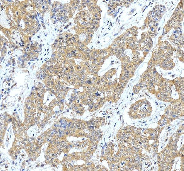
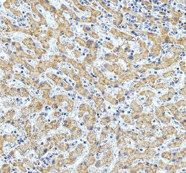
A10380-1 has IHC images from paraffin-embedded human liver and liver cancer sections (catalog image captions); its listed reactivity is Human, Mouse and Rat (catalog: reactivity).

Real IHC data IHC analysis of ATP6V1C1 using anti-ATP6V1C1 antibody (A10380-1). ATP6V1C1 was detected in a paraffin-embedded section of human liver cancer tissue. Heat mediated antigen retrieval was performed in EDTA buffer (pH 8.0, epitope retrieval solution). The tissue section was blocked with 10% goat serum. The tissue section was then incubated with 2 μg/ml rabbit anti-ATP6V1C1 Antibody (A10380-1) overnight at 4°C. Peroxidase Conjugated Goat Anti-rabbit IgG was used as secondary antibody and incubated for 30 minutes at 37°C. The tissue section was developed using HRP Conjugated Rabbit IgG Super Vision Assay Kit (Catalog # SV0002) with DAB as the chromogen.
Anti-ATP6V1C1 Antibody
Cat # A10380-1

A10380-1 has IHC images from paraffin-embedded human liver and liver cancer sections (catalog image captions). Its listed applications are IHC, ELISA and WB, and its listed reactivity is Human, Mouse and Rat (catalog: applications and reactivity).

Which to pick: For tissue IHC, choose A10380-1: its IHC images use paraffin-embedded human liver sections, EDTA retrieval at pH 8.0 and DAB detection (A10380-1 image captions); the fixative is unreported (A10380-1 image captions). For IF/ICC, A10380-1 has no listed IF/ICC application or IF image (catalog: applications and IF images). For cross-species work, A10380-1 lists Mouse and Rat reactivity, but the supplied IHC images show human tissue only (catalog: reactivity; A10380-1 image captions).

Each figure is that product's own IHC / IF validation image from its datasheet.

References

  1. UniProt Consortium. UniProt entry P21283 (VATC1_HUMAN, V-type proton ATPase subunit C 1).
  2. Human Protein Atlas. ATP6V1C1 tissue IHC expression (reliability: Approved).
  3. Human Protein Atlas. ATP6V1C1 subcellular location (ICC-IF): Mainly localized to the nucleoplasm. In addition localized to the cytosol..
  4. Human Protein Atlas. ATP6V1C1 antibody validation summary (2 antibodies).
  5. A novel four-gene of iron metabolism-related and methylated for prognosis prediction of hepatocellular carcinoma. Bioengineered 2021 — PMC8806199.
  6. Silencing of atp6v1c1 prevents breast cancer growth and bone metastasis. International journal of biological sciences 2013 — PMC3805834.
  7. Targeting Atp6v1c1 Prevents Inflammation and Bone Erosion Caused by Periodontitis and Reveals Its Critical Function in Osteoimmunology. PloS one 2015 — PMC4537256.
  8. Atp6v1c1 is an essential component of the osteoclast proton pump and in F-actin ring formation in osteoclasts. The Biochemical journal 2009 — PMC2773039.
  9. PubMed PMID:8250920 — UniProt-cited evidence.
  10. PubMed PMID:11707601 — UniProt-cited evidence.
  11. PubMed PMID:15489334 — UniProt-cited evidence.