ATXN3 / Ataxin-3 · IHC design guide

Design Immunohistochemistry for ATXN3

Plan ATXN3 staining in paraffin sections using the catalog antibody’s IHC protocol, with tissue controls informed by the nuclear and cytoplasmic pattern reported in most tissues (HPA tissue IHC; datasheet PB9423). Interpret staining cautiously because the tissue evidence is uncertain and may include protein from more than one gene (HPA tissue IHC).

Evidence assembled Oct 2026 · For research use; verify linked source records and product datasheet before use
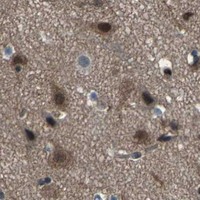
Immunohistochemistry protocol sheet for ATXN3 (IHC for ATXN3): expected localisation Nuclear and cytoplasmic tissue staining (HPA tissue IHC), antibody PB9423, validated IHC image, and IHC protocol steps
Printable ATXN3 IHC protocol sheet — expected localisation Nuclear and cytoplasmic tissue staining (HPA tissue IHC), antibody PB9423, controls and protocol steps. Open the full ATXN3 IHC guide →

ATXN3 Immunohistochemistry Experimental Design Guide

Expected localisation, validated protocols, controls and antibodies — the at-a-glance facts below, then the full design guide.

Must know before staining
Expected localisation Nuclear and cytoplasmic tissue staining (HPA tissue IHC)
Staining pattern Nuclear and cytoplasmic staining in most tissues (HPA tissue IHC)
Antigen retrieval Citrate pH 6 HIER, heat-mediated (datasheet PB9423)
Positive control ⓘ Adipose tissue+4 more · see all
Negative control ⓘ Liver+1 more · see all
Important caveats
Reasons your staining may differ from the expected pattern.
Fixation Keep fixation consistent across sections. (standard IHC practice; not target-specific)
Caveat Staining may include protein from more than one gene (HPA tissue IHC)
Regulation Expression regulation is unreported (UniProt)
Isoform / epitope Five isoforms; epitope coverage is unreported (UniProt; datasheet PB9423)
Section 1

Recommended ATXN3 IHC & IF Protocols

The catalog antibody’s IHC-P protocol is paired with 3 published ATXN3 chromogenic IHC protocols (PMC11254720; PMC4642276; PMC4624233).

Recommended immunohistochemistry (IHC-P) protocol parameters
SampleParaffin-embedded Human Lung Cancer tissue; fixative not specified (datasheet PB9423)
FixationImage fixative and duration unreported (datasheet PB9423); verify before use.
Sectioning4–5 µm sections on charged slides (standard)
DeparaffinisationXylene, graded ethanol series to water (standard)
Antigen retrievalHeat retrieval: Citrate pH 6, 20 min (datasheet PB9423)
Peroxidase block3% H2O2, 10 min, room temperature (standard)
Blocking10% goat serum (datasheet PB9423)
Primary antibodyRabbit anti-ATXN3, 0.5-1μg/ml (datasheet PB9423)
Primary incubationOvernight at 4 °C (datasheet PB9423)
DetectionStreptavidin-biotin complex (SABC), DAB chromogen (datasheet PB9423)
CounterstainHematoxylin, blue, dehydrate and mount (standard)
Expected resultATXN3-positive staining in adipocytes of adipose tissue (HPA tissue IHC: High). HPA tissue profile: Cytoplasmic and nuclear expression in most tissues. No signal in the no-primary control.
💡Decision noteStart with heat-mediated citrate retrieval at pH 6 (datasheet: PB9423); adjust conditions for the tissue and antibody used in each published protocol.
Section 2

What Is the Expected ATXN3 Staining Pattern?

ATXN3 is predominantly nuclear, including the inner nuclear matrix, but is not exclusively nuclear (UniProt P54252). HPA reports cytoplasmic and nuclear staining in most tissues, with high staining in several listed cell types (HPA tissue IHC). Interpret tissue patterns cautiously: HPA rates its IHC evidence uncertain and warns that the staining may reflect proteins from more than one gene (HPA tissue IHC). ATXN3 has no transmembrane segment (UniProt P54252 topology).

What am I looking at on my slide?
Nuclear staining with some cytoplasmic staining in adipocytes or adrenal glandular cells (HPA tissue IHC).This fits the reported compartment pattern and high staining in those cells (HPA tissue IHC). It supports a plausible positive result, although HPA rates the tissue IHC evidence uncertain and flags possible staining of proteins from more than one gene (HPA tissue IHC).
Signal is confined to extracellular deposits or tissue edges, without a recognizable cellular pattern.That distribution does not match the reported nuclear and cytoplasmic tissue pattern (HPA tissue IHC). Treat it as a possible staining artefact (standard IHC practice); review morphology and detection controls before attributing it to ATXN3.
Strong staining appears in liver cholangiocytes or smooth muscle cells.HPA reports ATXN3 as not detected in those respective cell types (HPA tissue IHC). Possible cross-reactivity or endogenous chromogenic activity needs investigation (standard IHC practice). The HPA result is an observed pattern, not proof that these cells can never express ATXN3.
Chromogen covers many structures evenly and obscures nuclei and cell boundaries.Diffuse background prevents a reliable compartment call (standard IHC practice). Compare the section with a detection control and assess blocking and development conditions (standard IHC practice); HPA’s uncertain IHC reliability makes an apparent widespread positive particularly hard to assign to ATXN3 (HPA tissue IHC).
No signal is visible in an otherwise interpretable adipose or adrenal gland section.Those sections contain cell types reported with high staining: adipocytes and glandular cells, respectively (HPA tissue IHC). Check whether those cells are present and preserved, then review the assay controls (standard IHC practice). A single negative section cannot settle target absence given HPA’s uncertain IHC reliability (HPA tissue IHC).
💡Expected ATXN3 appearanceA plausible positive is clear nuclear staining, with possible cytoplasmic staining, in adipocytes or adrenal glandular cells reported as High (HPA tissue IHC; UniProt P54252); uniform extracellular colour is a possible artefact (standard IHC practice).
How each factor affects the staining
Compartment assignment (UniProt P54252; HPA tissue IHC)UniProt describes ATXN3 as predominantly nuclear but not exclusively so; HPA observes both nuclear and cytoplasmic tissue staining (UniProt P54252; HPA tissue IHC). Score compartments separately rather than requiring every positive cell to show nuclear signal alone.
Choice of tissue and cell type (HPA tissue IHC)HPA reports High staining in adipocytes, adrenal glandular cells and bone marrow hematopoietic cells, but Low staining in cerebellar granular-layer cells and skeletal myocytes (HPA tissue IHC). Select controls by the scored cell type, and avoid treating a Low population as an equally strong positive.
Specificity of the tissue signal (HPA tissue IHC; HPA antibodies)HPA rates the overall tissue IHC profile Uncertain, notes possible staining of proteins from more than one gene, and lists HPA024123 and CAB021976 as Uncertain for IHC (HPA tissue IHC; HPA antibodies). Cell pattern and assay controls therefore matter when assigning chromogen to ATXN3.
Isoform coverage (UniProt P54252)UniProt lists five ATXN3 isoforms and a Josephin domain plus three ubiquitin-interacting motifs (UniProt P54252). The supplied record gives no IHC antibody epitope, so isoform coverage cannot be inferred; check the antibody’s documented immunogen before explaining a missing signal as isoform-specific.
IF/ICC localisation Q&A (HPA subcellular; UniProt P54252)What should IF/ICC show? HPA reports approved nucleoplasm and nucleoli localisation, with additional approved plasma-membrane localisation (HPA subcellular). UniProt also records recruitment to lysosomal membranes during amino acid deprivation (UniProt P54252). These observations do not establish an IHC-P staining intensity or an IF protocol.
Why is my staining missing, weak or wrong?
SituationLikely causeNext action
No chromogenic signal in adipocytes or adrenal glandular cells reported as High (HPA tissue IHC).A missed positive cell population or failed staining run is possible (standard IHC practice); the supplied sources do not establish ATXN3-specific fixation sensitivity.Verify tissue identity and morphology, then review the run’s positive and detection controls, retrieval setting and primary-antibody use (standard IHC practice). Record the negative result without converting HPA’s High observation into a guarantee.
Nuclei look negative while cytoplasm stains in cells expected to be positive (HPA tissue IHC).The pattern is incomplete relative to UniProt’s predominantly nuclear localisation, although HPA also reports cytoplasmic staining (UniProt P54252; HPA tissue IHC).Confirm nuclear boundaries with the counterstain and score nuclear and cytoplasmic signal separately (standard IHC practice). Review controls before declaring cytoplasmic signal false; HPA’s tissue IHC reliability is Uncertain (HPA tissue IHC).
Cholangiocytes or smooth muscle cells show conspicuous chromogen.Both are reported as Not detected in the specified HPA tissue observations (HPA tissue IHC). Cross-reactivity or endogenous detection activity is possible (standard IHC practice).Compare a detection control and the cell morphology, then evaluate antibody specificity with an independent control where available (standard IHC practice). Report the discrepancy as an observed result rather than assigning it automatically to ATXN3.
Colour is diffuse across the section and masks cellular localisation.Nonspecific background or excessive detection development can obscure a specific pattern (standard IHC practice); the appearance alone cannot identify which step caused it.Inspect detection controls and review blocking, primary-antibody dilution, washes and chromogen development against the established assay conditions (standard IHC practice). Reassess whether nuclei and cytoplasm can be scored after background is resolved.
Only low staining is seen in cerebellar granular-layer cells, hippocampal glia or skeletal myocytes.Low staining is the reported HPA level for these populations (HPA tissue IHC); weak signal there does not by itself show assay failure.Use a cell population reported as High, such as adipocytes or bone marrow hematopoietic cells, to assess assay performance (HPA tissue IHC). Compare sections under the same scoring criteria (standard IHC practice).
A nuclear, nucleolar or membrane IF/ICC image appears different from the IHC-P section.HPA’s approved IF/ICC locations include nucleoplasm, nucleoli and additional plasma membrane, while its tissue IHC profile is nuclear and cytoplasmic and rated Uncertain (HPA subcellular; HPA tissue IHC).Interpret each assay against its own reported localisation and controls (standard IHC/IF practice). Do not transfer IF/ICC compartment approval into a claim that a matching membrane pattern must appear in chromogenic IHC-P.

Sample controls for ATXN3 IHC & IF

🧪Run adipose tissue first and expect staining in adipocytes (HPA: High in adipocytes). Use smooth muscle as the negative tissue, with smooth muscle cells expected to be unstained (HPA: Not detected in smooth muscle cells); HPA does not identify a validated internal negative cell type on the adipose slide, so assess unstained neighboring cells as background checks only.
Positive control tissue: Adipose tissue (Adipocytes, HPA High)
Negative control tissue: Liver (HPA Not detected)
ICC-IF cell lines (HPA subcellular resource): HPA ICC-IF images show ATXN3 in MCF-7, Rh30, U2OS, with annotated localisation: Nucleoplasm (approved), Nucleoli (approved) (HPA subcellular).
Technical controls: Include no-primary (secondary-only) and concentration-matched rabbit immunoglobulin isotype controls (selected PB9423 caption: rabbit primary and goat anti-rabbit IgG secondary), plus ATXN3 knockout tissue as a biological negative where available. For DAB detection, block endogenous peroxidase and assess endogenous biotin because the selected protocol uses a streptavidin–biotin detection complex (selected PB9423 caption: SABC with DAB).
⚠️Feasibility: A target-specific fixation window or fixation effect is unreported, and the exact PB9423 tissue-IHC caption does not state a fixative; its paraffin section does not establish fixation. The reported IHC example used citrate retrieval at pH 6 for 20 minutes, but whether ATXN3 staining depends on that retrieval is unreported (selected PB9423 caption). Relative ease of frozen-section or IF staining is unreported; in paraffin-processed adipose tissue, lipid extraction can leave empty adipocyte vacuoles, so assess staining in preserved cellular structures rather than the vacuoles (standard IHC practice). The selected PB9423 tissue-IHC caption documents paraffin sections, but does not specify the fixative (selected-SKU IHC image PB9423).

HPA tissue IHC evidence for ATXN3

Comprehensive Human Protein Atlas IHC scoring per tissue (reliability: Uncertain — Medium consistency between antibody staining and RNA expression data. Caution, targets protein from more than one gene. Pending external verification.). Rows are taken directly from the HPA tissue chart — click any row's HPA link to view the source.

Positive expression · recommended positive controls

TissueCell typeLevelEvidenceSource
Adipose tissue Adipocytes High Protein (IHC) HPA →
Adrenal gland Glandular cells High Protein (IHC) HPA →
Appendix Glandular cells High Protein (IHC) HPA →
Bone marrow Hematopoietic cells High Protein (IHC) HPA →
Breast Adipocytes High Protein (IHC) HPA →

Undetected expression · recommended negative controls

TissueCell typeLevelEvidenceSource
Liver Cholangiocytes Not detected Protein (IHC) HPA →
Smooth muscle Smooth muscle cells Not detected Protein (IHC) HPA →
Section 3

Advanced ATXN3 IHC Tips

Troubleshoot ATXN3 staining in paraffin sections by checking retrieval, controls, compartment, and cell type before comparing signal intensity.

What retrieval conditions should I try first for ATXN3 IHC?
Start with heat-mediated citrate retrieval at pH 6 for paraffin sections (datasheet PB9423). The PB9423 tissue-IHC example heated sections in this buffer for 20 minutes before staining human lung cancer tissue (PB9423 tissue-IHC caption). If signal is weak, first check that heating, cooling, and subsequent washes were consistent across slides; uneven retrieval can create misleading regional differences (standard IHC practice). Only then compare an alternative retrieval condition on matched sections, keeping antibody concentration and detection constant (standard IHC practice). Record nuclear and cytoplasmic staining separately because both are plausible ATXN3 locations (UniProt P54252 subcellular location; HPA tissue IHC).
Could fixation be causing weak or uneven ATXN3 staining?
Target-specific fixation sensitivity is unknown: the PB9423 paraffin-section caption does not state a fixative (PB9423 tissue-IHC caption). Record the actual fixative, time in fixative, processing schedule, and section age for each specimen before attributing a weak result to ATXN3 biology (standard IHC practice). Compare matched sections processed under documented conditions, then hold citrate pH 6 retrieval and antibody concentration constant while assessing signal and morphology (datasheet PB9423; standard IHC practice). Excessive background or lost tissue architecture can make intensity comparisons unreliable regardless of the antigen (standard IHC practice). Neither tissue staining patterns nor ATXN3 topology establishes a fixation effect for this target (HPA tissue IHC; UniProt P54252 topology).
Should ATXN3 staining be nuclear, cytoplasmic, or membrane associated?
Assess nuclei and cytoplasm independently: ATXN3 is predominantly nuclear but not exclusively so (UniProt P54252 subcellular location), and tissue IHC reports both compartments in most tissues (HPA tissue IHC). Nuclear matrix localisation supports a nuclear signal, while amino acid deprivation can recruit ATXN3 to lysosomal membranes (UniProt P54252 subcellular location). A membrane-like outline alone needs caution because ATXN3 has no transmembrane segment, and plasma membrane localisation comes from a separate subcellular imaging dataset (UniProt P54252 topology; HPA subcellular). Check whether the pattern follows intact cell boundaries and repeats across adjacent sections (standard IHC practice). Report compartment-specific scores rather than calling every brown deposit equivalent ATXN3 signal (standard IHC practice).
How might the antibody epitope change which ATXN3 forms I detect?
Confirm the catalog antibody's mapped epitope before interpreting differences between specimens; the supplied PB9423 tissue-IHC caption does not identify that epitope (PB9423 tissue-IHC caption). ATXN3 has five listed isoforms, a Josephin domain at residues 1–180, and three ubiquitin-interacting motifs farther toward the C terminus (UniProt P54252 isoforms and domains). An epitope absent from a particular isoform could change its detectability, but isoform recognition cannot be assigned here without an epitope map (UniProt P54252 isoforms; standard antibody validation practice). Phosphoserines are reported at residues 219, 265, 272, and 328; their effect on this antibody is unknown (UniProt P54252 modified residues). Compare staining with an independently validated reagent if isoform-specific conclusions matter (standard IHC practice).
How should I investigate an ATXN3 pattern with multiplex IF?
Use IF as a separate validation experiment while interpreting this page's chromogenic paraffin IHC results (standard assay practice). Pair ATXN3 with a validated marker for the expected cell population; respiratory epithelial cells in bronchus are one high-staining example, although the tissue-IHC evidence has uncertain reliability (HPA tissue IHC). Select spectrally separated fluorophores after inspecting tissue autofluorescence and include single-label controls for channel bleed-through (standard IF practice). ATXN3 is predominantly nuclear and has no transmembrane segment, but its membrane-associated epitope orientation is unspecified; test permeabilisation against the antibody's mapped epitope and desired compartment (UniProt P54252 subcellular location and topology; standard IF practice). Compare nuclear and extranuclear signal with the chromogenic pattern without treating the two detection methods as interchangeable (standard assay practice).
How can I reduce diffuse brown staining without losing ATXN3 signal?
Inspect a no-primary control and the tissue's unstained appearance to distinguish antibody-dependent signal from pigment or detection background (standard IHC practice). The PB9423 example used 10% goat serum, 1 μg/ml primary overnight at 4°C, a biotinylated secondary, and DAB development (PB9423 tissue-IHC caption). In a peroxidase-based chromogenic workflow, optimise the peroxidase block and development time, and check for background from the biotin-based detection system (standard IHC practice). Titrate primary concentration on matched sections while keeping retrieval at citrate pH 6 and detection settings fixed (datasheet PB9423; standard IHC practice). Preserve plausible nuclear and cytoplasmic staining while reducing staining in no-primary controls (UniProt P54252 subcellular location; standard IHC practice).
What is a defensible way to score ATXN3 IHC across samples? ⚠ ANSWER MARKED FOR VERIFICATION
Define the cell population and compartment before scoring because ATXN3 can appear in nuclei and cytoplasm (UniProt P54252 subcellular location; HPA tissue IHC). For each compartment, report the percentage of positive eligible cells and an intensity-based H-score using a prespecified 0–3 intensity scale (standard IHC scoring practice). If the question concerns discrete positive cells, report density per mm² of viable tissue and state how cells were identified (standard image-analysis practice). Normalise counts to eligible cells or viable tissue area, and apply identical retrieval, exposure, threshold, and exclusion rules across samples (standard IHC practice). Record cell type because reported ATXN3 staining varies among sampled populations and HPA rates its tissue-IHC reliability uncertain (HPA tissue IHC).
When is a strong ATXN3 stain more likely artefact than true signal?
Treat a reproducible nuclear or cytoplasmic cellular pattern as plausible, while checking its cell type and morphology (UniProt P54252 subcellular location; HPA tissue IHC; standard IHC practice). HPA reports high staining in bronchial respiratory epithelial cells but no detected staining in liver cholangiocytes; its tissue-IHC reliability is uncertain, so these are contextual observations rather than absolute controls (HPA tissue IHC). Staining limited to section edges, damaged areas, or necrosis warrants repeat assessment on intact tissue (standard IHC practice). Brown signal persisting without primary antibody can reflect endogenous enzyme activity or detection chemistry rather than ATXN3 (standard IHC practice). Investigate a dominant membrane-only pattern particularly carefully because ATXN3 lacks a transmembrane segment, although membrane-associated localisation is reported (UniProt P54252 topology and subcellular location; HPA subcellular).
Boster reagents

Best ATXN3 / Ataxin-3 IHC Antibodies

PB9423 has real IHC data from human lung cancer paraffin sections and IF data from MCF-7 cells; catalog reactivity lists human and rat (IHC and IF captions; catalog reactivity).

Real IHC data IHC analysis of Ataxin 3 using anti-Ataxin 3 antibody (PB9423). Ataxin 3 was detected in paraffin-embedded section of Human Lung Cancer Tissue. Heat mediated antigen retrieval was performed in citrate buffer (pH6, epitope retrieval solution) for 20 mins. The tissue section was blocked with 10% goat serum. The tissue section was then incubated with 1μg/ml rabbit anti-Ataxin 3 Antibody (PB9423) overnight at 4°C. Biotinylated goat anti-rabbit IgG was used as secondary antibody and incubated for 30 minutes at 37°C. The tissue section was developed using Strepavidin-Biotin-Complex (SABC)(Catalog # SA1022) with DAB as the chromogen.
Anti-Ataxin 3/ATXN3 Antibody ®
Cat # PB9423

PB9423 will render with IHC evidence from human lung cancer paraffin sections (PB9423 IHC caption). PB9423 also has IF evidence from MCF-7 cells and is listed for IHC, IF, and ICC in human samples; rat is listed under reactivity (PB9423 IF caption; catalog applications and reactivity).

Which to pick: Choose PB9423 for paraffin-section IHC: its IHC caption shows human lung cancer tissue, with the fixative unreported (PB9423 IHC caption). Choose PB9423 for IF/ICC because its IF caption shows MCF-7 cells and the catalog lists both applications (PB9423 IF caption; catalog applications). For rat samples, PB9423 lists rat reactivity, but the supplied IHC and IF figures show human samples; clonality is unreported (catalog reactivity and clone field; PB9423 IHC and IF captions).

Each figure is that product's own IHC / IF validation image from its datasheet.

References

  1. UniProt Consortium. UniProt entry P54252 (ATX3_HUMAN, Ataxin-3).
  2. Human Protein Atlas. ATXN3 tissue IHC expression (reliability: Uncertain).
  3. Human Protein Atlas. ATXN3 subcellular location (ICC-IF): Mainly localized to the nucleoplasm and nucleoli. In addition localized to the plasma membrane..
  4. Human Protein Atlas. ATXN3 antibody validation summary (3 antibodies).
  5. ATXN3 functions as a tumor suppressor through potentiating galectin-9-mediated apoptosis in human colon adenocarcinoma. The Journal of biological chemistry 2024 — PMC11254720.
  6. Rad23b exacerbates pathological aggregates through disrupting proteasome functions in Spinocerebellar ataxia type 3. Neurotherapeutics : the journal of the American Society for Experimental NeuroTherapeutics 2026 — PMC13224077.
  7. Differential recruitment of UBQLN2 to nuclear inclusions in the polyglutamine diseases HD and SCA3. Neurobiology of disease 2015 — PMC4642276.
  8. Limited Effect of Chronic Valproic Acid Treatment in a Mouse Model of Machado-Joseph Disease. PloS one 2015 — PMC4624233.
  9. PubMed PMID:7874163 — UniProt-cited evidence.
  10. PubMed PMID:9274833 — UniProt-cited evidence.
  11. PubMed PMID:11450850 — UniProt-cited evidence.