AURKB / Aurora kinase B · IHC design guide

Design Immunohistochemistry for AURKB

Plan chromogenic paraffin IHC for AURKB using its nuclear tissue staining profile (HPA tissue IHC) and the catalog antibody’s documented tonsil example (datasheet M00762-1). This guide covers fixation consistency, controls, and interpretation of cell cycle dependent expression (UniProt).

Evidence assembled Oct 2026 · For research use; verify linked source records and product datasheet before use
Immunohistochemistry protocol sheet for AURKB (IHC for AURKB): expected localisation Nuclear staining in tissues (HPA tissue IHC), antibody M00762-1, validated IHC image, and IHC protocol steps
Printable AURKB IHC protocol sheet — expected localisation Nuclear staining in tissues (HPA tissue IHC), antibody M00762-1, controls and protocol steps. Open the full AURKB IHC guide →

AURKB Immunohistochemistry Experimental Design Guide

Expected localisation, validated protocols, controls and antibodies — the at-a-glance facts below, then the full design guide.

Must know before staining
Expected localisation Nuclear staining in tissues (HPA tissue IHC)
Staining pattern Nuclei in lymphoid immune and GI endocrine cells (HPA tissue IHC)
Antigen retrieval EDTA pH 8.0 HIER, heat-mediated (datasheet M00762-1)
Positive control ⓘ Appendix+4 more · see all
Negative control ⓘ Adipose tissue+4 more · see all
Important caveats
Reasons your staining may differ from the expected pattern.
Fixation Keep paraffin-section fixation consistent across samples. (standard IHC practice; not target-specific)
Caveat Presumed off-target binding may mimic staining (HPA tissue IHC)
Regulation Expressed in S/G2-M; higher in cancer M phase (UniProt)
Isoform / epitope 5 isoforms; epitope impact is unknown (UniProt)
Section 1

Recommended AURKB IHC & IF Protocols

Start with the catalog antibody’s EDTA pH 8.0 retrieval protocol (datasheet M00762-1); the four published IHC protocols below provide tissue-specific comparisons (PMC7053608; PMC2732361; PMC7960835; PMC10023558).

Recommended immunohistochemistry (IHC-P) protocol parameters
SampleParaffin-embedded human bladder urothelial carcinoma tissue; fixative not specified (datasheet M00762-1)
FixationImage fixative and duration unreported (datasheet M00762-1); verify before use.
Sectioning4–5 µm sections on charged slides (standard)
DeparaffinisationXylene, graded ethanol series to water (standard)
Antigen retrievalHeat retrieval: EDTA pH 8.0 (datasheet M00762-1); 20 min, 95–100 °C (standard)
Peroxidase block3% H2O2, 10 min, room temperature (standard)
Blocking10% goat serum (datasheet M00762-1)
Primary antibodyRabbit monoclonal (clone FA-1) anti-AURKB, 1:50 (datasheet M00762-1)
Primary incubationOvernight at 4 °C (datasheet M00762-1)
DetectionHRP-conjugated secondary, DAB chromogen (datasheet M00762-1)
CounterstainHematoxylin, blue, dehydrate and mount (standard)
Expected resultAURKB-positive staining in endocrine cells of appendix (HPA tissue IHC: High). HPA tissue profile: Nuclear expression in several tissues, most abundant in immune cells of lymphoid tissues and gastrointestinal tract. No signal in the no-primary control.
💡Decision noteTry EDTA pH 8.0 first for the catalog antibody (datasheet M00762-1). Published alternatives use citrate or citric acid pH 6.0 (PMC2732361; PMC10023558).
Section 2

What Is the Expected AURKB Staining Pattern?

AURKB is a nuclear mitotic kinase that moves from chromosome arms and inner centromeres to the spindle midzone and midbody as division proceeds (UniProt Q96GD4). It has no transmembrane segment (UniProt Q96GD4 topology). In paraffin-section IHC, expect selective nuclear staining, especially in immune cells of lymphoid and gastrointestinal tissues (HPA tissue IHC). HPA rates its tissue profile Enhanced but notes presumed off-target binding that was disregarded (HPA tissue IHC).

What am I looking at on my slide?
Selective nuclear staining in lymphoid or gastrointestinal cells, with stronger staining in some individual cells.This fits HPA's nuclear tissue profile (HPA tissue IHC). Uneven staining can fit cell-cycle dependence: AURKB is expressed during S and G2/M, and its location changes through mitosis (UniProt Q96GD4). Do not interpret the proportion of stained cells as an antibody dilution readout alone.
A discrete chromosome-associated signal in a mitotic cell, or signal at a cleavage structure.Chromosome arms and inner centromeres are expected from prophase through metaphase; the spindle midzone and midbody are expected from anaphase through cytokinesis (UniProt Q96GD4). Assess these structures in the context of mitotic morphology rather than calling every non-nuclear focus nonspecific.
Broad cytoplasmic staining in cells without recognizable mitotic structures.That distribution does not match the dominant nuclear tissue pattern (HPA tissue IHC) or the defined mitotic locations (UniProt Q96GD4). Treat it as suspect; review morphology and the negative control before scoring it as AURKB.
Strong staining in adipocytes or cardiomyocytes.HPA reports AURKB as not detected in those specific cell populations (HPA: adipocytes; cardiomyocytes). Such staining warrants a cross-reactivity or endogenous detection-activity check. The HPA result does not establish that every cell in adipose or heart tissue is negative.
No staining in a section containing an expected positive cell population.First check that the relevant cells are present: HPA reports high staining in gastrointestinal endocrine cells and medium staining in bone-marrow hematopoietic cells and lymph-node non-germinal-center cells (HPA tissue IHC). A negative result may also reflect an IHC workflow or detection failure (general IHC practice).
💡Expected AURKB appearanceCall positive when staining is selective and predominantly nuclear in expected cells, potentially stronger in dividing cells (HPA tissue IHC; UniProt Q96GD4); strong, widespread cytoplasmic staining in morphologically quiescent cells is suspect (UniProt Q96GD4 localization).
How each factor affects the staining
Cell-cycle stage and compartmentAURKB shifts from chromosome arms and inner centromeres before anaphase to the spindle midzone and midbody afterward (UniProt Q96GD4). This makes mitotic morphology relevant when judging a focal signal; a midbody focus should not be scored as a failed nuclear pattern.
Choice of positive and low-signal cellsHPA reports high staining in appendix, colon, duodenum, rectum and small-intestine endocrine cells; medium staining in bone-marrow hematopoietic and lymph-node non-germinal-center cells; and no detection in adipocytes or cardiomyocytes (HPA tissue IHC). Choose and score the named cell populations, not whole tissues.
Strength of HPA antibody evidenceThe tissue profile is rated Enhanced, with medium agreement between staining and RNA data and presumed off-target binding disregarded (HPA tissue IHC). HPA lists IHC Enhanced for CAB005862 and ICC Supported for HPA037708 (HPA antibodies). Those ratings do not validate an unspecified catalog antibody.
Isoforms and assay interpretationUniProt lists five AURKB isoforms and a 77–327 protein kinase domain (UniProt Q96GD4). Neither source supplies the catalog antibody's epitope, so the observed stain cannot be assigned to a particular isoform from these records alone.
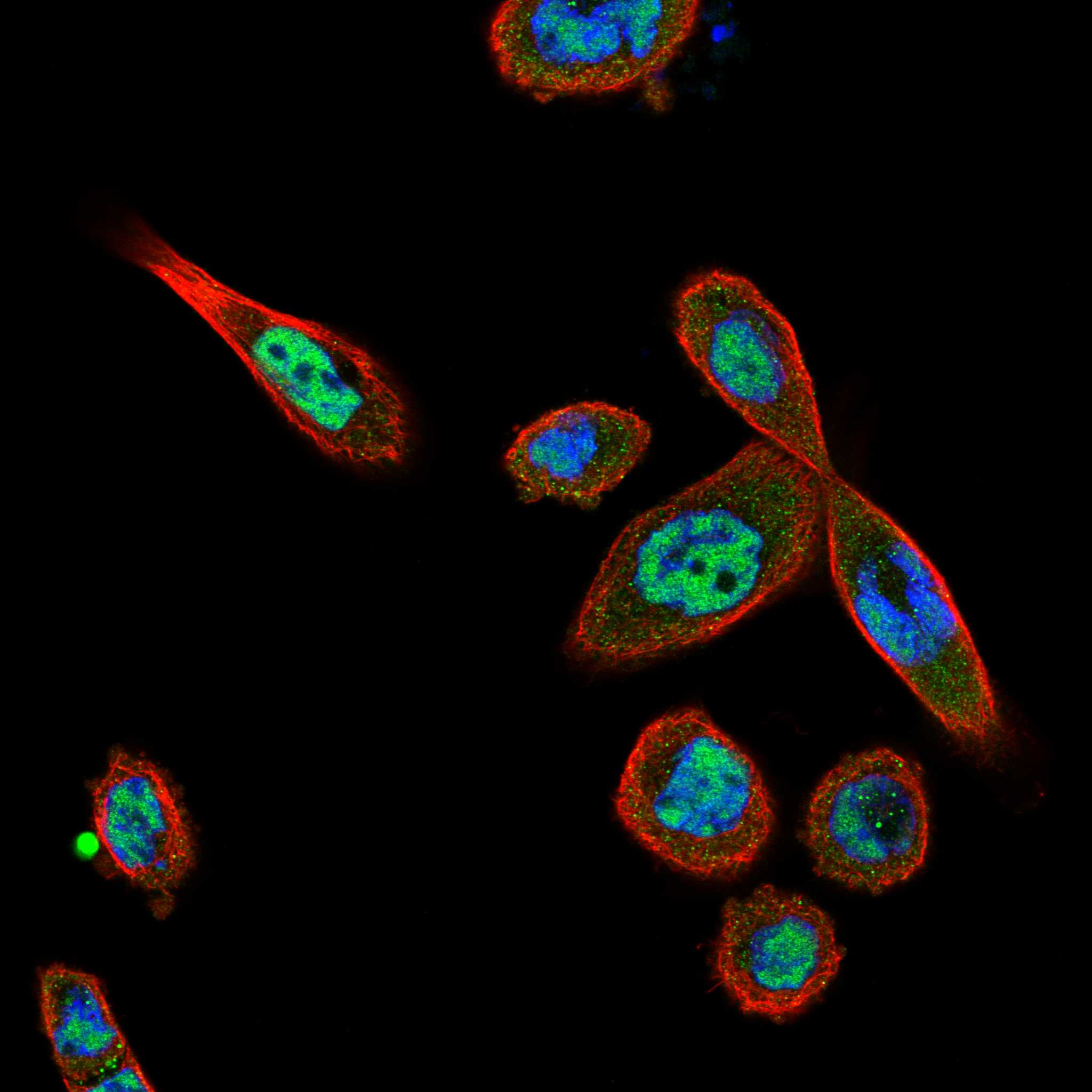
Does IF/ICC show the same location?HPA reports supported nucleoplasmic and midbody localization in ICC-IF, while kinetochore localization is uncertain (HPA subcellular). This is a useful localization comparison, but ICC-IF images do not establish the appearance or performance of a paraffin-section IHC assay.
Why is my staining missing, weak or wrong?
SituationLikely causeNext action
A known-positive cell population is unstained.The expected cells may be absent from the examined field (HPA tissue IHC), or an IHC processing or detection step may have failed (general IHC practice).Confirm cell identity and morphology, then review retrieval, primary incubation and detection against the antibody's IHC-P instructions; include a separately processed positive control (general IHC practice).
Signal is mostly diffuse across the section.A broad haze is inconsistent with HPA's selective nuclear profile (HPA tissue IHC). Inadequate blocking, washing or detection controls can produce nonspecific background (general IHC practice).Inspect the no-primary control, blocking and wash steps; adjust the primary concentration only within its documented IHC-P range (general IHC practice).
Many adipocytes or cardiomyocytes stain strongly.HPA reports no detection in these cell types (HPA tissue IHC). Cross-reactivity or endogenous detection activity should be considered (general IHC practice).Compare the no-primary and detection controls, then verify whether staining persists with an independently validated AURKB IHC antibody (general IHC practice).
A mitotic focus is scored as misplaced staining.AURKB changes location during mitosis and cytokinesis (UniProt Q96GD4); a midzone or midbody focus can be expected.Recheck the cell's mitotic morphology and score the focus by cell-cycle stage before classifying its compartment (UniProt Q96GD4).
Only a few cells stain in an otherwise suitable section.AURKB expression varies with cell-cycle phase (UniProt Q96GD4), and HPA's reported levels refer to particular cell populations (HPA tissue IHC).Count staining within the named positive population and assess mitotic morphology; use a positive-control section if the target population is sparse (general IHC practice).
Staining is weak despite an intact positive control.Target-specific fixation effects are not established by the supplied assay evidence. Verify with a matched IHC source before attributing a result to fixation.Check section quality and follow the IHC-validated antibody's documented retrieval and incubation conditions; record changes before comparing staining intensity (general IHC practice).

Sample controls for AURKB IHC & IF

🧪Run colon first: its endocrine cells should stain for AURKB (HPA: High in colon endocrine cells). Use adipose tissue as the negative tissue, focusing on adipocytes (HPA: Not detected in adipocytes); on the colon slide, compare endocrine cells with adjacent nonendocrine cells, which should lack comparable specific staining, while confirming that pattern experimentally.
Positive control tissue: Appendix (Endocrine cells, HPA High)
Negative control tissue: Adipose tissue (HPA Not detected)
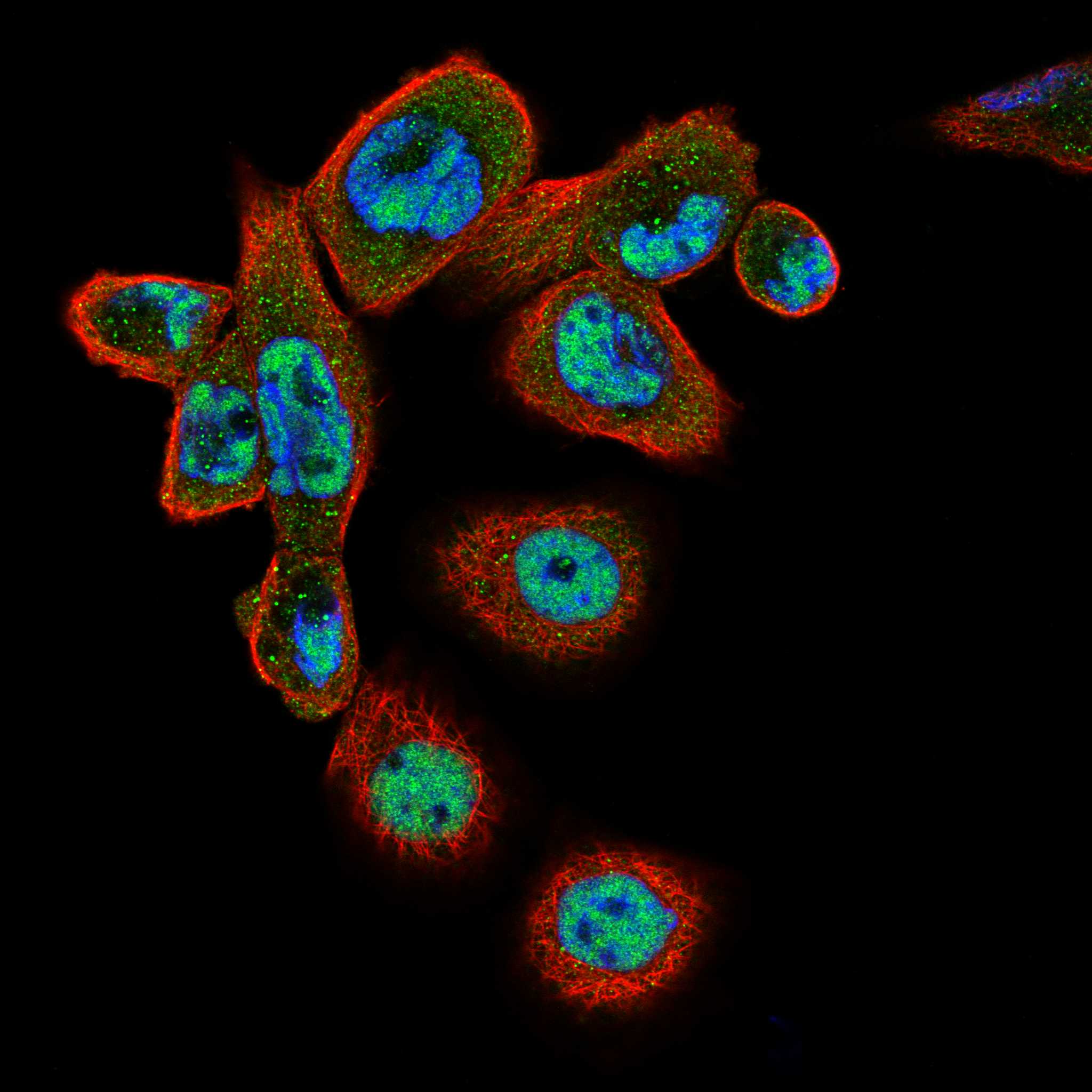
ICC-IF cell lines (HPA subcellular resource): HPA ICC-IF images show AURKB in A-431, HEK293, U2OS, ASC52telo, RPTEC/TERT1, hTERT-RPE1 (serum starved), KOLF2.1J, with annotated localisation: Nucleoplasm (supported) (HPA subcellular).
Technical controls: Include a no-primary, secondary-only control and a matched rabbit IgG isotype control, using the primary antibody’s clonality where applicable (selected tissue-IHC caption: rabbit anti-AURKB and peroxidase-conjugated goat anti-rabbit IgG). Use an AURKB knockout sample as a biological negative, and block endogenous peroxidase before chromogenic detection in colon sections (selected tissue-IHC caption: HRP/DAB detection).
⚠️Feasibility: A target-specific fixation window or fixation effect is unreported, and the selected M00762-1 paraffin-section caption does not state the fixative (selected tissue-IHC caption: fixative not stated). The caption uses heat retrieval in EDTA at pH 8.0, but does not establish that retrieval is required; the supplied evidence also does not establish whether frozen sections or IF/ICC would be easier (selected tissue-IHC caption: EDTA retrieval; HPA: ICC-IF images available). In colon, check that endogenous peroxidase or luminal background does not obscure the endocrine-cell signal (HPA: High in colon endocrine cells; selected tissue-IHC caption: HRP/DAB detection).

HPA tissue IHC evidence for AURKB

Comprehensive Human Protein Atlas IHC scoring per tissue (reliability: Enhanced — Medium consistency between antibody staining and RNA expression data. Presumed off target binding observed and disregarded.). Rows are taken directly from the HPA tissue chart — click any row's HPA link to view the source.

Positive expression · recommended positive controls

TissueCell typeLevelEvidenceSource
Appendix Endocrine cells High Protein (IHC) HPA →
Colon Endocrine cells High Protein (IHC) HPA →
Duodenum Endocrine cells High Protein (IHC) HPA →
Rectum Endocrine cells High Protein (IHC) HPA →
Skin Cells in basal layer Low Protein (IHC) HPA →

Undetected expression · recommended negative controls

TissueCell typeLevelEvidenceSource
Adipose tissue Adipocytes Not detected Protein (IHC) HPA →
Caudate Glial cells Not detected Protein (IHC) HPA →
Cerebellum Cells in granular layer Not detected Protein (IHC) HPA →
Cervix Glandular cells Not detected Protein (IHC) HPA →
Epididymis Glandular cells Not detected Protein (IHC) HPA →
Section 3

Advanced AURKB IHC Tips

Troubleshoot AURKB staining in paraffin sections by checking retrieval, controls, mitotic localisation and cell-specific scoring (datasheet M00762-1; UniProt Q96GD4; HPA tissue IHC).

How should I optimise retrieval when AURKB staining is weak?
Use heat-mediated antigen retrieval in EDTA at pH 8.0 for paraffin sections (datasheet M00762-1). Compare adjacent sections processed together, keeping retrieval conditions fixed while checking the documented 1:50 primary incubation overnight at 4°C and the 30-minute secondary incubation at 37°C (datasheet M00762-1; standard IHC practice). Assess signal in mitotic figures, where AURKB moves from chromosomes and inner centromeres to the spindle midzone and midbody as mitosis progresses (UniProt Q96GD4 localisation). If staining remains weak, test a different retrieval condition on matched sections and judge improvement against background and a no-primary control (standard IHC practice).
Can fixation explain absent AURKB staining in my paraffin sections?
The M00762-1 tissue caption identifies a paraffin-embedded section but does not state its fixative, so target-specific fixation sensitivity is unknown (datasheet M00762-1). Record the fixative and processing history for each specimen, and compare sections processed together before attributing a negative result to AURKB biology (standard IHC practice). Keep the documented EDTA retrieval at pH 8.0 and 1:50 primary incubation overnight at 4°C constant while examining the effect of fixation conditions (datasheet M00762-1; standard IHC practice). Include a processing control and inspect tissue preservation, because poor morphology makes a negative chromogenic stain difficult to interpret (standard IHC practice).
Where should convincing AURKB staining appear across mitotic stages?
Expect chromosome-arm and inner-centromere staining from prophase through metaphase, followed by spindle-midzone and midbody staining from anaphase through cytokinesis (UniProt Q96GD4 localisation). Use the counterstain to identify mitotic figures and compare chromogenic signal with their stage, because a single section samples cells at different points in division (standard IHC practice; UniProt Q96GD4 localisation). Nuclear staining outside obvious mitoses warrants cautious review: tissue IHC shows nuclear expression in several tissues, while ICC/IF reports mainly nucleoplasmic localisation (HPA tissue IHC; HPA subcellular). Check whether strong diffuse cytoplasmic colour also appears in no-primary sections before calling it AURKB (standard IHC practice).
Could an unknown epitope make some AURKB pools hard to detect?
AURKB has 5 annotated isoforms, but the supplied M00762-1 caption does not identify its epitope or establish recognition of every isoform (UniProt Q96GD4 isoforms; datasheet M00762-1). Its protein-kinase domain spans residues 77–327, and annotated modifications include phosphothreonine at 35 and phosphoserine at 227 (UniProt Q96GD4 domains and modified residues). Do not assign an absent chromogenic signal to a particular isoform or modification without epitope information and a suitable validation control (standard IHC practice). Compare anatomically matched sections under identical retrieval and detection conditions, and report the antibody and conditions alongside any negative finding (standard IHC practice).
How can IF help check an ambiguous chromogenic AURKB pattern?
Use IF as a separate localisation check, not as evidence that the catalog antibody's tissue IHC procedure validates IF staining (datasheet M00762-1; standard IHC practice). Multiplex AURKB with a marker for the expected cell population, such as the immune cells prominent in lymphoid tissue, and assess whether signals occur in the same cells (HPA tissue IHC; standard IF practice). Choose fluorophores away from strong tissue autofluorescence and include single-colour controls to assess bleed-through (standard IF practice). AURKB lacks a transmembrane segment and occupies intracellular nuclear and mitotic structures, so use permeabilisation suited to an intracellular epitope while preserving those structures (UniProt Q96GD4 topology and localisation; standard IF practice).
What should I check when DAB obscures rare AURKB-positive cells?
First inspect a no-primary section and the tissue edges for colour unrelated to specific primary-antibody binding (standard IHC practice). The documented tissue example uses a peroxidase-conjugated secondary and DAB, so include a peroxidase block and assess endogenous enzyme signal as general chromogenic IHC controls (datasheet M00762-1; standard IHC practice). Recheck blocking and washing before increasing signal amplification; the example used 10% goat serum and a 1:50 primary dilution overnight at 4°C (datasheet M00762-1; standard IHC practice). Treat widespread staining cautiously because the tissue atlas reports presumed off-target binding in its IHC assessment (HPA tissue IHC).
How should I score AURKB when only some cells are mitotic? ⚠ ANSWER MARKED FOR VERIFICATION
Define the counted cell population and tissue region before scoring, then record the percentage of positive cells and staining intensity for an H-score if intensity is reproducible (standard IHC practice). Alternatively, count AURKB-positive mitotic figures per mm² of viable tissue, or normalise their number to all mitotic figures in the same region (standard IHC practice). Keep chromogenic exposure, counterstaining, retrieval and positivity thresholds consistent across compared sections (standard IHC practice). Report mitotic-stage localisation separately where visible, because AURKB moves from chromosomes and centromeres to the spindle midzone and midbody during division (UniProt Q96GD4 localisation).
How can I distinguish true AURKB signal from artefact?
Give the greatest weight to staining that tracks chromosomes or inner centromeres in early mitosis and the midzone or midbody in late mitosis (UniProt Q96GD4 localisation). Check the stained cell population against tissue context: the atlas describes nuclear expression in several tissues, especially immune cells of lymphoid tissues and the gastrointestinal tract (HPA tissue IHC). Be cautious with staining restricted to section edges or necrotic areas, or colour persisting in a no-primary control, because these patterns can reflect processing or detection artefacts (standard IHC practice). Also check endogenous peroxidase before assigning unexpected DAB deposits to AURKB, and document discrepant patterns because presumed off-target binding was observed in the atlas assessment (standard IHC practice; HPA tissue IHC).
Boster reagents

Best AURKB / Aurora kinase B IHC Antibodies

Catalog antibodies have IHC images from human bladder urothelial carcinoma, liver carcinoma, and tonsil (catalog IHC captions); one has a HepG2 IF image (A00762T232 IF caption).

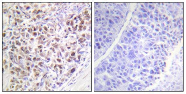
Real IHC data IHC analysis of AURKB using anti-AURKB antibody (M00762-1). AURKB was detected in a paraffin-embedded section of human bladder urothelial carcinoma tissue. Heat mediated antigen retrieval was performed in EDTA buffer (pH 8.0, epitope retrieval solution). The tissue section was blocked with 10% goat serum. The tissue section was then incubated with 1:50 rabbit anti-AURKB Antibody (M00762-1) overnight at 4°C. Peroxidase Conjugated Goat Anti-rabbit IgG was used as secondary antibody and incubated for 30 minutes at 37°C. The tissue section was developed using HRP Conjugated Rabbit IgG Super Vision Assay Kit (Catalog # SV0002) with DAB as the chromogen.
Anti-Aurora B AURKB Rabbit Monoclonal Antibody
Cat # M00762-1
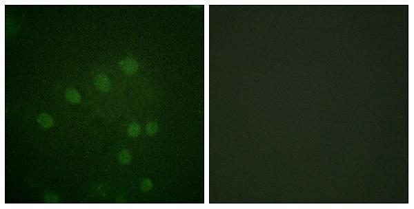
Real IHC data Immunohistochemistry analysis of paraffin-embedded human liver carcinoma, using AurB (Phospho-Thr232) Antibody. The picture on the right is blocked with the phospho peptide.
Anti-Phospho-ARK-2 (T232) AURKB Antibody
Cat # A00762T232
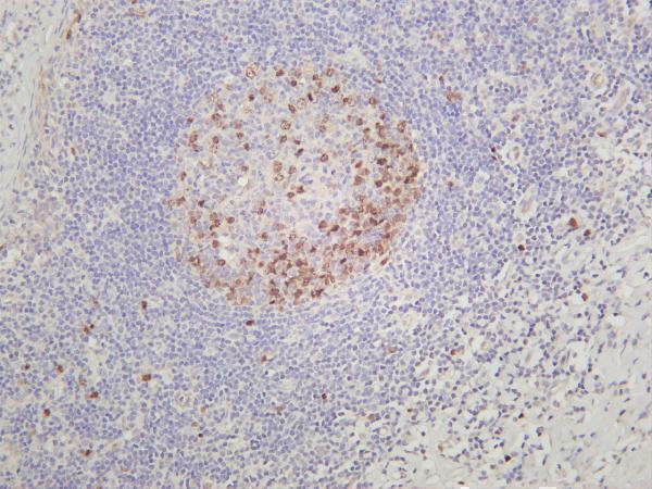
Real IHC data Human tonsil was stained with anti-Aurora B rabbit antibody
Anti-Aurora B Rabbit Monoclonal Antibody
Cat # M00762-5

M00762-1 shows IHC in paraffin-embedded human bladder urothelial carcinoma, and A00762T232 shows IHC in paraffin-embedded human liver carcinoma with a phospho-peptide block (respective IHC captions). M00762-5 shows IHC staining in human tonsil (M00762-5 IHC caption).

Which to pick: For tissue IHC, M00762-1 has a documented paraffin-section workflow with EDTA retrieval at pH 8.0 and a 1:50 primary dilution; the fixative is unreported (M00762-1 IHC caption). For IF/ICC, M00762-1 lists both applications, while A00762T232 has a HepG2 IF image with a phospho-peptide block (M00762-1 applications; A00762T232 IF caption). For work across species, A00762T232 lists Human, Monkey, Mouse, and Rat reactivity, although its IHC image documents only human tissue and does not report the fixative (A00762T232 catalog reactivity; A00762T232 IHC caption).

Each figure is that product's own IHC / IF validation image from its datasheet.

References

  1. UniProt Consortium. UniProt entry Q96GD4 (AURKB_HUMAN, Aurora kinase B).
  2. Human Protein Atlas. AURKB tissue IHC expression (reliability: Enhanced).
  3. Human Protein Atlas. AURKB subcellular location (ICC-IF): Mainly localized to the nucleoplasm. In addition localized to the kinetochore and midbody..
  4. Human Protein Atlas. AURKB antibody validation summary (2 antibodies).
  5. AURKB promotes gastric cancer progression via activation of CCND1 expression. Aging 2020 — PMC7053608.
  6. Gene expression changes are age-dependent and lobe-specific in the brown Norway rat model of prostatic hyperplasia. The Prostate 2009 — PMC2732361.
  7. Aurora Kinase B Expression, Its Regulation and Therapeutic Targeting in Human Retinoblastoma. Investigative ophthalmology & visual science 2021 — PMC7960835.
  8. Discovery of a new candidate drug to overcome cabazitaxel-resistant gene signature in castration-resistant prostate cancer by in silico screening. Prostate cancer and prostatic diseases 2023 — PMC10023558.
  9. PubMed PMID:9514916 — UniProt-cited evidence.
  10. PubMed PMID:9809983 — UniProt-cited evidence.
  11. PubMed PMID:9858806 — UniProt-cited evidence.