This website uses cookies to ensure you get the best experience on our website.


- Table of Contents
Plan AXIN2 staining in paraffin sections using the catalog antibody’s IHC protocol and the observed cytoplasmic and nuclear tissue pattern (HPA tissue IHC). Compare positive and low-staining cell populations, and interpret intensity with the reported medium consistency between antibody staining and RNA expression (HPA tissue IHC).

Expected localisation, validated protocols, controls and antibodies — the at-a-glance facts below, then the full design guide.
| Expected localisation | Cytoplasmic and nuclear tissue staining (HPA tissue IHC) | |
| Staining pattern | Glandular cells can stain; cytoplasmic and nuclear signal (HPA tissue IHC) | |
| Antigen retrieval | EDTA pH 8.0 HIER, heat-mediated (datasheet A01772-2) | |
| Positive control | Breast+4 more · see all | |
| Negative control | None in HPA (detected in all 45 tissues); use no-primary + isotype controls |
| Fixation | Keep fixation consistent across sections (standard IHC practice; not target-specific). Selected-image fixative and duration unreported (datasheet A01772-2); verify before use. | |
| Caveat | Staining has medium consistency with RNA expression (HPA tissue IHC) | |
| Regulation | Intensity regulation unreported (UniProt) | |
| Isoform / epitope | 0 listed isoforms; no processing reported (UniProt) |
The catalog antibody’s IHC-P protocol is paired with three published AXIN2 IHC protocols for rat lung, human ameloblastoma, and osteosarcoma sections (PMC3174135; PMC3934066; PMC8464802).
| Sample | Paraffin-embedded human renal carcinoma tissue; fixative not specified (datasheet A01772-2) |
| Fixation | Image fixative and duration unreported (datasheet A01772-2); verify before use. |
| Sectioning | 4–5 µm sections on charged slides (standard) |
| Deparaffinisation | Xylene, graded ethanol series to water (standard) |
| Antigen retrieval | Heat retrieval: EDTA pH 8.0 (datasheet A01772-2); 20 min, 95–100 °C (standard) |
| Peroxidase block | 3% H2O2, 10 min, room temperature (standard) |
| Blocking | 10% goat serum (datasheet A01772-2) |
| Primary antibody | Rabbit anti-AXIN2, 2-5 μg/ml (datasheet A01772-2) |
| Primary incubation | Overnight at 4 °C (datasheet A01772-2) |
| Detection | HRP-conjugated secondary, DAB chromogen (datasheet A01772-2) |
| Counterstain | Hematoxylin, blue, dehydrate and mount (standard) |
| Expected result | AXIN2-positive staining in adipocytes of breast (HPA tissue IHC: High). HPA tissue profile: General cytoplasmic and nuclear expression. No signal in the no-primary control. |
AXIN2 is a cytoplasmic protein with no transmembrane segment (UniProt Q9Y2T1). In paraffin-section IHC, expect cytoplasmic and sometimes nuclear staining in the relevant cells; HPA reports general cytoplasmic and nuclear expression, with high staining in several cell types (HPA tissue IHC). HPA rates its tissue pattern Approved, with medium consistency between staining and RNA expression (HPA tissue IHC).
| Cytoplasmic staining, with or without nuclear staining, in colon or duodenum glandular cells. | This fits HPA's general cytoplasmic and nuclear pattern and its high staining in these glandular cells (HPA tissue IHC). Assess signal within the cells rather than treating the entire gland as uniformly positive. |
| Staining confined to extracellular material, or a sharp membrane-only outline without convincing intracellular signal. | Reassess specificity: AXIN2 has no transmembrane segment and UniProt places it in the cytoplasm (UniProt Q9Y2T1). Membrane staining alone is not established as the expected paraffin-section pattern; HPA's plasma-membrane ICC-IF location is uncertain (HPA subcellular). |
| Strong staining chiefly in a cell population recorded as low, while expected cells show little signal. | Consider nonspecific antibody binding or endogenous detection activity (general IHC practice). HPA records low staining in oral-mucosa and esophageal squamous cells, and in prostate and fallopian-tube glandular cells; low does not mean absent (HPA tissue IHC). |
| A diffuse wash of chromogen obscures cell boundaries across the section. | The distribution cannot support a compartment call. Review blocking, antibody concentration, washes and detection controls (general IHC practice) before comparing it with HPA's cytoplasmic and nuclear pattern (HPA tissue IHC). |
| No detectable signal in a well-preserved colon glandular-cell control. | Investigate the IHC workflow before calling the specimen negative: HPA reports high staining in colon glandular cells (HPA tissue IHC). Check the antibody's IHC-P validation, detection reagents, retrieval conditions and control-section handling (general IHC practice). |
| Compartment evidence | UniProt lists cytoplasm, while HPA tissue IHC reports general cytoplasmic and nuclear expression (UniProt Q9Y2T1; HPA tissue IHC). Score the observed compartments separately; nuclear color is compatible with HPA tissue staining, but a nucleus-only result needs controls. |
| Choice of positive tissue | HPA reports High staining in breast adipocytes, cerebral-cortex neurons, colon and duodenum glandular cells, lung macrophages and placental trophoblasts (HPA tissue IHC). Choose a control whose relevant cell population is identifiable on the section. |
| Interpretation of low-staining tissue | HPA lists low staining in oral mucosa, esophagus, prostate and fallopian tube, and reports low tissue RNA specificity overall (HPA tissue IHC). A weak signal in those sites is plausible; they are not validated blank controls. |
| Antibody-validation limit | HPA lists CAB012283 and CAB017783 as Approved for IHC; neither is listed as IHC Enhanced in the supplied record (HPA antibodies). Treat agreement with the tissue pattern as supporting evidence, while checking controls for the antibody actually used. |
| IF/ICC cross-check: should its location dictate the IHC call? | No. HPA ICC-IF supports cytosol but labels nucleoplasm and plasma membrane uncertain (HPA subcellular). Use those images as context; judge paraffin-section staining against the tissue IHC pattern and its controls (HPA tissue IHC; general IHC practice). |
| Situation | Likely cause | Next action |
|---|---|---|
| The positive tissue has no signal. | A failed staining or detection step is possible; HPA reports High staining in colon glandular cells (HPA tissue IHC). | Run a known-positive section alongside the sample; verify antibody suitability for IHC-P, retrieval settings and the detection system (general IHC practice). |
| Most cells show uniform brown haze. | Excess antibody, inadequate washing or insufficient blocking can raise background (general IHC practice). | Compare a no-primary control; adjust antibody concentration, blocking and washes, then reassess cell-level localization (general IHC practice). |
| Color appears mainly in blood-rich or inflammatory areas. | Endogenous detection activity or nonspecific binding can mimic target staining (general IHC practice). | Check the no-primary control and use blocking appropriate to the detection chemistry; evaluate lung macrophages as cells, since HPA reports them High (HPA tissue IHC; general IHC practice). |
| Only nuclei are stained, with little cytoplasmic signal. | HPA tissue IHC allows nuclear expression, but UniProt lists cytoplasm and HPA ICC-IF marks nucleoplasm uncertain (HPA tissue IHC; UniProt Q9Y2T1; HPA subcellular). | Confirm that nuclear color follows recognizable cells in a positive control; review counterstain and no-primary control before accepting a nucleus-only interpretation (general IHC practice). |
| Strong color appears only in a low-staining reference tissue. | Nonspecific signal is possible, but HPA's Low category does not establish absence of AXIN2 (HPA tissue IHC). | Compare the same run with a High reference cell population and a no-primary control; report the observed cell type and compartment rather than calling the low tissue negative (HPA tissue IHC; general IHC practice). |
| A dominant membrane outline conflicts with the expected intracellular pattern. | UniProt places AXIN2 in cytoplasm without a transmembrane segment; HPA's ICC-IF membrane assignment is uncertain (UniProt Q9Y2T1; HPA subcellular). | Inspect a no-primary control and an IHC positive section, and check whether any cytoplasmic or nuclear signal accompanies the outline (HPA tissue IHC; general IHC practice). |
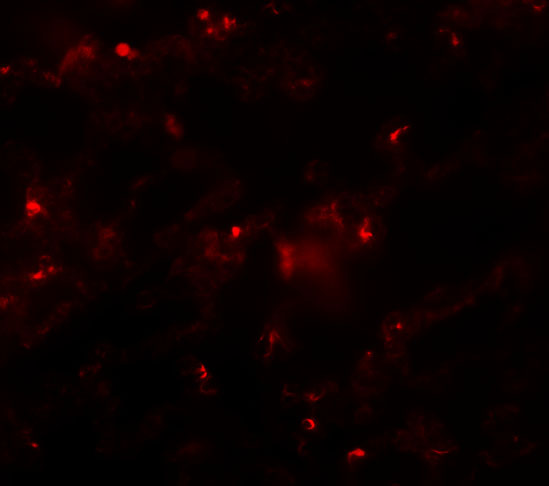
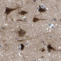

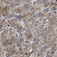


Comprehensive Human Protein Atlas IHC scoring per tissue (reliability: Approved — Medium consistency between antibody staining and RNA expression data.). Rows are taken directly from the HPA tissue chart — click any row's HPA link to view the source.
| Tissue | Cell type | Level | Evidence | Source |
|---|---|---|---|---|
| Breast | Adipocytes | High | Protein (IHC) | HPA → |
| Cerebral cortex | Neuronal cells | High | Protein (IHC) | HPA → |
| Colon | Glandular cells | High | Protein (IHC) | HPA → |
| Duodenum | Glandular cells | High | Protein (IHC) | HPA → |
| Epididymis | Glandular cells | High | Protein (IHC) | HPA → |
| Tissue | Cell type | Level | Evidence | Source |
|---|---|---|---|---|
| None in HPA: AXIN2 is detected in all 45 scored tissues. Use a no-primary (secondary-only) and an isotype control instead. | ||||
Troubleshoot AXIN2 staining in paraffin sections by checking retrieval, compartment, controls and scoring before interpreting signal as Wnt pathway activity.
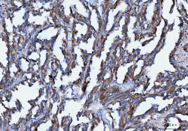
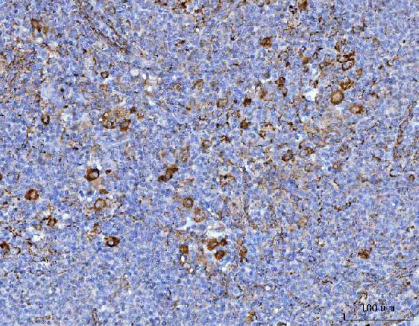
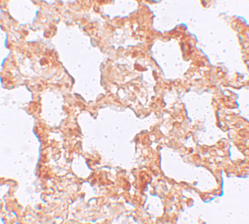
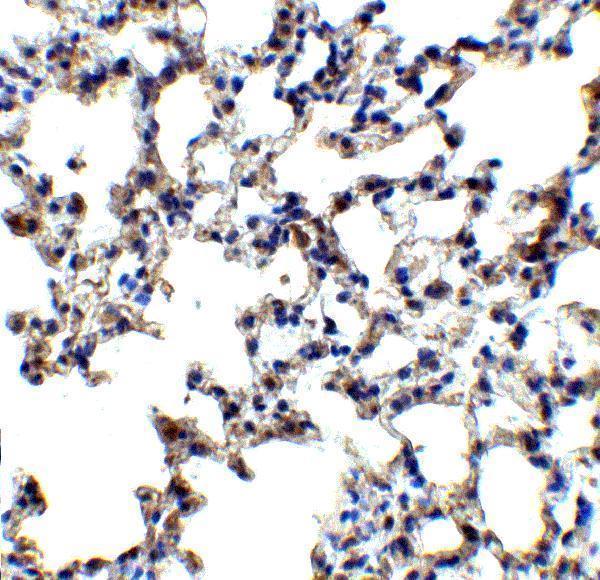
The catalog antibodies have IHC images from human renal carcinoma, spleen and thyroid cancer sections (catalog images: A01772-2), and rat and mouse lung tissue, plus IF images from rat lung cells, mouse lung tissue and HepG2 cells (catalog images: A01772).
A01772-2 has IHC images from paraffin-embedded human renal carcinoma, spleen and thyroid cancer sections (catalog images: A01772-2). A01772 has IHC images from rat and mouse lung tissue and IF images from rat lung cells, mouse lung tissue and HepG2 cells (catalog images: A01772).
Which to pick: For human paraffin-section IHC, choose A01772-2: its caption documents EDTA retrieval at pH 8.0 and staining at 2 μg/ml; the fixative is unreported (catalog IHC caption: A01772-2). For IF/ICC work, choose A01772 for its IF images and 20 μg/mL starting concentration; ICC validation is unreported (catalog IF images and datasheet: A01772). For mouse or rat tissue IHC, choose A01772: its IHC images show lung tissue from both species at 5 μg/mL; its listed reactivity also includes human (catalog IHC images and reactivity: A01772).