This website uses cookies to ensure you get the best experience on our website.


- Table of Contents
Plan BACE1 staining in paraffin sections using the observed granular cytoplasmic tissue pattern (HPA tissue IHC). This guide covers fixation, staining controls, and interpretation alongside BACE1’s trans-Golgi localization and membrane topology (UniProt).

Expected localisation, validated protocols, controls and antibodies — the at-a-glance facts below, then the full design guide.
| Expected localisation | Granular cytoplasm (HPA tissue IHC); mainly trans-Golgi network (UniProt) | |
| Staining pattern | Granular cytoplasm; high in cerebellar GLUC cells (HPA tissue IHC) | |
| Antigen retrieval | Citrate pH 6 HIER, heat-mediated (datasheet A00322) | |
| Positive control | Cerebellum+4 more · see all | |
| Negative control | Adipose tissue+4 more · see all |
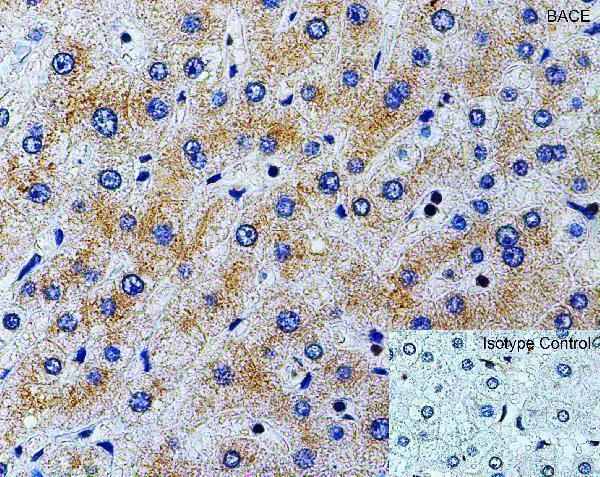
| Fixation | Formaldehyde-fixed human liver was used for IHC (datasheet A00322). (selected-SKU IHC image A00322) | |
| Caveat | Staining has low consistency with RNA expression (HPA tissue IHC) | |
| Regulation | No expression regulator specified (UniProt) | |
| Isoform / epitope | 6 isoforms; check whether the epitope lies in the ectodomain or cytoplasmic tail (UniProt) |
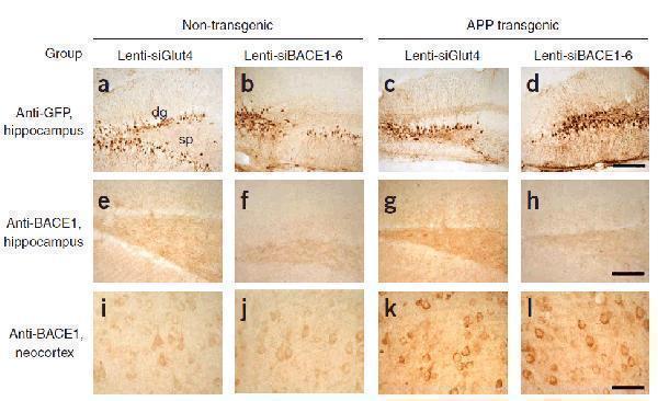
The catalog antibody’s citrate pH 6 IHC-P protocol (datasheet A00322) is followed by four published BACE1 chromogenic IHC workflows (PMC4829010; PMC2970759; PMC6073981; PMC10969905).
| Sample | Formaldehyde-fixed, paraffin-embedded human liver tissue (datasheet A00322) |
| Fixation | Image formalin-fixed; duration unreported (datasheet A00322); verify before use. |
| Sectioning | 4–5 µm sections on charged slides (standard) |
| Deparaffinisation | Xylene, graded ethanol series to water (standard) |
| Antigen retrieval | Heat retrieval: Citrate pH 6 (datasheet A00322); 20 min, 95–100 °C (standard) |
| Peroxidase block | 3% H2O2, 10 min, room temperature (standard) |
| Blocking | 10% serum (datasheet A00322) |
| Primary antibody | Rabbit anti-BACE1, 1-2 μg/mL (datasheet A00322) |
| Primary incubation | Overnight at 4 °C (standard) |
| Detection | HRP-polymer secondary, DAB chromogen 5–10 min (standard) |
| Counterstain | Hematoxylin, blue, dehydrate and mount (standard) |
| Expected result | BACE1-positive staining in gLUC cells - cytoplasm/membrane of cerebellum (HPA tissue IHC: High). HPA tissue profile: Granular cytoplasmic expression in several tissues. No signal in the no-primary control. |
In paraffin-section IHC, expect granular cytoplasmic staining in selected neural cells, with possible membrane-associated signal (HPA tissue IHC: granular cytoplasmic expression; UniProt P56817: membrane and intracellular locations). BACE1 has an extracellular region, one transmembrane segment and a short cytoplasmic tail (UniProt P56817 topology). HPA rates its tissue IHC evidence Approved while reporting low consistency between antibody staining and RNA expression (HPA tissue IHC: reliability). Interpret staining with that limitation in mind.
| Granular cytoplasmic staining in cerebellar GLUC cells; staining in cortical neurons or caudate glia. | This fits the reported tissue pattern: cerebellar GLUC cells are High, while cortical neuronal cells and caudate glial cells are Medium (HPA tissue IHC). Golgi and endosomal localization also supports intracellular signal (UniProt P56817 subcellular location). |
| Predominantly nuclear staining, with little granular cytoplasmic signal in expected positive cells. | A nuclear-dominant pattern is discordant with the reported granular cytoplasmic IHC and membrane-associated localization (HPA tissue IHC; UniProt P56817 subcellular location). Treat it as suspect; morphology alone cannot establish the cause. |
| Strong staining in adipocytes, cervical squamous epithelium or salivary glandular cells. | These cell types are reported Not detected (HPA tissue IHC). Check for nonspecific antibody binding or endogenous chromogenic activity (general IHC practice), particularly if expected positive cells on the same run stain poorly. |
| Uniform color across cells and extracellular spaces, obscuring granules and cell boundaries. | That distribution is difficult to reconcile with granular cytoplasmic expression (HPA tissue IHC). Consider background from antibody concentration, blocking, washing or detection chemistry (general IHC practice); do not score haze as cellular positivity. |
| No staining in cerebellar GLUC cells or cortical neurons, despite interpretable tissue morphology. | These are reported High and Medium, respectively (HPA tissue IHC). A blank result raises a run-performance concern, but HPA’s low staining-to-RNA consistency limits conclusions about any individual specimen (HPA tissue IHC: reliability). |
| Which compartment should guide scoring? | HPA describes granular cytoplasmic tissue IHC, while UniProt places BACE1 predominantly in the later Golgi/trans-Golgi network and also in endosomes and at the cell surface (HPA tissue IHC; UniProt P56817 subcellular location). Score the observed cell pattern, not an assumed single organelle. |
| Does epitope position matter? | The mature chain spans residues 46–501; residues 22–457 are extracellular and 479–501 cytoplasmic across a 458–478 transmembrane segment (UniProt P56817 processing and topology). The antibody epitope is unspecified here, so this record cannot predict its accessibility or an optimal retrieval condition. |
| Can processing or isoforms change interpretation? | UniProt lists a cleaved signal peptide, a propeptide and 6 isoforms (UniProt P56817 processing and isoforms). Antibody recognition across those forms is unspecified; neither altered staining nor a particular staining intensity can be assigned to one form from morphology alone. |
| How strong is antibody support? | CAB016358 is Approved for IHC and Supported for ICC, while tissue IHC has low consistency with RNA expression (HPA antibody validation; HPA tissue IHC: reliability). Use the tissue examples as guides, not guarantees for every specimen or proof of antibody specificity. |
| What does retrieval establish? | Antigen retrieval is a routine paraffin IHC variable (general IHC practice). No target-specific fixation or retrieval response is supplied for BACE1, so optimize and document the chosen condition against positive and negative tissue controls without claiming a predicted gain. |
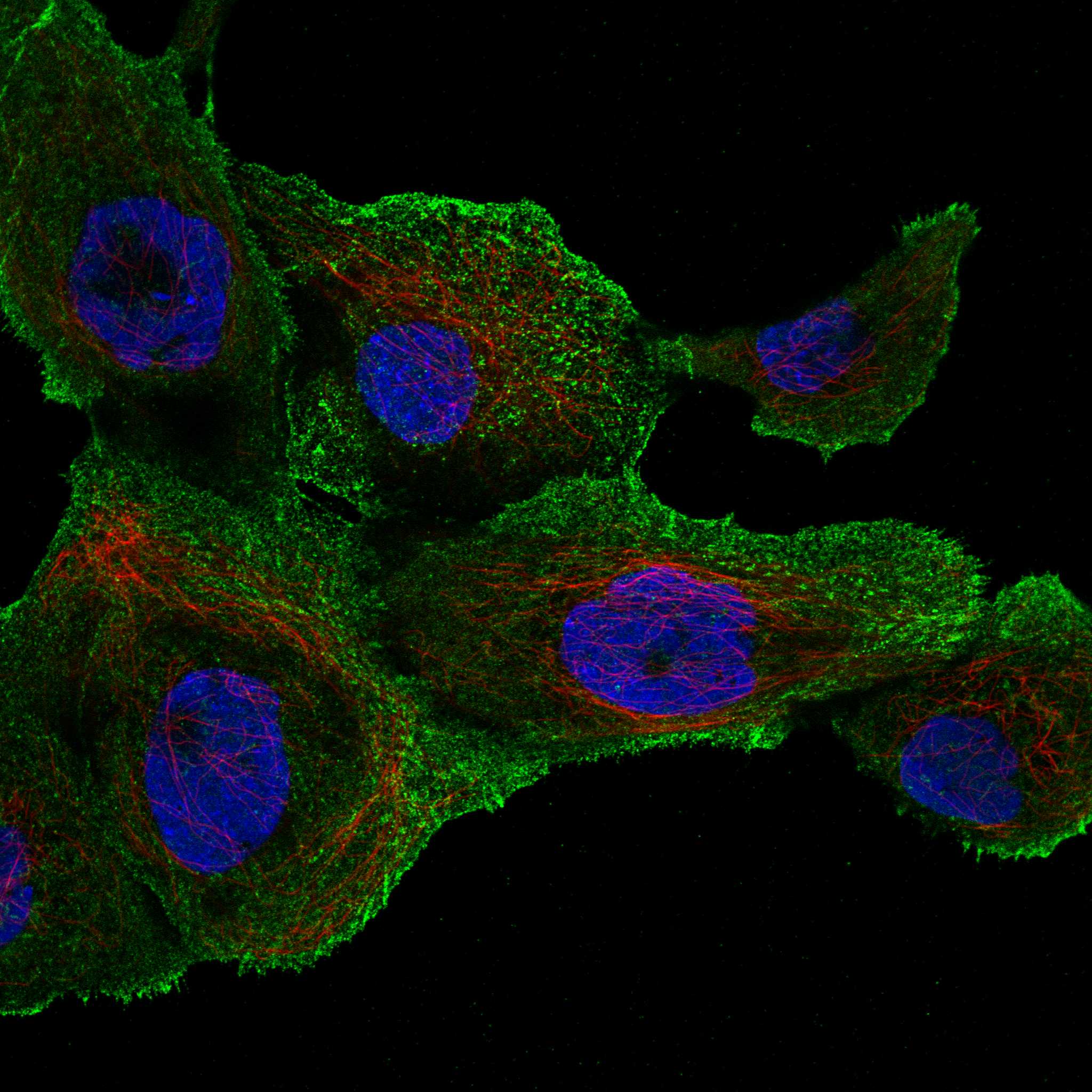
| IF/ICC Q: Should plasma-membrane staining be expected? | A: Plasma membrane is the supported main ICC-IF location, with images listed for A-431 and U-251MG (HPA subcellular ICC-IF). That observation informs localization but does not replace the granular cytoplasmic pattern reported for tissue IHC (HPA tissue IHC). |
| Situation | Likely cause | Next action |
|---|---|---|
| The known-positive tissue is blank. | The reported High cerebellar GLUC-cell or Medium cortical neuronal signal is absent (HPA tissue IHC); a failed detection step or unsuitable working conditions are possible (general IHC practice). | Verify tissue preservation and run controls, then review retrieval, primary-antibody application and chromogen development in that order (general IHC practice). Recheck a second reported positive tissue before calling a specimen negative (HPA tissue IHC). |
| Background covers most of the section. | Diffuse signal does not match granular cytoplasmic expression (HPA tissue IHC). Excess primary or detection reagent, inadequate blocking or insufficient washes can produce background (general IHC practice). | Compare a no-primary control, strengthen appropriate blocking and washing, and titrate the primary and detection reagents (general IHC practice). Judge improvement by recovery of discrete cellular granules (HPA tissue IHC). |
| Color appears in negative control cell types. | Adipocytes, cervical squamous cells and salivary glandular cells are reported Not detected (HPA tissue IHC); nonspecific binding or endogenous chromogenic activity may account for color (general IHC practice). | Check no-primary and detection-only controls, suppress endogenous enzyme activity when applicable, and compare the suspect cells with a reported positive tissue (general IHC practice; HPA tissue IHC). |
| Nuclei dominate the stain. | Nuclear-dominant staining conflicts with HPA’s granular cytoplasmic tissue pattern and UniProt’s membrane and organelle locations (HPA tissue IHC; UniProt P56817 subcellular location). | Review counterstain and chromogen separately, inspect a no-primary control and reassess whether positive cells also show the expected cytoplasmic granules (general IHC practice; HPA tissue IHC). |
| Only a thin cell-edge signal is visible. | Cell-surface localization is possible (UniProt P56817 subcellular location), but HPA’s tissue IHC profile emphasizes granular cytoplasm (HPA tissue IHC). Edge staining alone leaves the tissue interpretation uncertain. | Inspect a reported positive cell population for intracellular granules and compare a negative cell population on the same run; avoid scoring edge-only staining as definitive BACE1 positivity (HPA tissue IHC). |
| A specimen disagrees with an RNA-based expectation. | HPA explicitly reports low consistency between antibody staining and RNA expression, despite its Approved tissue IHC rating (HPA tissue IHC: reliability). RNA enrichment in brain and pancreas does not specify staining in every cell (HPA tissue IHC: RNA specificity). | Record the actual stained cell type, compartment and intensity; compare them with HPA’s cell-specific IHC examples and controls before interpreting the discrepancy (HPA tissue IHC; general IHC practice). |
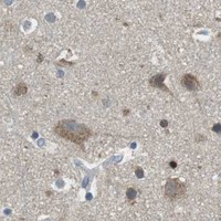


Comprehensive Human Protein Atlas IHC scoring per tissue (reliability: Approved — Low consistency between antibody staining and RNA expression data.). Rows are taken directly from the HPA tissue chart — click any row's HPA link to view the source.
| Tissue | Cell type | Level | Evidence | Source |
|---|---|---|---|---|
| Cerebellum | GLUC cells - cytoplasm/membrane | High | Protein (IHC) | HPA → |
| Adrenal gland | Glandular cells | Medium | Protein (IHC) | HPA → |
| Appendix | Glandular cells | Medium | Protein (IHC) | HPA → |
| Bronchus | Respiratory epithelial cells | Medium | Protein (IHC) | HPA → |
| Caudate | Glial cells | Medium | Protein (IHC) | HPA → |
| Tissue | Cell type | Level | Evidence | Source |
|---|---|---|---|---|
| Adipose tissue | Adipocytes | Not detected | Protein (IHC) | HPA → |
| Breast | Adipocytes | Not detected | Protein (IHC) | HPA → |
| Cervix | Squamous epithelial cells | Not detected | Protein (IHC) | HPA → |
| Ovary | Ovarian stroma cells | Not detected | Protein (IHC) | HPA → |
| Salivary gland | Glandular cells | Not detected | Protein (IHC) | HPA → |
Use matched controls and compartment-aware scoring when troubleshooting chromogenic BACE1 IHC in paraffin sections.
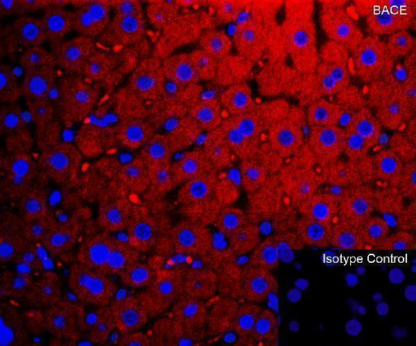
The IHC-validated BACE1 antibody A00322 has paraffin-section IHC data from human liver and IF data from mouse liver (IHC and IF image captions).
A00322 is listed for IHC-P and IF, with human IHC and mouse IF validation (catalog applications; catalog validation note). Its images show formaldehyde-fixed, paraffin-embedded human liver IHC and 4% paraformaldehyde-fixed mouse liver IF (IHC and IF image captions).
Which to pick: For tissue IHC, choose A00322; its human liver image documents paraffin sections, formaldehyde fixation and heat retrieval in citrate buffer at pH 6 (IHC image caption). For IF, A00322 has mouse liver data; ICC validation is unreported (IF image caption; catalog validation note). For work spanning human and mouse samples, A00322 is a rabbit polyclonal with both species listed as reactive, while its documented IHC validation is human and IF validation is mouse (catalog host, clonality and reactivity; catalog validation note).