This website uses cookies to ensure you get the best experience on our website.


- Table of Contents
Plan BAG1 staining in paraffin sections using the predominantly nuclear tissue pattern (HPA tissue IHC). Compare positive and negative cell populations while accounting for tissue-dependent isoform expression (UniProt).

Expected localisation, validated protocols, controls and antibodies — the at-a-glance facts below, then the full design guide.
| Expected localisation | Nuclear staining in most tissues (HPA tissue IHC) | |
| Staining pattern | Nuclear staining across most tissues (HPA tissue IHC) | |
| Antigen retrieval | EDTA pH 8.0 HIER, heat-mediated (datasheet A02423-2) | |
| Positive control | Appendix+4 more · see all | |
| Negative control | Adipose tissue+4 more · see all |
| Fixation | Matched tissue-IHC evidence does not establish the fixation claim. Validate the specimen-specific method before use. (selected-SKU IHC image A02423-2) | |
| Caveat | Isoform 4 expression varies by tissue (UniProt) | |
| Regulation | Tissue-dependent isoform expression (UniProt) | |
| Isoform / epitope | 4 isoforms; verify antibody epitope coverage (UniProt) |
Compare the catalog antibody’s IHC-P protocol (datasheet: A02423-2) with two published lung cancer tissue protocols (PMC4734146; PMC8247369).
| Sample | Paraffin-embedded human breast cancer tissue; fixative not specified (datasheet A02423-2) |
| Fixation | Image fixative and duration unreported (datasheet A02423-2); verify before use. |
| Sectioning | 4–5 µm sections on charged slides (standard) |
| Deparaffinisation | Xylene, graded ethanol series to water (standard) |
| Antigen retrieval | Heat retrieval: EDTA pH 8.0 (datasheet A02423-2); 20 min, 95–100 °C (standard) |
| Peroxidase block | 3% H2O2, 10 min, room temperature (standard) |
| Blocking | 10% goat serum (datasheet A02423-2) |
| Primary antibody | Rabbit anti-BAG1, 2-5μg/ml (datasheet A02423-2) |
| Primary incubation | Overnight at 4 °C (datasheet A02423-2) |
| Detection | HRP-conjugated secondary, DAB chromogen (datasheet A02423-2) |
| Counterstain | Hematoxylin, blue, dehydrate and mount (standard) |
| Expected result | BAG1-positive staining in endocrine cells of appendix (HPA tissue IHC: High). HPA tissue profile: Nuclear expression in most tissues. No signal in the no-primary control. |
BAG1 should show predominantly nuclear staining in many tissue cell types, with cytoplasmic signal also biologically plausible (HPA: nuclear expression in most tissues; UniProt Q99933: nucleus and cytoplasm). Prioritize the listed high-staining cell populations, such as colon endocrine cells and lung alveolar type II cells (HPA: High). The tissue IHC profile has Supported reliability, with medium consistency between staining and RNA data (HPA: Supported). BAG1 has no transmembrane segment (UniProt Q99933 topology).
| Distinct nuclear staining in the expected cells, possibly with weaker cytoplasmic staining. | This agrees with the dominant tissue pattern (HPA: nuclear expression in most tissues) and reported cytoplasmic localization (UniProt Q99933). Compare cells within the same section; nuclear signal in the expected population is more persuasive than uniform color across every compartment (general IHC interpretation). |
| Predominantly membrane outlining, with little convincing nuclear staining. | A membrane-dominant pattern conflicts with BAG1's reported nuclear and cytoplasmic locations and lack of a transmembrane segment (UniProt Q99933). Review morphology, antibody specificity and chromogen deposits before scoring it as BAG1 (general IHC interpretation). Cytoplasmic staining alone is less decisive because that compartment is reported (UniProt Q99933). |
| Strong staining in a cell population listed as not detected, such as adipocytes, while expected positive cells are unstained. | That distribution disagrees with the reference profile (HPA: adipocytes Not detected; colon endocrine cells High). Consider cross-reactivity or endogenous detection activity, particularly if the signal ignores cell boundaries (general IHC interpretation). A single discordant field does not establish either cause; assess controls and tissue morphology (general IHC practice). |
| Diffuse color covers nuclei, cytoplasm, extracellular spaces and slide background. | A continuous haze cannot establish BAG1 localization (general IHC interpretation). Check whether the negative reagent control shares the haze, then assess blocking, washing and detection background (general IHC practice). HPA's tissue pattern describes cellular nuclear expression; it does not validate diffuse extracellular staining (HPA: tissue IHC profile). |
| No visible signal in a listed high-staining population, such as gallbladder glandular cells. | This fails the selected positive-control expectation (HPA: gallbladder glandular cells High). Check that the glandular cells are present, then review antibody and detection performance, retrieval conditions and section quality (general IHC practice). A failed control makes negative results in other specimens difficult to interpret (general IHC interpretation). |
| Which cells provide a reference pattern? | Use the specified cell population, not an entire organ, as the comparator: appendix and duodenum endocrine cells and cervix glandular cells are High, while adipocytes and smooth muscle cells are Not detected (HPA: tissue IHC). These levels describe observed staining, not universal positive or negative status for every cell in those tissues (HPA: tissue IHC). |
| What does an apparent cytoplasmic component mean? | Cytoplasmic staining can fit BAG1 localization (UniProt Q99933: cytoplasm), although nuclear expression predominates in tissue IHC (HPA: tissue IHC). The distinct ICC-IF reference places BAG1 mainly in the nucleoplasm and additionally in the cytosol (HPA: supported subcellular localization). |
| Can isoforms change interpretation? | Four BAG1 isoforms are listed; isoform 1 is predominantly nuclear (UniProt Q99933). Isoform 4 has broad reported expression with specified exceptions (UniProt Q99933). The supplied record does not map an IHC antibody epitope to those isoforms, so a staining difference cannot be assigned to one isoform from this evidence alone. |
| Is retrieval or epitope behavior predictable? | No BAG1-specific retrieval condition or fixation sensitivity is supplied. Retrieval optimization against a known-positive section is general IHC practice. The listed ubiquitin-like and BAG domains and phosphoserine 223 do not, by themselves, predict epitope accessibility in paraffin sections (UniProt Q99933: domains and modified residue). |
| IF/ICC: where should BAG1 fluorescence appear? | Expect mainly nucleoplasmic fluorescence with additional cytosolic signal (HPA: supported ICC-IF subcellular localization). This is a localization cross-check, not an IHC-P protocol or an IF preparation recommendation; compare with the tissue IHC pattern separately (HPA: nuclear expression in most tissues). |
| Situation | Likely cause | Next action |
|---|---|---|
| Positive-control nuclei are blank. | The run has not reproduced a listed high-staining population (HPA: gallbladder glandular cells High). | Confirm cell identity and section quality; inspect primary antibody, retrieval and detection steps against the chosen antibody's IHC-P instructions (general IHC practice). Resolve the positive control before interpreting study negatives (general IHC interpretation). |
| A tissue expected to contain positive cells looks uniformly negative. | The relevant cell population may be absent from the section, or the run may have failed (general IHC interpretation). HPA assigns High to particular cells, such as colon endocrine cells, rather than to every colon cell (HPA: tissue IHC). | Locate the specified cells on a counterstained section and compare a separate listed high-staining control processed in the same run (general IHC practice). |
| Color appears in negative-control and test sections alike. | Shared signal suggests background from detection chemistry or insufficient blocking or washing (general IHC interpretation). It does not confirm BAG1, even where the tissue profile predicts staining (HPA: tissue IHC). | Inspect the negative reagent control and review detection chemistry, blocking and washes using general IHC controls. Score BAG1 only where cell-associated signal separates from that background (general IHC practice). |
| Strong staining outlines cell membranes, with weak or absent nuclear signal. | A membrane-dominant result disagrees with the reported localization and topology (UniProt Q99933: nucleus, cytoplasm and no transmembrane segment). | Check for edge deposits and nonspecific staining, then compare a listed high-staining tissue and the chosen antibody's validation information (general IHC practice; HPA: tissue IHC). Do not use membrane outlines alone to call BAG1 positive (general IHC interpretation). |
| Cytoplasmic staining seems stronger than nuclear staining. | Cytoplasmic BAG1 is plausible, but tissue IHC is chiefly nuclear (UniProt Q99933: cytoplasm; HPA: nuclear expression in most tissues). Relative compartment intensity alone cannot identify an isoform without epitope information (UniProt Q99933: four isoforms). | Compare well-preserved cells and controls, record nuclear and cytoplasmic components separately, and check whether the antibody's epitope is documented before proposing an isoform explanation (general IHC practice). |
| A proposed negative-control population stains strongly. | The observation conflicts with a cell-specific reference if the population is correctly identified (HPA: adipocytes, smooth muscle cells and soft-tissue fibroblasts Not detected). Cross-reactivity or endogenous detection activity is possible, not established (general IHC interpretation). | Confirm cell identity and compare negative reagent and positive tissue controls. Reassess detection background and antibody validation before treating the unexpected signal as BAG1 (general IHC practice). |
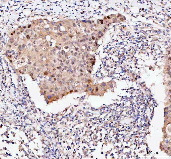
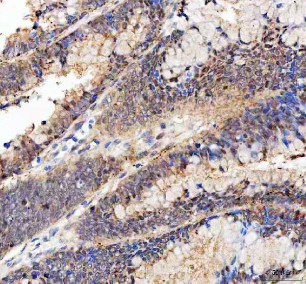
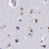


Comprehensive Human Protein Atlas IHC scoring per tissue (reliability: Supported — Medium consistency between antibody staining and RNA expression data.). Rows are taken directly from the HPA tissue chart — click any row's HPA link to view the source.
| Tissue | Cell type | Level | Evidence | Source |
|---|---|---|---|---|
| Appendix | Endocrine cells | High | Protein (IHC) | HPA → |
| Cervix | Glandular cells | High | Protein (IHC) | HPA → |
| Colon | Endocrine cells | High | Protein (IHC) | HPA → |
| Duodenum | Endocrine cells | High | Protein (IHC) | HPA → |
| Epididymis | Glandular cells | High | Protein (IHC) | HPA → |
| Tissue | Cell type | Level | Evidence | Source |
|---|---|---|---|---|
| Adipose tissue | Adipocytes | Not detected | Protein (IHC) | HPA → |
| Adrenal gland | Glandular cells | Not detected | Protein (IHC) | HPA → |
| Bone marrow | Hematopoietic cells | Not detected | Protein (IHC) | HPA → |
| Smooth muscle | Smooth muscle cells | Not detected | Protein (IHC) | HPA → |
| Soft tissue | Fibroblasts | Not detected | Protein (IHC) | HPA → |
Troubleshoot BAG1 staining in paraffin sections by checking retrieval, compartment, background and cell specific scoring before interpreting intensity.
A02423-2 has BAG1 IHC images from human breast cancer, colorectal adenocarcinoma and tonsil, and mouse colon paraffin sections (catalog IHC captions). M02423-2 lists ICC/IF without an image (catalog applications; image fields).
A02423-2 has IHC images from human and mouse paraffin sections and lists human, mouse and rat reactivity (catalog IHC captions; reactivity). M02423-2 lists IHC and ICC/IF for human, mouse and rat, but has no IHC or IF images in the payload (catalog applications; reactivity; image fields).
Which to pick: For paraffin-section tissue IHC, choose A02423-2: its captions document EDTA retrieval at pH 8.0, a 2 μg/ml primary antibody incubation and HRP/DAB detection; the fixative is unreported (A02423-2 IHC captions). For IF/ICC, M02423-2 is the listed option: it is rabbit monoclonal clone 28B16 with a listed IF dilution of 1:50, though no IF image is supplied (M02423-2 catalog). For cross-species work, both list human, mouse and rat reactivity; A02423-2 has IHC images from human and mouse sections, while neither SKU has a rat image in the payload (catalog reactivity; image fields).