This website uses cookies to ensure you get the best experience on our website.
- Table of Contents
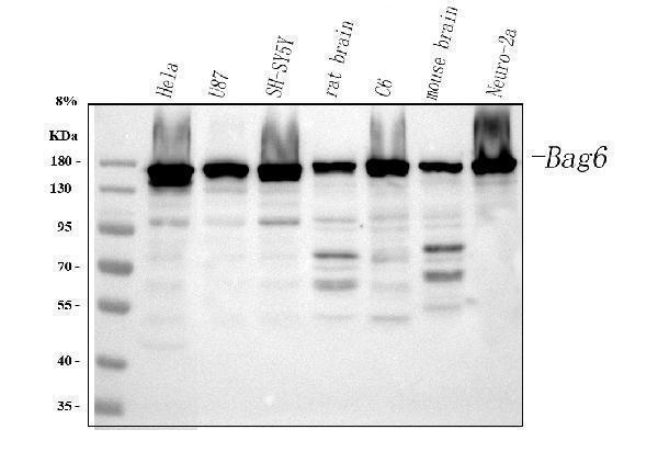
Facts about Large proline-rich protein BAG6.
Recruited to ribosomes, it interacts with the transmembrane region of newly synthesized tail-anchored proteins and collectively with SGTA and ASNA1 mediates their delivery to the endoplasmic reticulum (PubMed:20516149, PubMed:20676083, PubMed:28104892, PubMed:25535373). Client proteins which can't be correctly delivered to the endoplasmic reticulum are ubiquitinated by RNF126, an E3 ubiquitin-protein ligase associated with BAG6 and are sorted to the proteasome (PubMed:24981174, PubMed:28104892, PubMed:27193484).
Human | |
---|---|
Gene Name: | BAG6 |
Uniprot: | P46379 |
Entrez: | 7917 |
Belongs to: |
---|
No superfamily |
BAG6; BAT3; BAT3HLA-B associated transcript-3; BCL2-associated athanogene 6BAG-6; D6S52E; D6S52EHLA-B-associated transcript 3; G3BAG family molecular chaperone regulator 6; HLA-B associated transcript 3; large proline-rich protein BAT3; Protein G3; Protein Scythe; Scythe
Mass (kDA):
119.409 kDA
Human | |
---|---|
Location: | 6p21.33 |
Sequence: | 6; NC_000006.12 (31639028..31660900, complement) |
Expressed by immature dendritic cells (at protein level).
Cytoplasm, cytosol. Nucleus. Secreted, extracellular exosome. Normally localized in cytosol and nucleus, it can also be released extracellularly, in exosomes, by tumor and myeloid dendritic cells (PubMed:18055229, PubMed:18852879). Cytoplasmic retention is due to interaction with GET4 (PubMed:29042515).
The BAG6 marker is a useful tool in many applications, including protein transfer efficiency. You can test this marker in various ways: using membrane staining, autoradiography film ECL chemiluminescent detector, DAB chromogenic detection, and lateral flow immunoassay. Boster scientists can submit their findings to earn product credits. These benefits are available to scientists around the world.
To test membrane-stained gels attach the sample comb to the gel. Make sure you place the samples in an uniform line. The samples should be incubated at 37degC for 30-60 minutes until the gel solution is fully solid and polymerized. Carefully remove the sample comb out of the gel. Mix the protein sample and the Boster 4X Dual Color Protein loading buffer in a ratio of 3:1. Once this is complete, observe the gels in a dish containing transfer buffer.
Prepare the gels and test the efficiency of protein transfer using the appropriate chemicals. Gold in colloids is sensitive. There are a variety of commercial ready-to-use staining agents. The higher concentrations of proteins are visible as reddish-colored bands on a pink background. To maximize the transfer of protein efficiency, the gel concentration should be adjusted prior to staining. The higher the concentration of the gel, the longer the duration of staining.
The efficiency of transfer of proteins was determined by using the appropriate NC membrane based on the molecular weight. The lower molecular weight of the protein, the better the binding efficiency. If the protein is over 20KD, use an NC membrane with 0.45 um pores. Proteins with lower molecular weights must be treated with a PVDF membrane that is a high affinity and sensitivity Reagent.
The sensitivity of the membrane staining is affected by the loss of protein and transfer efficiency. If the membrane isn't fully saturated with the protein Try using a buffer that has detergents like Tween 20. Then, rinse the membrane with TBS Wash Buffer three times. It is possible to stain the membrane using specific antibodies after washing. The blots are then assessed and a digital image is provided.
The BAG6 marker can be used to detect interactions between proteins and proteins. This molecule is a part of the chimeric virus and is found in numerous tissues and cells. It is widely used in research, drug development, and many other applications. The BAG6 marker has a variety of uses. It is extremely specific. To make it more precise, you can add a chimeric antigen receptor (CAR) domain to it. The C-terminal domain is an essential characteristic in biochemical analysis.
ECL Chemistry is a widely used method for detecting abundance of proteins on membranes. However, this method has its limitations. One of these limitations is the inability to simultaneously capture two targets with the same molecular mass. It's therefore difficult to accurately quantify the results. It is important to understand the limitations of the system and how it functions. We'll show you how Boster Bio's ECL chemiluminescent detection device can help with your research.
ECL also referred to by ECL, is a luminol-based substrate used for Western Blot applications. This reagent is sensitive antigens, and its Luminescence is proportional to the quantity of antibodies labeled with HRP. The ECL signal can be assessed in pictograms that allow for indirect measurements of proteins. Researchers can also employ imaging instruments to identify antigens. However it is essential to avoid prolonged exposure to the sun.
ECL Chemistry also has a high sensitivity. It is able to detect small amounts of material without sacrificing sensitivity. It is also Boster Bio's ECL chemiluminescent detection technology can be used for many different research applications. A protein-containing sample with a Luminol molecules, for instance, will show a tenfold increase in Luminol's detection sensitivity.
Based on the research goals, ECL can be used to detect low levels of target proteins. It is essential to use a detection system that can detect low levels of target proteins for quantitative analysis. With a higher dilution factor for the primary antibody an impressive signal can be achieved. The list of components can be found under the Documents tab. If you're not sure which kit to choose, consult the instructions in the manual.
The Boster Bio DAB chromogenic staining system enables researchers to observe the expression of antigens in tissue sections cells, tissues, and other samples. This method employs an enzyme to convert the soluble substrate into an insoluble colored product that is then deposited at the site of antigen expression. The enzymes typically used in this system of detection are alkaline phosphatase and horseradish peroxidase.
Steve Boster passed away on Sunday June 26, 2022 at the age of 67. He was born in Joliet, IL. He was a retired retail sales manager and a veteran of the U.S. Army. Steve was an active member of the Concordia Hall in Staunton. He leaves behind two daughters who were Natosha Peck as well as Crystal Boster. His family includes his brothers Jack Boster, his sister Tammy, and many nieces and nephews.
His first product, which became known as a household name was invented in 1993. Steven Boster had already developed numerous products by the late 1990s including IHC and a plethora of other primary antibodies. By the end of the decade Boster's company had expanded to become the largest catalog antibody manufacturer in China. PicoKine was his exclusive ELISA platform and he also developed high-sensitivity ELISA kit kits.
PMID: 2156268 by Banerji J., et al. A gene pair from the human major histocompatibility complex encodes large proline-rich proteins with multiple repeated motifs and a single ubiquitin-like domain.
PMID: 14960581 by Wu Y.-H., et al. Ricin triggers apoptotic morphological changes through caspase-3 cleavage of BAT3.