This website uses cookies to ensure you get the best experience on our website.


- Table of Contents
Plan BAG6 chromogenic IHC in paraffin sections using the cytoplasmic and nuclear tissue pattern, with strong testis staining as a reference (HPA tissue IHC). The catalog antibody's IHC dilution is 2–5 μg/mL (datasheet A00967-1).

Expected localisation, validated protocols, controls and antibodies — the at-a-glance facts below, then the full design guide.
| Expected localisation | Cytoplasmic and nuclear staining (HPA tissue IHC) | |
| Staining pattern | Cytoplasmic/nuclear in most tissues; abundant in testis (HPA tissue IHC) | |
| Antigen retrieval | EDTA pH 8.0 HIER, heat-mediated (datasheet A00967-1) | |
| Positive control | Lung+4 more · see all | |
| Negative control | None in HPA (detected in all 45 tissues); use no-primary + isotype controls |
| Fixation | Keep fixation consistent across sections. (standard IHC practice; not target-specific) | |
| Caveat | Tumor and myeloid cells can release BAG6 in exosomes (UniProt) | |
| Regulation | No staining-intensity regulator established (UniProt) | |
| Isoform / epitope | 5 isoforms; epitope coverage is unspecified (UniProt) |
The catalog antibody protocol (datasheet: A00967-1) and two published BAG6 IHC protocols (PMC6052567; PMC8748226) provide starting points for paraffin sections.
| Sample | Paraffin-embedded human lung cancer tissue; fixative not specified (datasheet A00967-1) |
| Fixation | Image fixative and duration unreported (datasheet A00967-1); verify before use. |
| Sectioning | 4–5 µm sections on charged slides (standard) |
| Deparaffinisation | Xylene, graded ethanol series to water (standard) |
| Antigen retrieval | Heat retrieval: EDTA pH 8.0 (datasheet A00967-1); 20 min, 95–100 °C (standard) |
| Peroxidase block | 3% H2O2, 10 min, room temperature (standard) |
| Blocking | 10% goat serum (datasheet A00967-1) |
| Primary antibody | Rabbit anti-BAG6, 2-5μg/ml (datasheet A00967-1) |
| Primary incubation | Overnight at 4 °C (datasheet A00967-1) |
| Detection | HRP-conjugated secondary, DAB chromogen (datasheet A00967-1) |
| Counterstain | Hematoxylin, blue, dehydrate and mount (standard) |
| Expected result | BAG6-positive staining in alveolar cells type II of lung (HPA tissue IHC: High). HPA tissue profile: Cytoplasmic and nuclear expression in most tissues, highly abundant in testis. No signal in the no-primary control. |
BAG6 is normally found in the cytosol and nucleus and has no transmembrane segment (UniProt P46379). In paraffin-section IHC, expect cytoplasmic and nuclear staining across many tissues, with particularly abundant staining in testis (HPA tissue IHC: Enhanced reliability). High-staining examples are spermatogonia, lung alveolar type II cells and placental cytotrophoblasts (HPA tissue IHC: High in each cell type).
| Nuclear and cytoplasmic staining in the expected cells, with strong spermatogonial signal. | This fits BAG6’s reported compartments (UniProt P46379: cytosol and nucleus) and the tissue pattern (HPA tissue IHC: cytoplasmic and nuclear expression in most tissues; highly abundant in testis). Judge intensity within the relevant cell population, since other tissues and cell types have different reported levels (HPA tissue IHC: High, Medium and Low categories). |
| Signal appears confined to an unexpected compartment, such as a sharp cell-surface rim. | A membrane-rim pattern alone does not match the reported intracellular pattern (UniProt P46379: cytosol and nucleus; no transmembrane segment). Consider nonspecific staining or a detection artefact before assigning it to BAG6. Extracellular release in exosomes is reported, but it does not establish a diagnostic membrane-rim IHC pattern (UniProt P46379: extracellular exosomes). |
| A cell population stains strongly where the HPA profile reports low staining. | For example, strong signal in skeletal myocytes or adipocytes conflicts with their reported low staining (HPA tissue IHC: Low in skeletal muscle myocytes and adipose tissue adipocytes). Check cell identification and controls; cross-reactivity or endogenous detection activity may explain the result (standard IHC practice). An HPA Low designation does not mean every cell must be blank. |
| Color spreads across tissue structures and obscures cell boundaries. | Diffuse background prevents a reliable nuclear-versus-cytoplasmic call (UniProt P46379: nucleus and cytosol; HPA tissue IHC: cytoplasmic and nuclear pattern). It can reflect nonspecific antibody binding or detection background (standard IHC practice). Interpret the slide only after comparison with an appropriate detection control and a defined cell-level pattern. |
| Expected cells lack visible signal in a putative positive section. | Absence of staining in testis spermatogonia, lung alveolar type II cells or placental cytotrophoblasts is inconsistent with their reported High levels (HPA tissue IHC: High in each). First verify that the named cells are present and identifiable. A failed stain remains possible; a negative-looking field without those cells cannot assess the expected positive pattern (standard IHC practice). |
| Tissue and cell selection | The HPA profile has low tissue specificity, yet individual cell levels vary (HPA tissue IHC: Low tissue specificity; High, Medium and Low cell categories). A section containing High-staining cells makes an interpretable positive reference; Low-staining cells alone are a weak failure check (HPA tissue IHC: named High and Low cells). |
| Antibody validation | The tissue profile has Enhanced reliability, meaning high consistency between antibody staining and RNA expression data (HPA tissue IHC: Enhanced). Individual antibodies have separate IHC statuses: HPA045116 and HPA053291 are Enhanced; CAB020704 is Supported (HPA antibody validation). These labels support interpretation but do not guarantee every staining run. |
| Isoforms and epitope | Five BAG6 isoforms are listed, but the supplied record does not map an IHC antibody epitope to them (UniProt P46379: isoforms 1–5). If stains disagree, epitope coverage is a question to resolve from the antibody’s documentation; the isoform count alone cannot predict which cells or compartments an antibody will reveal. |
| Topology and extracellular release | BAG6 has no transmembrane segment and no signal peptide or propeptide (UniProt P46379). Release in exosomes from tumor and myeloid dendritic cells is reported (UniProt P46379: extracellular exosomes). These facts do not justify treating a surface-only chromogenic pattern as the expected tissue result. |
| IF/ICC Q&A: Where should signal appear? | Mainly in the nucleoplasm, with additional vesicular and cytosolic localization (HPA subcellular ICC-IF: nucleoplasm Supported; vesicles Approved; cytosol Supported). This is evidence from ICC-IF images; use the tissue IHC profile to judge paraffin-section staining (HPA tissue IHC: cytoplasmic and nuclear expression). |
| Detection background | Endogenous enzyme activity and nonspecific antibody binding can add color in chromogenic IHC (standard IHC practice). They are possible explanations for background, not documented BAG6-specific properties; assess them with suitable detection controls before changing a biological interpretation. |
| Situation | Likely cause | Next action |
|---|---|---|
| No staining in a positive reference section | The expected cells may be absent from the examined field, or the IHC workflow may have failed (HPA tissue IHC: High in spermatogonia, alveolar type II cells and cytotrophoblasts; standard IHC practice). | Confirm cell identity and section quality, then review retrieval, antibody use and detection against the IHC-validated antibody’s instructions (standard IHC practice). Do not infer a BAG6-specific retrieval requirement from the HPA tissue profile. |
| Only a membrane-like rim stains | A surface-only pattern conflicts with the reported nuclear and cytosolic localization and lack of a transmembrane segment (UniProt P46379). | Check a detection control and inspect whether the same rim appears in unrelated cells (standard IHC practice). Seek reproducible nuclear or cytoplasmic signal in HPA High cells before calling the rim BAG6 (HPA tissue IHC: High cell examples). |
| Unexpected cell types stain more strongly than expected cells | Cell misidentification, cross-reactivity or endogenous detection activity can mislead interpretation (standard IHC practice); HPA reports different staining levels by cell type (HPA tissue IHC: High, Medium and Low categories). | Recheck morphology and compare named High and Low cell populations on suitable sections (HPA tissue IHC: cell-level categories). Use an appropriate detection control to examine nonspecific color (standard IHC practice). |
| Diffuse color masks nuclei and cytoplasm | Nonspecific binding or detection background can obscure the reported cellular distribution (standard IHC practice; HPA tissue IHC: cytoplasmic and nuclear profile). | Review blocking, antibody concentration, washes and chromogen development using the validated IHC workflow (standard IHC practice). Reassess localization only when cell boundaries and the counterstain are readable. |
| A Low-staining tissue looks negative | Low is a relative HPA staining category, not a guaranteed negative control (HPA tissue IHC: Low in oral mucosa squamous cells, skeletal myocytes and other listed cells). | Compare the run with a section containing an HPA High cell population, such as testis spermatogonia (HPA tissue IHC: High in spermatogonia). Interpret the Low tissue only in that context. |
| IF/ICC looks more nuclear than tissue IHC | ICC-IF reports a main nucleoplasmic location, while tissue IHC reports both cytoplasmic and nuclear expression (HPA subcellular ICC-IF; HPA tissue IHC). | Score each application against its own reported pattern and antibody validation status (HPA subcellular ICC-IF; HPA antibody validation). Do not use an ICC-IF image alone to reject an otherwise consistent paraffin-section IHC result. |
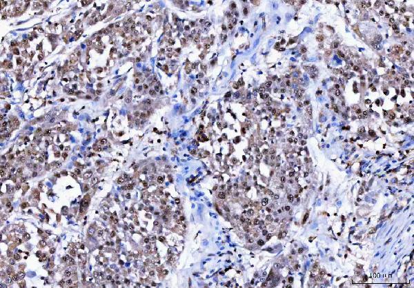
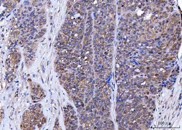
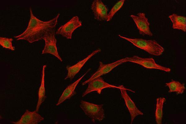


Comprehensive Human Protein Atlas IHC scoring per tissue (reliability: Enhanced — High consistency between antibody staining and RNA expression data.). Rows are taken directly from the HPA tissue chart — click any row's HPA link to view the source.
| Tissue | Cell type | Level | Evidence | Source |
|---|---|---|---|---|
| Lung | Alveolar cells type II | High | Protein (IHC) | HPA → |
| Placenta | Cytotrophoblasts | High | Protein (IHC) | HPA → |
| Testis | Spermatogonia cells | High | Protein (IHC) | HPA → |
| Adrenal gland | Glandular cells | Medium | Protein (IHC) | HPA → |
| Appendix | Glandular cells | Medium | Protein (IHC) | HPA → |
| Tissue | Cell type | Level | Evidence | Source |
|---|---|---|---|---|
| None in HPA: BAG6 is detected in all 45 scored tissues. Use a no-primary (secondary-only) and an isotype control instead. | ||||
Use the documented paraffin-section staining conditions as a starting point, then assess BAG6 signal by compartment, cell type and matched controls.
A00967-1 has pictured human paraffin-section IHC and PC3 cell IF data (catalog image captions); A00967-1 and M00967 list human, mouse, and rat reactivity (catalog reactivity fields).
The A00967-1 card renders with IHC data from a human lung cancer paraffin section (card image caption). Its catalog also documents IHC in human ovarian serous adenocarcinoma paraffin sections and IF in PC3 cells (catalog image captions).
Which to pick: For tissue IHC, choose A00967-1: its own human paraffin-section captions document EDTA retrieval at pH 8.0 and 2 μg/ml primary antibody; the fixative is unreported (catalog IHC image captions). For IF/ICC, A00967-1 has pictured PC3 cell IF at 5 μg/ml, while M00967 lists IF/ICC without a supplied image (catalog IF caption; M00967 application and image fields). For mouse or rat samples, both SKUs list reactivity, but the supplied IHC/IF images support human samples only; M00967 is a rabbit monoclonal, clone 25B57 (catalog reactivity, image and clone fields).