BCAM / Basal cell adhesion molecule · IHC design guide

Design Immunohistochemistry for BCAM

Plan BCAM paraffin IHC around membranous staining in basal membranes and endothelial cells (HPA tissue IHC). Use kidney Bowman's capsule as a high-staining reference (HPA tissue IHC) and start with 0.5–1 μg/mL catalog antibody (datasheet A03148-1).

Evidence assembled Oct 2026 · For research use; verify linked source records and product datasheet before use
Immunohistochemistry protocol sheet for BCAM (IHC for BCAM): expected localisation Membranous at basal membranes and endothelium (HPA tissue IHC), antibody A03148-1, validated IHC image, and IHC protocol steps
Printable BCAM IHC protocol sheet — expected localisation Membranous at basal membranes and endothelium (HPA tissue IHC), antibody A03148-1, controls and protocol steps. Open the full BCAM IHC guide →

BCAM Immunohistochemistry Experimental Design Guide

Expected localisation, validated protocols, controls and antibodies — the at-a-glance facts below, then the full design guide.

Must know before staining
Expected localisation Membranous at basal membranes and endothelium (HPA tissue IHC)
Staining pattern Basal membranes and endothelial cells stain membranously (HPA tissue IHC)
Antigen retrieval EDTA pH 8.0 HIER, heat-mediated (datasheet A03148-1)
Positive control ⓘ Cervix+4 more · see all
Negative control ⓘ None in HPA (detected in all 45 tissues); use no-primary + isotype controls
Important caveats
Reasons your staining may differ from the expected pattern.
Fixation Keep fixation conditions consistent across sections. (standard IHC practice; not target-specific)
Caveat Antibody staining and RNA show medium consistency (HPA tissue IHC)
Regulation Expression regulation not specified (UniProt)
Isoform / epitope 0 isoforms listed; epitope side matters (UniProt)
Section 1

Recommended BCAM IHC & IF Protocols

The catalog antibody protocol uses EDTA pH 8.0 retrieval (datasheet: A03148-1). The published BCAM IHC examples below cover prostate, periapical, ovarian and renal specimens (PMC12599034; PMC8883837; PMC9842900; PMC9361577).

Recommended immunohistochemistry (IHC-P) protocol parameters
SampleParaffin-embedded human thyroid cancer tissue; fixative not specified (datasheet A03148-1)
FixationImage fixative and duration unreported (datasheet A03148-1); verify before use.
Sectioning4–5 µm sections on charged slides (standard)
DeparaffinisationXylene, graded ethanol series to water (standard)
Antigen retrievalHeat retrieval: EDTA pH 8.0 (datasheet A03148-1); 20 min, 95–100 °C (standard)
Peroxidase block3% H2O2, 10 min, room temperature (standard)
Blocking10% goat serum (datasheet A03148-1)
Primary antibodyRabbit anti-BCAM, 0.5-1μg/ml (datasheet A03148-1)
Primary incubationOvernight at 4 °C (datasheet A03148-1)
DetectionStreptavidin-biotin complex (SABC), DAB chromogen (datasheet A03148-1)
CounterstainHematoxylin, blue, dehydrate and mount (standard)
Expected resultBCAM-positive staining in glandular cells of cervix (HPA tissue IHC: High). HPA tissue profile: Membranous expression in basal membranes and endothelial cells. No signal in the no-primary control.
💡Decision noteStart with heat-mediated EDTA pH 8.0 for the catalog antibody (datasheet: A03148-1); the published citrate and Trilogy conditions used other reported procedures (PMC8883837; PMC9842900).
Section 2

What Is the Expected BCAM Staining Pattern?

BCAM is a cell membrane protein with an extracellular region at residues 32–547 and a transmembrane segment at 548–568 (UniProt P50895 topology). In paraffin section IHC, expect membranous staining in basal epithelial structures and vascular endothelial cells (HPA tissue IHC: membranous profile). HPA rates the tissue staining evidence “Enhanced,” while reporting medium consistency between staining and RNA expression (HPA tissue IHC: reliability description).

What am I looking at on my slide?
Membranous staining outlines basal cells or vascular endothelial cells, with adjacent structures less prominent.This matches BCAM’s membrane topology (UniProt P50895 topology) and the basal membrane and endothelial pattern observed in tissue IHC (HPA tissue IHC: profile). Judge the cell and compartment together; overall section darkness alone does not establish a correct result.
A high signal appears in kidney Bowman’s capsule, nasopharyngeal basal cells or urinary bladder urothelial cells.These are reported high staining sites (HPA tissue IHC: Kidney, Nasopharynx, Urinary bladder). Their expected cellular distribution helps interpret a positive control; a high signal elsewhere needs its own morphological assessment.
Nuclear or broadly cytoplasmic staining dominates the IHC section without a convincing membrane pattern.That distribution does not match the reported tissue IHC profile (HPA tissue IHC: membranous expression) or membrane topology (UniProt P50895 topology). Treat it as suspect and review morphology, controls and detection background before assigning BCAM positivity.
Strong color appears in cell types outside the expected basal epithelial or endothelial pattern.The tissue IHC profile supports basal membranes and endothelial cells (HPA tissue IHC: profile). Unexpected staining can reflect cross-reactivity or endogenous detection activity (general IHC practice); it is not, by itself, proof of either cause.
A known high staining structure has no detectable signal.A blank kidney Bowman’s capsule or nasopharyngeal basal compartment conflicts with the reported high staining pattern (HPA tissue IHC: Kidney, Nasopharynx). Check section identity, antibody and detection controls, and the paraffin section workflow (general IHC practice) before interpreting other negatives.
💡Expected BCAM appearanceCall an IHC result positive when cell membranes stain in the expected basal epithelial or endothelial distribution (HPA tissue IHC: profile), with conspicuous signal in a reported high staining site (HPA tissue IHC: high sites); diffuse color without that cellular pattern is suspect (general IHC practice).
How each factor affects the staining
Topology and expected compartmentBCAM has an extracellular region, one transmembrane segment and a cytoplasmic tail (UniProt P50895 topology). This supports membrane localization as the primary IHC interpretation; topology alone does not establish how a particular antibody epitope behaves after processing.
Tissue and cell contextHigh staining is reported in several defined cell populations, including cervical glandular cells and skin sebaceous glands (HPA tissue IHC: Cervix, Skin). Low staining in selected neuronal, glial and endocrine cells (HPA tissue IHC: low sites) is a comparison, not a universal negative control.
Strength of tissue IHC evidenceThe listed antibody, HPA005654, has “Enhanced” IHC validation (HPA antibodies: HPA005654), while the tissue summary notes medium agreement between staining and RNA (HPA tissue IHC: reliability description). Use the reported distribution as a guide and scrutinize unannotated structures.
IF/ICC Q: should nucleolar signal change the IHC scoring rule?A: HPA reports an approved nucleoli fibrillar center location in ICC-IF (HPA subcellular: main location). Its tissue IHC profile is membranous (HPA tissue IHC: profile). Record these as application-specific observations; the ICC-IF result does not establish a nucleolar IHC positive pattern.
Why is my staining missing, weak or wrong?
SituationLikely causeNext action
No staining in an expected high staining structure.The result conflicts with a reported high site, such as kidney Bowman’s capsule (HPA tissue IHC: Kidney); the record does not identify which workflow step failed.Confirm the structure and tissue identity, then review antibody, antigen retrieval and detection controls for the paraffin section run (general IHC practice). Avoid assigning BCAM absence until a positive control works.
Color is spread across the section and obscures cell borders.Diffuse background can arise from nonspecific binding or detection chemistry (general IHC practice); it cannot be attributed to BCAM from this appearance alone.Compare reagent omission controls and review blocking, washing, antibody concentration and chromogen development (general IHC practice). Reassess whether a distinct membrane outline remains.
Nuclear or diffuse cytoplasmic color is the dominant pattern.That pattern disagrees with membrane localization (UniProt P50895 topology) and the membranous tissue IHC profile (HPA tissue IHC: profile).Check cellular morphology and background controls, then score only defensible membrane staining in the expected cells (general IHC practice; HPA tissue IHC: profile).
Strong staining appears in an unexpected cell population.Cross-reactivity or endogenous detection activity is possible (general IHC practice); HPA’s tissue profile alone cannot distinguish those explanations (HPA tissue IHC: profile).Inspect the no-primary control and the expected basal or endothelial structures on the same run (general IHC practice; HPA tissue IHC: profile). Report the unexpected population separately if it persists.
A low staining comparison tissue shows a strong signal.Low staining is reported for selected cerebellar Purkinje, hippocampal neuronal and endocrine glandular cells (HPA tissue IHC: low sites), but low does not mean absent.Verify the specific cell type and membrane distribution before calling a discrepancy; compare it with a high staining control processed in the same run (general IHC practice; HPA tissue IHC: high sites).
The tissue IHC result appears inconsistent with a nucleolar ICC-IF image.HPA assigns different observed locations to tissue IHC and ICC-IF (HPA tissue IHC: profile; HPA subcellular: main location). The supplied sources do not explain the difference.Interpret the paraffin section against its membranous tissue IHC profile (HPA tissue IHC: profile). Document the ICC-IF observation separately without using it to redefine an IHC positive result.

Sample controls for BCAM IHC & IF

🧪Run cervix first and look for staining in glandular cells (HPA: High in cervix glandular cells). HPA lists no negative tissue and detects BCAM in all 45 scored tissues, so no-primary and isotype controls carry the negative comparison; any unstained cells on the positive slide should show counterstain without specific chromogen, but no cell type is established here as BCAM-negative (HPA: no negative rows; HPA: BCAM detected in all 45 scored tissues).
Positive control tissue: Cervix (Glandular cells, HPA High)
Negative control tissue: None in HPA: BCAM is detected in all 45 scored tissues. Use a no-primary (secondary-only) and an isotype control instead.
ICC-IF cell lines (HPA subcellular resource): HPA ICC-IF images show BCAM in A-431, U-251MG, U2OS, with annotated localisation: Nucleoli fibrillar center (approved) (HPA subcellular).
Technical controls: Include a no-primary, secondary-only control and a concentration-matched nonimmune rabbit IgG control for the rabbit primary; a BCAM knockout specimen or a validated immunogen-peptide block can provide a biological specificity control (selected-SKU caption: rabbit primary; standard IHC practice). Quench endogenous peroxidase and check endogenous biotin background when using the caption’s biotinylated secondary, streptavidin–biotin detection and DAB (selected-SKU caption: biotinylated secondary, SABC and DAB).
⚠️Feasibility: A target-specific fixation window and fixation effect are unreported, and the selected-SKU paraffin-section caption does not state a fixative (selected-SKU caption: fixative not stated). The caption uses heat retrieval in EDTA at pH 8.0, but does not establish that retrieval is required; the supplied evidence does not establish whether frozen sections or IF would be easier (selected-SKU caption: EDTA retrieval). For IF, validate localisation carefully: UniProt places BCAM at the cell membrane, while HPA reports an approved nucleoli fibrillar-center ICC-IF signal (UniProt P50895: cell membrane; HPA subcellular: nucleoli fibrillar center).

HPA tissue IHC evidence for BCAM

Comprehensive Human Protein Atlas IHC scoring per tissue (reliability: Enhanced — Medium consistency between antibody staining and RNA expression data. Antibody staining in cells/structures not annotated, view images.). Rows are taken directly from the HPA tissue chart — click any row's HPA link to view the source.

Positive expression · recommended positive controls

TissueCell typeLevelEvidenceSource
Cervix Glandular cells High Protein (IHC) HPA →
Fallopian tube Non-ciliated cells High Protein (IHC) HPA →
Kidney Bowman's capsule High Protein (IHC) HPA →
Nasopharynx Basal cells High Protein (IHC) HPA →
Skin Sebaceous glands High Protein (IHC) HPA →

Undetected expression · recommended negative controls

TissueCell typeLevelEvidenceSource
None in HPA: BCAM is detected in all 45 scored tissues. Use a no-primary (secondary-only) and an isotype control instead.
Section 3

Advanced BCAM IHC Tips

These questions focus on BCAM staining in paraffin sections with chromogenic detection; the immunofluorescence entry addresses the separate IF/ICC application.

What retrieval conditions should I start with for BCAM in paraffin sections?
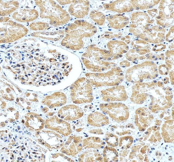
Use heat-mediated antigen retrieval in EDTA at pH 8.0 for BCAM paraffin-section IHC (datasheet A03148-1). The selected tissue image used that retrieval before overnight incubation with 1 μg/mL primary antibody at 4°C (image caption A03148-1). If staining is weak, first check that heating was uniform, sections stayed covered by buffer, and the same retrieval time was used across slides (standard IHC practice). Compare any longer heating or alternative buffer only on matched sections, because excessive retrieval can damage morphology and make membranous staining harder to judge (standard IHC practice; HPA: membranous expression).
How should I troubleshoot weak BCAM staining when fixation conditions vary?
Target-specific BCAM sensitivity to fixation is unknown from the supplied evidence; the selected paraffin-section caption does not state a fixative (image caption A03148-1). Record the fixative and fixation duration for each specimen, and compare staining only among sections processed in the same way (standard IHC practice). When staining varies, run matched sections through the documented EDTA pH 8.0 retrieval and identical antibody incubation before changing detection conditions (datasheet A03148-1; standard IHC practice). Inspect tissue preservation alongside signal: poor morphology, tissue loss, or uneven staining can confound an apparent loss of BCAM staining (standard IHC practice).
Where should convincing BCAM staining appear in a tissue section?
Expect predominantly membranous staining, especially along basal membranes and in endothelial cells (HPA: tissue IHC profile; UniProt P50895: cell membrane). BCAM has an extracellular region at residues 32–547, one transmembrane segment at 548–568, and a cytoplasmic region at 569–628 (UniProt P50895 topology). Use the hematoxylin counterstain and tissue architecture to decide whether chromogen follows cell borders rather than pooling over nuclei or debris (standard IHC practice). HPA reports a nucleolar fibrillar-center location in its separate subcellular dataset, so record nuclear staining separately and avoid treating it as established tissue-IHC localisation (HPA: subcellular; HPA: tissue IHC profile).
How can epitope position affect interpretation of BCAM IHC?
Check the antibody's stated immunogen or mapped epitope before interpreting an unusual pattern; the supplied A03148-1 caption does not identify its epitope (image caption A03148-1). An extracellular epitope would fall within residues 32–547, whereas a cytoplasmic epitope would fall within 569–628 (UniProt P50895 topology). Five annotated glycosylation sites lie in the extracellular region, and annotated phosphorylation sites lie in the cytoplasmic tail; these features make epitope accessibility a reasonable troubleshooting question, without establishing an effect for this antibody (UniProt P50895 modifications). The supplied record lists 0 isoforms, which provides no basis here for assigning distinct IHC patterns to specific BCAM isoforms (UniProt P50895 isoforms).
How should an IF follow-up test a surprising BCAM IHC pattern?
Use IF/ICC as a separate follow-up and compare its cell-border signal with the chromogenic IHC pattern; BCAM is annotated at the cell membrane (UniProt P50895 localisation). Multiplex BCAM with a validated marker for the expected basal or endothelial cell population, checking that the marker's staining pattern is established for the specimen being examined (HPA: tissue IHC profile; standard IF practice). Choose a fluorophore channel after checking tissue autofluorescence and include single-label controls when interpreting apparent overlap (standard IF practice). If the antibody epitope is extracellular, test surface access without permeabilisation; if it is cytoplasmic, test a permeabilised condition, since the A03148-1 epitope is unspecified here (UniProt P50895 topology; image caption A03148-1).
What should I check when BCAM chromogenic staining looks diffuse?
Compare the stained section with a no-primary control and inspect whether DAB appears over tissue edges, folds, debris, or broad extracellular areas (standard IHC practice). The selected image used 10% goat serum blocking, a biotinylated secondary, a streptavidin-biotin complex, and DAB, so each detection step deserves scrutiny when background rises (image caption A03148-1). Check peroxidase blocking and assess endogenous biotin if background persists with this detection chemistry; use matched controls to locate the contributing step (standard IHC practice; image caption A03148-1). True BCAM staining should be evaluated against its reported membranous basal and endothelial distribution, while retaining tissue-specific differences (HPA: tissue IHC profile).
How should I score BCAM staining across paraffin sections? ⚠ ANSWER MARKED FOR VERIFICATION
Define the scored compartment before measuring: the HPA tissue-IHC profile describes BCAM at basal membranes and endothelial cells (HPA: tissue IHC profile). Within a prespecified cell population, report the percentage of positive cells and staining intensity, or calculate an H-score using percentages at each intensity level (standard IHC practice). For vessel-associated staining, count positive structures per mm² of evaluable tissue and consider the proportion of vessels positive, rather than using raw DAB area alone (standard IHC practice). Keep retrieval, detection, counterstain, imaging, and threshold settings consistent, and normalise counts to evaluable tissue area or the relevant cell or vessel denominator (standard IHC practice).
How can I distinguish a true BCAM signal from artefact?
A plausible positive follows cell borders in the expected basal or endothelial distribution and remains interpretable against preserved tissue architecture (HPA: tissue IHC profile; standard IHC practice). High staining has been reported in nasopharyngeal basal cells and kidney Bowman's capsule, while thyroid glandular cells were reported low; these are context checks, not universal cutoffs (HPA: tissue IHC). Treat isolated nuclear deposits, strong section-edge staining, and signal concentrated in necrotic material as reasons to inspect controls and morphology before calling a positive (standard IHC practice). If DAB appears in a no-primary control, investigate endogenous enzyme activity or detection background before assigning the signal to BCAM (standard IHC practice).
Boster reagents

Best BCAM / Basal cell adhesion molecule IHC Antibodies

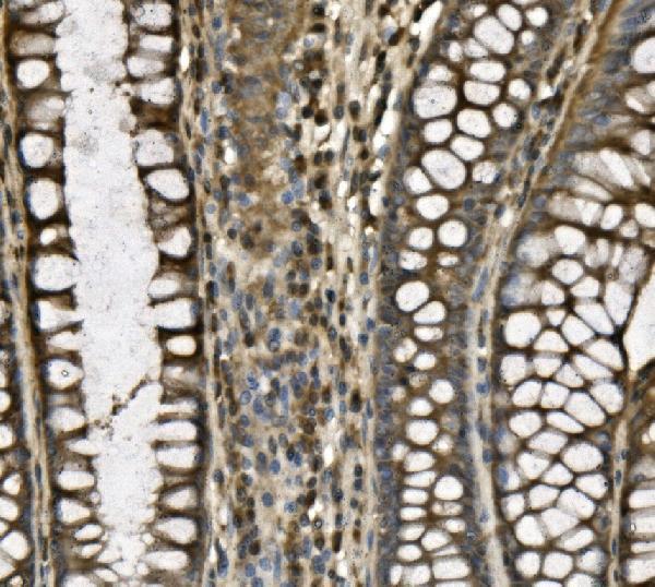
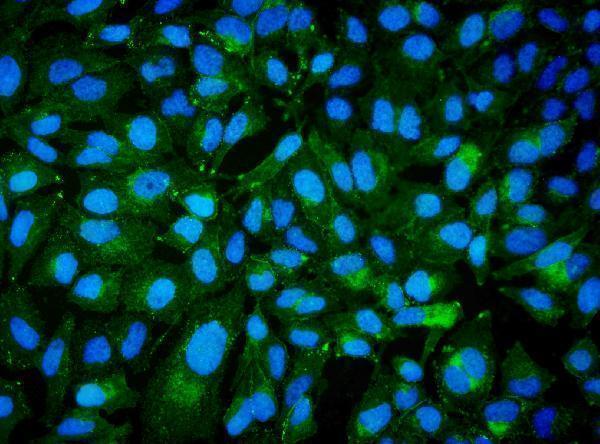
A03148-1 has pictured IHC in human paraffin sections and IF in HeLa cells (A03148-1 image captions); its listed reactivity includes human, mouse and rat (catalog: A03148-1 reactivity).

Real IHC data IHC analysis of CD239/BCAM using anti-CD239/BCAM antibody (A03148-1). CD239/BCAM was detected in paraffin-embedded section of human thyroid cancer tissue. Heat mediated antigen retrieval was performed in EDTA buffer (pH8.0, epitope retrieval solution). The tissue section was blocked with 10% goat serum. The tissue section was then incubated with 1μg/ml rabbit anti-CD239/BCAM Antibody (A03148-1) overnight at 4°C. Biotinylated goat anti-rabbit IgG was used as secondary antibody and incubated for 30 minutes at 37°C. The tissue section was developed using Strepavidin-Biotin-Complex (SABC) (Catalog # SA1022) with DAB as the chromogen.
Anti-CD239/BCAM Antibody ®
Cat # A03148-1

A03148-1 is the only SKU with an IHC figure for a rendered card: its captions show human thyroid cancer and kidney paraffin sections, plus IF in HeLa cells (A03148-1 image captions). M03148 lists IHC and human, mouse and rat reactivity, but has no supplied IHC or IF image (catalog: M03148 applications, reactivity and image captions).

Which to pick: For tissue IHC, choose A03148-1 when a pictured human paraffin-section example is useful; its captions do not report the fixative (A03148-1 IHC captions). For IF/ICC, choose A03148-1, which lists both applications and has a HeLa IF image (catalog: A03148-1 applications; A03148-1 IF caption). For mouse or rat work, both SKUs list reactivity, but the supplied IHC images document human tissue only; M03148 is the monoclonal option, clone 22B46 (catalog: A03148-1 and M03148 reactivity; A03148-1 IHC captions; catalog: M03148 clone).

Each figure is that product's own IHC / IF validation image from its datasheet.

References

  1. UniProt Consortium. UniProt entry P50895 (BCAM_HUMAN, Basal cell adhesion molecule).
  2. Human Protein Atlas. BCAM tissue IHC expression (reliability: Enhanced).
  3. Human Protein Atlas. BCAM subcellular location (ICC-IF): Localized to the nucleoli fibrillar center..
  4. Human Protein Atlas. BCAM antibody validation summary (1 antibodies).
  5. Unveiling prognostic biomarkers and immunotherapeutic insights in prostate cancer through multi-omics and machine learning. European journal of medical research 2025 — PMC12599034.
  6. Single-Cell Sequencing Unveils the Heterogeneity of Nonimmune Cells in Chronic Apical Periodontitis. Frontiers in cell and developmental biology 2021 — PMC8883837.
  7. Basal cell adhesion molecule promotes metastasis-associated processes in ovarian cancer. Clinical and translational medicine 2023 — PMC9842900.
  8. Integrated multi-omics analyses reveal that BCAM is associated with epigenetic modification and tumor microenvironment subtypes of clear cell renal cell carcinoma. Clinical epigenetics 2022 — PMC9361577.
  9. PubMed PMID:7777537 — UniProt-cited evidence.
  10. PubMed PMID:15057824 — UniProt-cited evidence.
  11. PubMed PMID:15489334 — UniProt-cited evidence.