BCKDK / Branched-chain alpha-ketoacid dehydrogenase kinase · IHC design guide

Design Immunohistochemistry for BCKDK

Plan chromogenic paraffin IHC for BCKDK using granular cytoplasmic staining as a tissue benchmark (HPA tissue IHC). Start with the catalog antibody’s 2–5 μg/mL IHC range (datasheet A07798-2), and compare colon glandular cells with skeletal muscle myocytes (HPA tissue IHC).

Evidence assembled Oct 2026 · For research use; verify linked source records and product datasheet before use
Immunohistochemistry protocol sheet for BCKDK (IHC for BCKDK): expected localisation Granular cytoplasm (HPA tissue IHC); mitochondrial matrix expected (UniProt), antibody A07798-2, validated IHC image, and IHC protocol steps
Printable BCKDK IHC protocol sheet — expected localisation Granular cytoplasm (HPA tissue IHC); mitochondrial matrix expected (UniProt), antibody A07798-2, controls and protocol steps. Open the full BCKDK IHC guide →

BCKDK Immunohistochemistry Experimental Design Guide

Expected localisation, validated protocols, controls and antibodies — the at-a-glance facts below, then the full design guide.

Must know before staining
Expected localisation Granular cytoplasm (HPA tissue IHC); mitochondrial matrix expected (UniProt)
Staining pattern Granular cytoplasm in glandular and respiratory epithelial cells (HPA tissue IHC)
Antigen retrieval EDTA pH 8.0 HIER, heat-mediated (datasheet A07798-2)
Positive control ⓘ Adrenal gland+4 more · see all
Negative control ⓘ Adipose tissue+4 more · see all
Important caveats
Reasons your staining may differ from the expected pattern.
Fixation Keep fixation consistent across sections. (standard IHC practice; not target-specific)
Caveat Skeletal muscle myocytes may show no staining (HPA tissue IHC)
Regulation Refeeding raises the BCKDK:PPM1K ratio (UniProt)
Isoform / epitope 3 isoforms; confirm epitope coverage of the mature chain, aa 31–412 (UniProt)
Section 1

Recommended BCKDK IHC & IF Protocols

The catalog antibody has an IHC-P protocol (datasheet A07798-2). Published BCKDK IHC methods from three PMC articles provide additional staining conditions (PMC10637060; PMC6506390; PMC11336342).

Recommended immunohistochemistry (IHC-P) protocol parameters
SampleParaffin-embedded human gall bladder adenosquamous carcinoma tissue; fixative not specified (datasheet A07798-2)
FixationImage fixative and duration unreported (datasheet A07798-2); verify before use.
Sectioning4–5 µm sections on charged slides (standard)
DeparaffinisationXylene, graded ethanol series to water (standard)
Antigen retrievalHeat retrieval: EDTA pH 8.0 (datasheet A07798-2); 20 min, 95–100 °C (standard)
Peroxidase block3% H2O2, 10 min, room temperature (standard)
Blocking10% goat serum (datasheet A07798-2)
Primary antibodyRabbit anti-BCKDK, 2-5 μg/ml (datasheet A07798-2)
Primary incubationOvernight at 4 °C (datasheet A07798-2)
DetectionStreptavidin-biotin complex (SABC), DAB chromogen (datasheet A07798-2)
CounterstainHematoxylin, blue, dehydrate and mount (standard)
Expected resultBCKDK-positive staining in glandular cells of adrenal gland (HPA tissue IHC: High). HPA tissue profile: General cytoplasmic expression with a granular pattern. No signal in the no-primary control.
💡Decision noteStart with heat-mediated EDTA pH 8.0 retrieval for the catalog antibody (datasheet A07798-2); citrate retrieval is a published alternative for liver sections (PMC6506390).
Section 2

What Is the Expected BCKDK Staining Pattern?

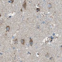
BCKDK is primarily a mitochondrial matrix protein with no transmembrane segment (UniProt O14874: localization and topology). In paraffin-section IHC, expect granular cytoplasmic staining, particularly in colon and duodenal glandular cells, pancreatic exocrine cells, respiratory epithelium and lung macrophages (HPA: tissue IHC). HPA rates its tissue pattern Supported, with medium consistency between staining and RNA data (HPA: reliability). UniProt also reports BCKDK in the cytosolic compartment of liver cells (UniProt O14874: localization).

What am I looking at on my slide?
Granular cytoplasmic staining in colon glandular cells or lung macrophages (HPA: tissue IHC).This fits HPA's general granular cytoplasmic profile and High staining in those cells (HPA: tissue IHC). Interpret the granules as compatible with mitochondrial localization, while recognizing that chromogenic IHC alone does not resolve individual mitochondria (UniProt O14874: matrix localization; general IHC practice).
Predominantly nuclear or cell-surface staining, with little cytoplasmic signal.This conflicts with the mitochondrial matrix assignment and absence of a transmembrane segment (UniProt O14874: localization and topology). Reassess specificity and detection background before calling it BCKDK; a less granular cytosolic pattern in liver cells is a documented exception (UniProt O14874: liver localization).
Strong signal in adipocytes or cardiomyocytes while expected positive cells are unstained.HPA reports BCKDK as Not detected in adipocytes and cardiomyocytes, but High in several glandular cell groups and lung macrophages (HPA: tissue IHC). Consider cross-reactivity or endogenous chromogenic activity, then compare cell identity and controls; an isolated unexpected stain does not establish BCKDK expression (general IHC practice).
Uniform haze across cells, stroma and cell-free areas obscures cytoplasmic granules.HPA describes a granular cytoplasmic pattern rather than uniform field-wide staining (HPA: tissue IHC). Diffuse deposition may reflect nonspecific detection or inadequate washing; inspect a no-primary control and the tissue architecture before scoring cellular signal (general IHC practice).
No stain in colon glandular cells or lung macrophages on a technically intact section.Both are reported High and can serve as reference positive cell populations, although HPA's Supported rating has only medium staining–RNA consistency (HPA: tissue IHC and reliability). Check run controls and section quality before interpreting the sample as biologically negative (general IHC practice).
💡Expected BCKDK appearanceCall a result consistent with BCKDK when High granular cytoplasmic staining occurs in HPA-positive glandular cells or lung macrophages; dominant nuclear, cell-surface or tissue-wide haze warrants review as possible false signal (HPA: tissue IHC; UniProt O14874: localization; general IHC practice).
How each factor affects the staining
Reference cell populationChoose identifiable colon or duodenal glandular cells, pancreatic exocrine cells, respiratory epithelium, or lung macrophages reported High by HPA; compare the specified cells rather than treating a whole organ as uniformly positive (HPA: tissue IHC).
Subcellular interpretationThe mitochondrial matrix assignment and lack of a transmembrane segment favor intracellular granular signal; UniProt's reported cytosolic liver compartment prevents treating every less granular liver-cell pattern as automatically false (UniProt O14874: localization and topology).
Antibody evidenceHPA017995 has Supported IHC validation, and the tissue-level assessment is Supported with medium staining–RNA consistency; these ratings support pattern comparison without proving specificity in every specimen (HPA: antibody validation and reliability).
Processing and isoformsUniProt lists a mature chain at residues 31–412 and 3 isoforms (UniProt O14874: processing and isoforms). Without an epitope map, these facts do not establish which forms a particular IHC antibody detects (general interpretation).
IF/ICC: what should be seen?A mitochondrial signal is the supported IF/ICC expectation; HPA lists A-431, U-251MG and U2OS images and rates HPA056067 Supported for ICC (HPA: subcellular and antibody validation). This observation does not supply an IHC protocol or establish that both antibodies perform identically (HPA: antibody validation).
Why is my staining missing, weak or wrong?
SituationLikely causeNext action
Positive reference cells show no chromogenic signal.An unsuccessful IHC run or unsuitable detection conditions may leave even HPA High cells unstained (HPA: tissue IHC; general IHC practice).Verify that the run control stained, confirm the antibody and detection reagents were applied, and review retrieval and reagent conditions using the IHC-validated antibody's instructions (general IHC practice).
Signal appears mainly in nuclei or along plasma membranes.That distribution disagrees with mitochondrial matrix localization and no transmembrane segment (UniProt O14874: localization and topology).Compare with a known positive cell population and a no-primary control; review morphology and detection background before assigning BCKDK positivity (HPA: tissue IHC; general IHC practice).
Adipocytes or muscle cells stain strongly.HPA reports Not detected in adipocytes, cardiomyocytes and skeletal myocytes (HPA: tissue IHC); nonspecific binding or endogenous detection activity is possible (general IHC practice).Identify the stained cells, inspect a no-primary control and apply the appropriate endogenous-activity control for the chromogenic system; retain any unexpected biological finding as unconfirmed (general IHC practice).
Diffuse brown haze masks the granular pattern.Excess background from detection or insufficient washing can obscure a cellular pattern (general IHC practice); HPA describes general granular cytoplasmic expression (HPA: tissue IHC).Review a no-primary section, washing and blocking; score only clearly cellular signal with interpretable morphology (general IHC practice).
A weak signal occurs in a presumed negative area.HPA's Not detected entries apply to named cell types, and its tissue reliability has medium staining–RNA consistency (HPA: tissue IHC and reliability).Compare the exact cell population, nearby positive cells and controls before calling the area positive or negative; record weak or ambiguous staining separately (HPA: tissue IHC; general IHC practice).
A liver-cell stain looks diffuse rather than punctate.UniProt reports BCKDK in the cytosolic compartment of liver cells as well as in the mitochondrial matrix (UniProt O14874: localization).Evaluate cellular localization and controls together; do not reject the liver pattern solely for lacking granules, and do not infer specificity from diffuse staining alone (UniProt O14874: liver localization; general IHC practice).

Sample controls for BCKDK IHC & IF

🧪Run adrenal gland first: glandular cells should stain (HPA: High in adrenal gland glandular cells). Use adipose tissue as a negative comparator (HPA: Not detected in adipocytes), and assess adjacent nonglandular cells on the adrenal slide as an internal background reference without assuming they are BCKDK-negative.
Positive control tissue: Adrenal gland (Glandular cells, HPA High)
Negative control tissue: Adipose tissue (HPA Not detected)
ICC-IF cell lines (HPA subcellular resource): HPA ICC-IF images show BCKDK in A-431, U-251MG, U2OS, with annotated localisation: Mitochondria (supported) (HPA subcellular).
Technical controls: Include a no-primary (secondary-only) control, a nonimmune IgG control matched to the primary antibody’s host species and isotype, and a BCKDK knockout sample or validated peptide-block control. Check endogenous peroxidase activity and endogenous biotin background because the reported chromogenic method uses a biotinylated secondary antibody and streptavidin–biotin detection (selected-SKU IHC caption: A07798-2).
⚠️Feasibility: The selected-SKU paraffin-section caption does not report a fixative, and no target-specific fixation window or fixation effect is reported in the supplied evidence (selected-SKU IHC caption: fixative not stated). The reported IHC example uses heat retrieval in EDTA at pH 8.0; whether BCKDK staining requires that retrieval condition is unreported (selected-SKU IHC caption: A07798-2). Frozen-section performance and relative ease of IF/ICC are unreported; mitochondrial localisation provides an IF/ICC pattern to assess (HPA subcellular: mitochondria, supported).

HPA tissue IHC evidence for BCKDK

Comprehensive Human Protein Atlas IHC scoring per tissue (reliability: Supported — Medium consistency between antibody staining and RNA expression data.). Rows are taken directly from the HPA tissue chart — click any row's HPA link to view the source.

Positive expression · recommended positive controls

TissueCell typeLevelEvidenceSource
Adrenal gland Glandular cells High Protein (IHC) HPA →
Bronchus Respiratory epithelial cells High Protein (IHC) HPA →
Colon Glandular cells High Protein (IHC) HPA →
Duodenum Glandular cells High Protein (IHC) HPA →
Endometrium Glandular cells High Protein (IHC) HPA →

Undetected expression · recommended negative controls

TissueCell typeLevelEvidenceSource
Adipose tissue Adipocytes Not detected Protein (IHC) HPA →
Heart muscle Cardiomyocytes Not detected Protein (IHC) HPA →
Ovary Ovarian stroma cells Not detected Protein (IHC) HPA →
Skeletal muscle Myocytes Not detected Protein (IHC) HPA →
Smooth muscle Smooth muscle cells Not detected Protein (IHC) HPA →
Section 3

Advanced BCKDK IHC Tips

Troubleshoot BCKDK staining by comparing the antibody’s documented paraffin-section conditions with expected cellular and subcellular patterns.

What retrieval conditions should I try first for weak BCKDK staining in paraffin sections?
Start with heat-mediated retrieval in EDTA at pH 8.0 for paraffin sections (datasheet A07798-2). The selected image pairs that retrieval with 2 μg/ml primary antibody overnight at 4°C; keep those conditions together for the first comparison (datasheet A07798-2). Include a no-primary control and a section containing an expected positive cell population, such as colon glandular cells (HPA: High in colon glandular cells; standard IHC practice). If signal remains weak, compare heating durations while keeping the buffer constant and checking tissue morphology (standard IHC practice). Trial another retrieval buffer only as a fallback, with otherwise matched sections and detection conditions (standard IHC practice).
Could fixation explain weak or uneven BCKDK staining?
The selected paraffin-section caption does not state its fixative, so BCKDK-specific fixation sensitivity is unknown for this antibody (datasheet A07798-2). Record the actual fixative, fixation duration, tissue thickness and processing history before comparing sections (standard IHC practice). If material permits, stain sections from differently processed blocks together using EDTA at pH 8.0 and the same antibody incubation (datasheet A07798-2; standard IHC practice). Evaluate preservation of morphology alongside staining, since damaged or unevenly processed regions can distort chromogenic interpretation (standard IHC practice). Do not infer a BCKDK-specific fixation effect from tissue expression patterns or protein features alone (HPA tissue IHC; UniProt O14874).
Where should convincing BCKDK signal appear in a chromogenic section?
Look for granular cytoplasmic staining: tissue IHC reports that pattern, while subcellular evidence supports mitochondrial localisation (HPA tissue IHC; HPA subcellular). BCKDK is annotated in the mitochondrial matrix and has no transmembrane segment, so a crisp plasma-membrane outline would be unexpected (UniProt O14874). Interpret staining within identified cells; lung macrophages and colon glandular cells are reported as High, whereas cardiomyocytes are reported as Not detected (HPA tissue IHC). Liver cells require particular care because cytosolic BCKDK has also been detected there (UniProt O14874). Compare serial sections or suitable cell-type markers when morphology alone cannot identify the stained population (standard IHC practice).
Could isoforms or epitope processing change what this antibody detects?
BCKDK has 3 annotated isoforms, and its processed chain spans residues 31–412 (UniProt O14874). The supplied antibody caption does not map its epitope, so these facts alone cannot establish isoform coverage or recognition of the processed protein (datasheet A07798-2; UniProt O14874). Annotated modifications include phosphoserine at residue 31 and acetyllysines at residues 192 and 233, but their effects on this stain are unknown (UniProt O14874). If samples disagree, check the antibody’s immunogen or epitope documentation before attributing the difference to an isoform or modification (standard IHC practice). Keep retrieval and detection conditions matched while evaluating that possibility (standard IHC practice).
How can I investigate the chromogenic pattern with multiplex immunofluorescence?
For a separate IF experiment, pair BCKDK with a marker identifying the cell population under study, such as lung macrophages, and assess signal within those cells (HPA: High in lung macrophages; standard IF practice). Choose fluorophores after inspecting unstained tissue for autofluorescence; a spectrally separated far-red channel may improve discrimination where shorter wavelengths are bright (standard IF practice). Mitochondrial matrix localisation means intracellular access matters, but the supplied antibody epitope is unmapped (UniProt O14874; datasheet A07798-2). Optimize permeabilisation for the antibody and specimen rather than assuming access from a surface-staining workflow (standard IF practice). Compare the IF distribution with the granular cytoplasmic IHC pattern, using appropriate single-stain controls (HPA tissue IHC; standard IF practice).
How do I distinguish BCKDK staining from diffuse brown background?
The selected chromogenic example used 10% goat-serum blocking and a biotinylated secondary with streptavidin–biotin detection and DAB (datasheet A07798-2). Include a no-primary section to reveal signal from detection reagents, endogenous enzyme activity or nonspecific deposition (standard IHC practice). Apply an appropriate peroxidase block for DAB detection and assess endogenous biotin interference when using a biotin-based system (standard IHC practice). Compare background in reported negative populations, such as adipocytes or cardiomyocytes, with granular cytoplasmic staining in an expected positive population (HPA tissue IHC). If background persists, titrate the primary around the documented 2 μg/ml condition while holding retrieval and development constant (datasheet A07798-2; standard IHC practice).
How should I score BCKDK across sections with different cell populations? ⚠ ANSWER MARKED FOR VERIFICATION
Define the cell population and viable tissue region before scoring, then apply the same definitions to every section (standard IHC practice). Report the percentage of positive cells and intensity categories, or calculate an H-score by summing each intensity category multiplied by its percentage, on a 0–300 scale (standard IHC practice). Normalize counts to the number of eligible cells; for spatial density, report positive cells per mm² of annotated viable tissue (standard IHC practice). Keep macrophages and glandular cells in separate scoring strata because both can show High staining in reported tissues (HPA tissue IHC). Exclude folds, edges and necrotic areas using consistent rules (standard IHC practice).
Which findings support a true BCKDK-positive result rather than an artefact?
A credible result shows granular cytoplasmic staining in an identifiable expected cell population, consistent with the reported tissue pattern and mitochondrial localisation (HPA tissue IHC; HPA subcellular). High staining is reported in lung macrophages and colon glandular cells, while cardiomyocytes are reported as Not detected (HPA tissue IHC). Predominantly nuclear or plasma-membrane staining warrants scrutiny because BCKDK is annotated to the mitochondrial matrix without a transmembrane segment (UniProt O14874). Check whether apparent positives cluster at section edges, folds or necrosis, and whether they persist in a no-primary control (standard IHC practice). With DAB and biotin-based detection, investigate endogenous peroxidase or biotin as possible signal sources (datasheet A07798-2; standard IHC practice).
Boster reagents

Best BCKDK / Branched-chain alpha-ketoacid dehydrogenase kinase IHC Antibodies

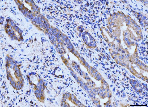
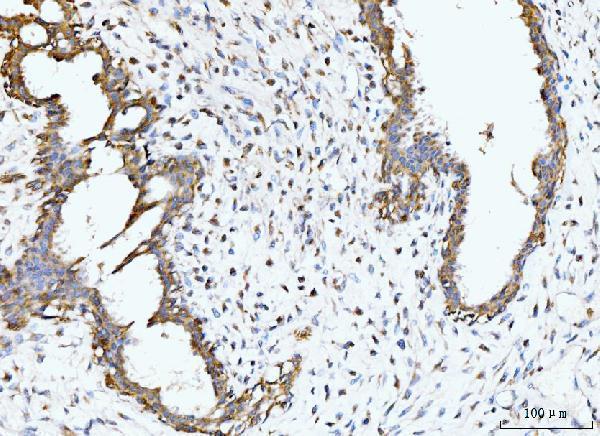
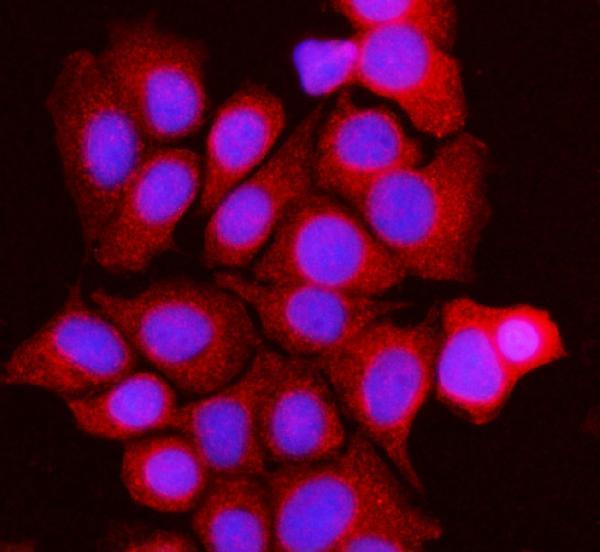
A07798-2 has IHC images from human paraffin-embedded carcinoma sections and an IF image from MCF-7 cells (catalog image captions). Human, Mouse, and Rat reactivity is listed (catalog reactivity).

Real IHC data IHC analysis of BCKDK using anti-BCKDK antibody (A07798-2). BCKDK was detected in a paraffin-embedded section of human gall bladder adenosquamous carcinoma tissue. Heat mediated antigen retrieval was performed in EDTA buffer (pH 8.0, epitope retrieval solution). The tissue section was blocked with 10% goat serum. The tissue section was then incubated with 2 μg/ml rabbit anti-BCKDK Antibody (A07798-2) overnight at 4°C. Biotinylated goat anti-rabbit IgG was used as secondary antibody and incubated for 30 minutes at 37°C. The tissue section was developed using Strepavidin-Biotin-Complex (SABC) (Catalog # SA1022) with DAB as the chromogen.
Anti-BCKDK Antibody ®
Cat # A07798-2

A07798-2 has IHC images from human gall bladder adenosquamous carcinoma, breast cancer, colonic adenocarcinoma, and gastric adenocarcinoma paraffin sections (A07798-2 IHC captions). Its IF image shows MCF-7 cells (A07798-2 IF caption), and its listed applications include IHC, ICC, and IF (catalog applications).

Which to pick: For tissue IHC, choose A07798-2: its paraffin-section caption documents EDTA retrieval at pH 8.0 and 2 μg/ml primary antibody; the fixative is unreported (A07798-2 IHC caption). For IF/ICC, A07798-2 has an MCF-7 IF image using 5 μg/ml primary antibody (A07798-2 IF caption). For cross-species work, A07798-2 lists Human, Mouse, and Rat reactivity, while its supplied IHC and IF images show human samples (catalog reactivity; A07798-2 image captions).

Each figure is that product's own IHC / IF validation image from its datasheet.

References

  1. UniProt Consortium. UniProt entry O14874 (BCKD_HUMAN, Branched-chain alpha-ketoacid dehydrogenase kinase).
  2. Human Protein Atlas. BCKDK tissue IHC expression (reliability: Supported).
  3. Human Protein Atlas. BCKDK subcellular location (ICC-IF): Localized to the mitochondria..
  4. Human Protein Atlas. BCKDK antibody validation summary (2 antibodies).
  5. Branched-chain keto-acid dehydrogenase kinase regulates vascular permeability and angiogenesis to facilitate tumor metastasis in renal cell carcinoma. Cancer science 2023 — PMC10637060.
  6. Loss of BCAA Catabolism during Carcinogenesis Enhances mTORC1 Activity and Promotes Tumor Development and Progression. Cell metabolism 2019 — PMC6506390.
  7. MAZ-mediated up-regulation of BCKDK reprograms glucose metabolism and promotes growth by regulating glucose-6-phosphate dehydrogenase stability in triple-negative breast cancer. Cell death & disease 2024 — PMC11258276.
  8. FYN-mediated phosphorylation of BCKDK at Y151 promotes GBM proliferation by increasing the oncogenic metabolite N-acetyl-L-alanine. Heliyon 2024 — PMC11336342.
  9. PubMed PMID:14702039 — UniProt-cited evidence.
  10. PubMed PMID:15616553 — UniProt-cited evidence.
  11. PubMed PMID:15489334 — UniProt-cited evidence.