This website uses cookies to ensure you get the best experience on our website.


- Table of Contents
Plan BCL2L13 chromogenic IHC using its selective cytoplasmic tissue profile (HPA tissue IHC). Compare staining with appropriate tissue controls and interpret weak signals cautiously because staining reliability is uncertain (HPA tissue IHC).

Expected localisation, validated protocols, controls and antibodies — the at-a-glance facts below, then the full design guide.
| Expected localisation | Selective cytoplasmic tissue staining (HPA tissue IHC) | |
| Staining pattern | Cytoplasmic staining in glands, hepatocytes and muscle (HPA tissue IHC) | |
| Antigen retrieval | Tris-EDTA pH 9.0 HIER, 95–98 °C, 20 min (rule: nuclear antigen) | |
| Positive control | Bone marrow+4 more · see all | |
| Negative control | Adipose tissue+4 more · see all |
| Fixation | Keep fixation consistent across paraffin sections. (standard IHC practice; not target-specific) | |
| Caveat | Staining reliability is uncertain (HPA tissue IHC) | |
| Regulation | Highest expression in heart, placenta and pancreas (UniProt) | |
| Isoform / epitope | 3 isoforms; check epitope coverage across variants (UniProt) |
The catalog antibody protocol is followed by two published BCL2L13 IHC protocols using paraffin sections and chromogenic detection (PMC10475114; PMC8247248).
| Sample | Tissue sections; selected-image fixative not specified (standard IHC workflow) |
| Fixation | Image fixative and duration unreported (datasheet A06151); verify before use. |
| Sectioning | 4–5 µm sections on charged slides (standard) |
| Deparaffinisation | Xylene, graded ethanol series to water (standard) |
| Antigen retrieval | Heat-induced epitope retrieval in Tris-EDTA buffer, pH 9.0, 20 min at 95–98 °C (standard rule: nuclear antigen) |
| Peroxidase block | 3% H2O2, 10 min, room temperature (standard) |
| Blocking | 10% normal serum of the secondary host, 30 min, room temperature (standard) |
| Primary antibody | Rabbit anti-BCL2L13, 10 μg/mL (datasheet A06151) |
| Primary incubation | Overnight at 4 °C (standard) |
| Detection | HRP-polymer secondary, DAB chromogen 5–10 min (standard) |
| Counterstain | Hematoxylin, blue, dehydrate and mount (standard) |
| Expected result | BCL2L13-positive staining in hematopoietic cells of bone marrow (HPA tissue IHC: Medium). HPA tissue profile: Selective cytoplasmic expression in salivary glands, exocrine pancreas, hepatocytes, muscle tissues and a subsets of lymphoid tissues. No signal in the no-primary control. |
BCL2L13 is annotated at the mitochondrial membrane, with a transmembrane segment at residues 460–480; UniProt also lists the nucleus (UniProt Q9BXK5 topology and subcellular location). In tissue IHC, expect selective cytoplasmic staining, including in exocrine pancreas, hepatocytes, muscle tissues and subsets of lymphoid tissues (HPA: tissue IHC profile). Treat that pattern as provisional: HPA rates tissue IHC reliability Uncertain because staining and RNA expression have medium consistency (HPA: reliability).
| Selective cytoplasmic staining in cardiomyocytes or skeletal myocytes, with adjacent cells differing in intensity. | This fits the reported tissue distribution: both cell types show Medium staining (HPA: heart muscle cardiomyocytes; skeletal muscle myocytes). Evaluate cell identity and compartment together; intensity alone does not establish specificity, especially with the Uncertain tissue IHC rating (HPA: reliability). |
| Predominantly extracellular staining, or a uniform deposit over tissue regardless of cell boundaries. | This does not fit the selective cytoplasmic tissue pattern (HPA: tissue IHC profile). Check section quality, chromogen deposition and wash performance as general IHC practice. Nuclear staining needs separate review: UniProt also annotates the nucleus, while HPA's tissue IHC profile emphasizes cytoplasm (UniProt Q9BXK5 subcellular location; HPA: tissue IHC profile). |
| Strong signal in adipocytes or bronchial respiratory epithelium while the expected cytoplasmic cell pattern is absent. | Both cell populations are reported Not detected in the supplied tissue IHC record (HPA: adipose tissue adipocytes; bronchus respiratory epithelial cells). Consider antibody cross-reactivity or endogenous detection activity; compare primary-omission and detection controls as general IHC practice before assigning the signal to BCL2L13. |
| Low-level color covers many cell types and tissue spaces, obscuring cell borders. | Diffuse background prevents a defensible cell-level call against HPA's selective cytoplasmic profile (HPA: tissue IHC profile). Review blocking, washing, detection exposure and section handling as general IHC practice. Do not interpret a broad haze as evidence of ubiquitous protein staining simply because UniProt describes broad tissue expression (UniProt Q9BXK5 tissue specificity). |
| No signal in heart-muscle cardiomyocytes under conditions that stain other targets. | HPA reports Medium cardiomyocyte staining, making this a useful comparison tissue, but its BCL2L13 IHC evidence remains Uncertain (HPA: heart muscle cardiomyocytes; reliability). Check the assay's positive control and detection steps first; a negative result alone cannot prove BCL2L13 is absent (general IHC practice). |
| Tissue and cell selection | HPA reports selective cytoplasmic staining in salivary glands, exocrine pancreas, hepatocytes, muscle tissues and subsets of lymphoid tissues, despite low tissue RNA specificity (HPA: tissue IHC profile; RNA specificity). Choose comparison regions by identified cell type; whole-section averages can conceal that selectivity (general IHC practice). |
| Membrane topology and compartment | The 460–480 transmembrane segment supports a membrane-associated mitochondrial expectation (UniProt Q9BXK5 topology and subcellular location). HPA's tissue IHC description is cytoplasmic, without resolving mitochondria in paraffin sections (HPA: tissue IHC profile). Do not require visible mitochondrial puncta to score chromogenic IHC. |
| Isoforms and epitope coverage | UniProt lists 3 isoforms and one annotated chain spanning residues 1–485 (UniProt Q9BXK5 isoforms and processing). The payload gives no antibody epitope, isoform-specific staining or tissue-level processing result; check the antibody's epitope documentation before making any isoform claim. |
| Strength of tissue IHC evidence | Both antibodies with a reported IHC status here, HPA030994 and CAB002052, are rated Uncertain (HPA: antibody validation). HPA describes medium consistency between staining and RNA data (HPA: reliability). Use morphology and independent controls when interpreting a weak or unexpected stain (general IHC practice). |
| IF/ICC Q&A: should puncta be expected? | Yes for IF/ICC interpretation: HPA reports enhanced mitochondrial localization and lists approved ICC evidence for HPA030994 and HPA050377 (HPA: subcellular summary; antibody validation). That supports a mitochondrial IF expectation; it does not upgrade the Uncertain tissue IHC rating or set an IHC protocol (HPA: reliability). |
| Situation | Likely cause | Next action |
|---|---|---|
| Cardiomyocytes remain blank in a completed chromogenic run. | The reported comparison population has Medium staining, but IHC reliability is Uncertain; a blank slide does not identify the failed step (HPA: heart muscle cardiomyocytes; reliability). | Confirm tissue identity, a working detection control, primary incubation and reagent condition; then review retrieval against the antibody's IHC instructions (general IHC practice). Do not assign a BCL2L13-specific retrieval requirement from these sources. |
| Color appears across the slide rather than within selected cells. | The distribution conflicts with selective cytoplasmic tissue staining (HPA: tissue IHC profile); excess detection or incomplete washing can produce background (general IHC practice). | Compare a primary-omission control; review blocking, washes and chromogen development (general IHC practice). Reassess only signal that follows identifiable cells and compartments (HPA: tissue IHC profile). |
| Adipocytes or bronchial respiratory epithelial cells stain strongly. | Those populations are reported Not detected (HPA: adipose tissue adipocytes; bronchus respiratory epithelial cells). Cross-reactivity or endogenous detection activity is possible (general IHC practice). | Inspect a primary-omission control and the detection system's endogenous-activity control; compare with a cell population reported positive in the same run (general IHC practice; HPA: heart muscle cardiomyocytes). |
| A strong nuclear-only pattern appears without selective cytoplasmic staining. | UniProt annotates the nucleus, so nuclear signal cannot be dismissed categorically; HPA's tissue IHC profile instead describes selective cytoplasmic expression (UniProt Q9BXK5 subcellular location; HPA: tissue IHC profile). | Check whether nuclear staining follows plausible cell types and whether controls also stain nuclei (general IHC practice). Report the discrepancy explicitly; do not call nuclear-only tissue staining validated by HPA. |
| Signal is weaker in one tissue region than another. | HPA describes selective expression and reports different cell populations at Medium, Low or Not detected levels (HPA: tissue IHC profile; tissue cell-level entries). Region-wide comparison can mix distinct cell types. | Compare the same identified cell type across matched regions and score localization alongside intensity (general IHC practice). Use HPA's cell-level entries as context, while retaining its Uncertain reliability caveat (HPA: reliability). |
| IF/ICC shows mitochondrial signal, but chromogenic IHC looks broadly cytoplasmic. | HPA reports enhanced mitochondrial localization in ICC-IF and selective cytoplasmic staining in tissue IHC (HPA: subcellular summary; tissue IHC profile). The two observations have different spatial resolution (general microscopy practice). | Interpret each image within its application: assess cell-level cytoplasmic selectivity for tissue IHC and mitochondrial localization for IF/ICC (HPA: tissue IHC profile; subcellular summary). Keep the tissue IHC Uncertain rating in the report (HPA: reliability). |


Comprehensive Human Protein Atlas IHC scoring per tissue (reliability: Uncertain — Medium consistency between antibody staining and RNA expression data.). Rows are taken directly from the HPA tissue chart — click any row's HPA link to view the source.
| Tissue | Cell type | Level | Evidence | Source |
|---|---|---|---|---|
| Bone marrow | Hematopoietic cells | Medium | Protein (IHC) | HPA → |
| Breast | Myoepithelial cells | Medium | Protein (IHC) | HPA → |
| Cerebral cortex | Glial cells | Medium | Protein (IHC) | HPA → |
| Heart muscle | Cardiomyocytes | Medium | Protein (IHC) | HPA → |
| Lymph node | Non-germinal center cells | Medium | Protein (IHC) | HPA → |
| Tissue | Cell type | Level | Evidence | Source |
|---|---|---|---|---|
| Adipose tissue | Adipocytes | Not detected | Protein (IHC) | HPA → |
| Adrenal gland | Glandular cells | Not detected | Protein (IHC) | HPA → |
| Bronchus | Respiratory epithelial cells | Not detected | Protein (IHC) | HPA → |
| Caudate | Glial cells | Not detected | Protein (IHC) | HPA → |
| Cerebellum | Cells in granular layer | Not detected | Protein (IHC) | HPA → |
Troubleshoot BCL2L13 staining in paraffin sections by checking retrieval, controls, subcellular pattern and scoring before interpreting chromogenic signal.
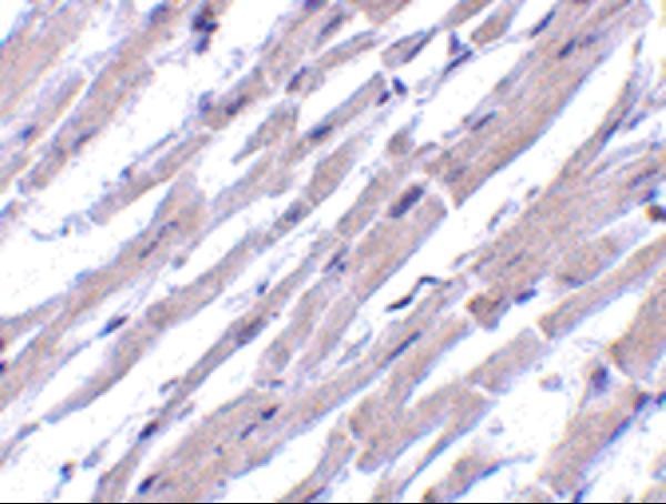
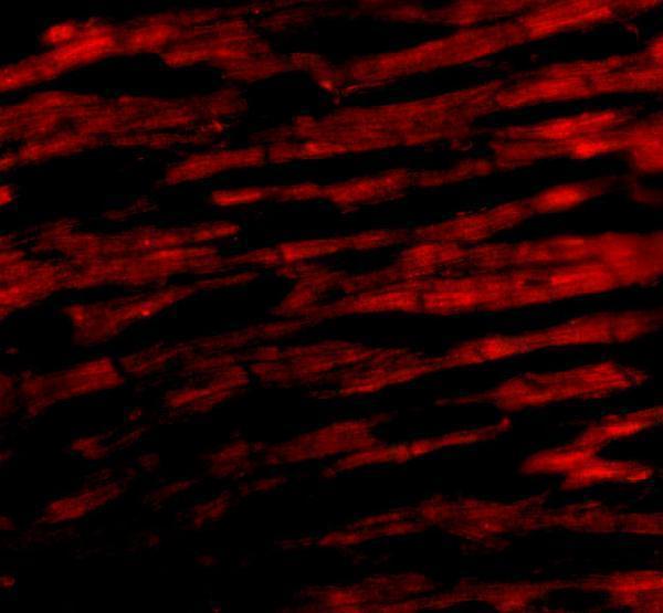
A06151 has human heart IHC and IF images (catalog image captions) and lists Human, Mouse and Rat reactivity (catalog applications and reactivity); the IHC specimen’s fixative is unreported (catalog IHC caption).
A06151 will render with an IHC image of human heart tissue at 10 μg/mL (catalog IHC caption). Its IF image shows human heart cells at 20 μg/mL (catalog IF caption), and its application list includes IHC-P and IF (catalog applications).
Which to pick: Choose A06151 for paraffin-section tissue IHC because IHC-P is listed and its own IHC caption shows human heart tissue at 10 μg/mL; the caption does not report the fixative (catalog applications; catalog IHC caption). For IF/ICC, start with A06151 because IF is listed and its own IF caption shows human heart cells at 20 μg/mL (catalog applications; catalog IF caption). For Mouse or Rat IHC, both A06151 and the polyclonal A06151-1 list those species and IHC among their applications, but the supplied IHC image documents Human tissue only for A06151, while A06151-1 has no supplied IHC image (catalog reactivity, applications, dilution details and image captions).