This website uses cookies to ensure you get the best experience on our website.


- Table of Contents
Plan BCL9L paraffin IHC using high-staining squamous epithelium or kidney tubules as candidate controls (HPA tissue IHC). Interpret nuclear and cytoplasmic staining cautiously because antibody staining shows low consistency with RNA expression (HPA tissue IHC).

Expected localisation, validated protocols, controls and antibodies — the at-a-glance facts below, then the full design guide.
| Expected localisation | Nuclear and cytoplasmic in several tissues (HPA tissue IHC) | |
| Staining pattern | High squamous epithelial staining; nuclear/cytoplasmic overall (HPA tissue IHC) | |
| Antigen retrieval | Tris-EDTA pH 9.0 HIER, 95–98 °C, 20 min (rule: nuclear antigen) | |
| Positive control | Adrenal gland+4 more · see all | |
| Negative control | Adipose tissue+4 more · see all |
| Fixation | Keep fixation consistent across sections (standard IHC practice; not target-specific) | |
| Caveat | Low staining–RNA concordance limits interpretation (HPA tissue IHC) | |
| Regulation | Expression regulators unspecified (UniProt) | |
| Isoform / epitope | 4 isoforms; verify epitope coverage (UniProt) |
The catalog antibody’s IHC-P protocol is accompanied by published BCL9L IHC methods for osteosarcoma (PMC7575756), bladder cancer (PMC9141496), and pancreatic cancer (PMC5342010).
| Sample | Tissue sections; selected-image fixative not specified (standard IHC workflow) |
| Fixation | Image fixative and duration unreported (datasheet A05905-1); verify before use. |
| Sectioning | 4–5 µm sections on charged slides (standard) |
| Deparaffinisation | Xylene, graded ethanol series to water (standard) |
| Antigen retrieval | Heat-induced epitope retrieval in Tris-EDTA buffer, pH 9.0, 20 min at 95–98 °C (standard rule: nuclear antigen) |
| Peroxidase block | 3% H2O2, 10 min, room temperature (standard) |
| Blocking | 10% normal serum of the secondary host, 30 min, room temperature (standard) |
| Primary antibody | Rabbit anti-BCL9L, 5 μg/mL (datasheet A05905-1) |
| Primary incubation | Overnight at 4 °C (standard) |
| Detection | HRP-polymer secondary, DAB chromogen 5–10 min (standard) |
| Counterstain | Hematoxylin, blue, dehydrate and mount (standard) |
| Expected result | BCL9L-positive staining in glandular cells of adrenal gland (HPA tissue IHC: High). HPA tissue profile: Nuclear and cytoplasmic expression in several tissues. No signal in the no-primary control. |
BCL9L is a nuclear protein with no transmembrane segment (UniProt Q86UU0). In tissue IHC, HPA reports nuclear and cytoplasmic staining in several tissues, including high staining in squamous epithelial cells, skin keratinocytes and kidney tubular cells (HPA tissue IHC). Interpret that pattern cautiously: HPA rates its tissue IHC reliability Uncertain because antibody staining has low consistency with RNA expression (HPA tissue IHC).
| Distinct nuclear staining in squamous epithelial cells or skin keratinocytes, with some cytoplasmic staining (HPA tissue IHC). | This fits the reported tissue pattern: these cell groups score High, and HPA describes both nuclear and cytoplasmic expression (HPA tissue IHC). Score nuclei and cytoplasm separately; the tissue IHC reliability remains Uncertain (HPA tissue IHC). |
| Membrane-only, extracellular or predominantly granular staining without a discernible nuclear component. | That pattern conflicts with the nuclear location and absence of a transmembrane segment (UniProt Q86UU0). Treat it as suspect, check morphology and detection controls, and seek independent confirmation before assigning it to BCL9L; the tissue IHC assessment is Uncertain (HPA tissue IHC). |
| Strong staining in adipocytes or bone-marrow hematopoietic cells, where HPA reports Not detected (HPA tissue IHC). | The cell identity conflicts with those HPA observations and warrants checks for cross-reactivity or endogenous chromogen-generating activity. It is not proof of either cause: HPA reports low consistency between antibody staining and RNA expression (HPA tissue IHC). |
| Broad chromogen haze across nuclei, cytoplasm and tissue-free areas, obscuring cell boundaries. | A field-wide deposit cannot be scored as cell-specific BCL9L. In a general chromogenic IHC workflow, inspect the no-primary control, background blocking, washes and detection chemistry; compare only interpretable cells with the reported nuclear and cytoplasmic pattern (HPA tissue IHC). |
| No discernible staining in adrenal glandular cells or kidney tubular cells, both scored High by HPA (HPA tissue IHC). | A failed run or limited assay sensitivity is possible. First verify tissue identity and the detection system with controls, then review the antibody's validated IHC-P conditions. HPA's Uncertain reliability means one negative section does not settle BCL9L expression (HPA tissue IHC). |
| Tissue and cell selection | HPA scores squamous epithelial cells in cervix, esophagus, oral mucosa, tonsil and vagina High, as well as skin keratinocytes and kidney tubular cells (HPA tissue IHC). Select and score the named cell population, because a tissue-wide average can hide a cell-specific result. |
| Interpretive confidence | HPA rates tissue IHC Uncertain due to low consistency between antibody staining and RNA expression; HPA049370 is IHC Uncertain (HPA tissue IHC; HPA antibody validation). Use the pattern as a comparison, and require corroboration for a consequential localization claim. |
| Isoforms and processing | UniProt lists four BCL9L isoforms and a full-length chain, with no signal peptide or propeptide (UniProt Q86UU0). The supplied record gives no antibody epitope, so it cannot establish which isoforms an IHC signal represents. |
| IF/ICC Q: what location should an image show? | A: HPA reports mainly nucleoplasmic signal, with additional nucleoli fibrillar-center localization; its listed ICC assessments are Supported (HPA subcellular; HPA antibody validation). That supports an IF/ICC localization comparison, while the separate tissue IHC assessment remains Uncertain (HPA tissue IHC). |
| Situation | Likely cause | Next action |
|---|---|---|
| A High-scoring HPA cell population is blank (HPA tissue IHC). | The run may lack sensitivity, or the sampled section may not contain the intended cells; tissue IHC reliability is Uncertain (HPA tissue IHC). | Confirm morphology, include a positive control and check chromogenic detection. As general IHC practice, review the IHC-P antibody instructions and retrieval conditions before changing them. |
| Signal is cytoplasmic only, with no clear nuclear staining. | Cytoplasmic staining is reported, but a purely cytoplasmic result does not show the nuclear location reported for BCL9L (HPA tissue IHC; UniProt Q86UU0). | Check nuclear counterstaining and focus through the section; score compartments separately. If the pattern persists, seek independent antibody or orthogonal evidence because HPA tissue IHC is Uncertain (HPA tissue IHC). |
| Cells scored Not detected by HPA stain strongly (HPA tissue IHC). | Cross-reactivity or endogenous detection activity is possible; an HPA Not detected call is an observation, not an absolute absence claim (HPA tissue IHC). | Identify the stained cells, run a no-primary detection control and review blocking. Compare the same cell type across sections before interpreting the result. |
| Diffuse brown background prevents cell-level scoring. | General chromogenic IHC causes include nonspecific antibody binding, residual detection activity or inadequate washing. | Inspect a no-primary control, refresh blocking and wash steps, and optimize antibody concentration within the antibody's validated IHC-P guidance. Score only signal with identifiable cellular boundaries. |
| Staining is concentrated at tissue edges or varies across one section. | Uneven reagent coverage or local processing variation can produce this general IHC artifact; the supplied sources establish no BCL9L-specific fixation effect. | Repeat with even reagent coverage and matched processing, then compare intact regions containing the same cell population. Do not infer a BCL9L-specific fixation mechanism from this pattern. |
| IHC and ICC/IF appear to disagree. | HPA supports nucleoplasmic ICC localization, while its tissue IHC assessment is Uncertain (HPA subcellular; HPA tissue IHC). The assays also show different preparations. | Compare the nuclear compartment and cell identity in each assay, verify each assay's controls, and report the disagreement without treating Supported ICC validation as tissue IHC validation (HPA antibody validation). |


Comprehensive Human Protein Atlas IHC scoring per tissue (reliability: Uncertain — Low consistency between antibody staining and RNA expression data.). Rows are taken directly from the HPA tissue chart — click any row's HPA link to view the source.
| Tissue | Cell type | Level | Evidence | Source |
|---|---|---|---|---|
| Adrenal gland | Glandular cells | High | Protein (IHC) | HPA → |
| Cervix | Squamous epithelial cells | High | Protein (IHC) | HPA → |
| Esophagus | Squamous epithelial cells | High | Protein (IHC) | HPA → |
| Kidney | Cells in tubules | High | Protein (IHC) | HPA → |
| Oral mucosa | Squamous epithelial cells | High | Protein (IHC) | HPA → |
| Tissue | Cell type | Level | Evidence | Source |
|---|---|---|---|---|
| Adipose tissue | Adipocytes | Not detected | Protein (IHC) | HPA → |
| Appendix | Glandular cells | Not detected | Protein (IHC) | HPA → |
| Bone marrow | Hematopoietic cells | Not detected | Protein (IHC) | HPA → |
| Caudate | Glial cells | Not detected | Protein (IHC) | HPA → |
| Cerebral cortex | Endothelial cells | Not detected | Protein (IHC) | HPA → |
Use compartment-aware controls and matched tissue processing to troubleshoot BCL9L staining in chromogenic paraffin-section IHC.
Two anti-BCL9L antibodies have IHC images from human tissues; the catalog also shows rat kidney IHC and human cell IF/ICC images (catalog image captions).
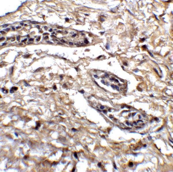
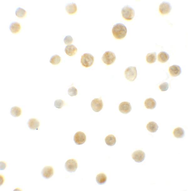
A05905-1 is the rendered card, with human breast tissue IHC, human breast tissue IF, and HeLa cell ICC images (A05905-1 image captions). A05905 has IHC images from human lung, ovarian and oesophageal cancer tissues and rat kidney, plus IF/ICC images from A431 cells (A05905 image captions).
Which to pick: For human tissue IHC, A05905-1 has a paraffin-section application listing and a breast tissue image at 5 μg/mL (A05905-1 application list and IHC caption); its image caption does not report the fixative (A05905-1 IHC caption). For IF/ICC, A05905-1 lists both applications and shows human breast tissue IF and HeLa cell ICC (A05905-1 application list and image captions). For cross-species work, A05905 lists human, mouse and rat reactivity, with paraffin-section IHC images from human tissues and rat kidney; mouse IHC is not shown, and the captions do not report the fixative (A05905 reactivity and IHC captions).