This website uses cookies to ensure you get the best experience on our website.


- Table of Contents
Plan chromogenic BDKRB2 IHC on paraffin sections using the catalog antibody’s reported workflow (datasheet M03117). Assess cytoplasmic staining in CNS neuronal cells while accounting for the receptor’s annotated cell membrane localisation (HPA tissue IHC; UniProt).

Expected localisation, validated protocols, controls and antibodies — the at-a-glance facts below, then the full design guide.
| Expected localisation | Cytoplasmic tissue staining; cell membrane localisation annotated (HPA tissue IHC; UniProt) | |
| Staining pattern | Cytoplasmic staining in CNS neuronal cells (HPA tissue IHC) | |
| Antigen retrieval | EDTA pH 8.0 HIER, heat-mediated (datasheet M03117) | |
| Positive control | Caudate+3 more · see all | |
| Negative control | Adipose tissue+4 more · see all |
| Fixation | Keep fixation consistent (standard IHC practice; not target-specific); Matched tissue-IHC evidence does not establish the fixation claim. Validate the specimen-specific method before use. (selected-SKU IHC image M03117) | |
| Caveat | Antibody staining has low consistency with tissue RNA data (HPA tissue IHC) | |
| Regulation | Expression regulation is not specified (UniProt) | |
| Isoform / epitope | 2 isoforms; verify extracellular or cytoplasmic epitope coverage (UniProt) |
The catalog antibody’s IHC-P protocol is accompanied by published chromogenic IHC methods for human amnion, gastric cancer specimens, and rat paraffin sections (PMC9130483; PMC3208739; PMC8275397).
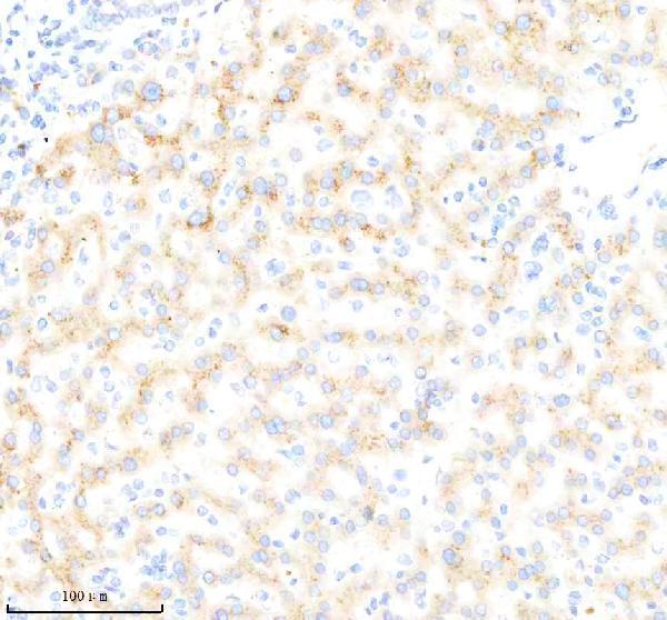
| Sample | Paraffin-embedded human liver cancer tissue; fixative not specified (datasheet M03117) |
| Fixation | Image fixative and duration unreported (datasheet M03117); verify before use. |
| Sectioning | 4–5 µm sections on charged slides (standard) |
| Deparaffinisation | Xylene, graded ethanol series to water (standard) |
| Antigen retrieval | Heat retrieval: EDTA pH 8.0 (datasheet M03117); 20 min, 95–100 °C (standard) |
| Peroxidase block | 3% H2O2, 10 min, room temperature (standard) |
| Blocking | 10% goat serum (datasheet M03117) |
| Primary antibody | Rabbit monoclonal (clone AFAG-2) anti-BDKRB2, 1:50 recommended; image 1:200 (datasheet M03117) |
| Primary incubation | Overnight at 4 °C (datasheet M03117) |
| Detection | HRP-conjugated secondary, DAB chromogen (datasheet M03117) |
| Counterstain | Hematoxylin, blue, dehydrate and mount (standard) |
| Expected result | BDKRB2-positive staining in neuronal cells of caudate (HPA tissue IHC: High). HPA tissue profile: Cytoplasmic expression in a group of tissues, including CNS. No signal in the no-primary control. |
BDKRB2 is a 7-pass cell-membrane receptor (UniProt P30411 topology). For paraffin-section IHC, HPA reports cytoplasmic staining in a group of tissues, with High staining in caudate and cerebral-cortex neuronal cells and Medium staining in hippocampal neuronal cells (HPA tissue IHC). HPA rates the tissue staining Approved but reports low consistency with RNA expression; interpret an isolated positive result cautiously (HPA tissue IHC).
| Caudate or cerebral-cortex neurons show a clear chromogenic signal. | This fits HPA's High neuronal staining in both tissues (HPA tissue IHC). Score the stained cells and compartment, not color intensity alone (general IHC practice). |
| Neuronal staining looks cytoplasmic, without a sharply outlined membrane. | HPA describes the tissue IHC pattern as cytoplasmic (HPA tissue IHC). Membrane localization is supported by UniProt and HPA ICC-IF, but a membrane outline is not required to match the reported paraffin-section pattern (UniProt P30411; HPA subcellular). |
| Strong signal is confined to nuclei instead of the reported cell compartment. | Predominantly nuclear staining is discordant with the reported cytoplasmic tissue pattern and membrane localization (HPA tissue IHC; UniProt P30411). Check a negative reagent control and the antibody's validated IHC conditions before assigning it to BDKRB2 (general IHC practice). |
| An HPA-negative cell population stains strongly, or color appears broadly across cells. | For example, HPA reports no detection in adipocytes of adipose tissue (HPA tissue IHC). Unexpected cell staining can reflect cross-reactivity; widespread color can reflect endogenous detection activity or nonspecific background (general IHC practice). |
| Known-positive caudate or cerebral-cortex neurons show no signal. | An absent signal conflicts with HPA's High staining in those neuronal cells (HPA tissue IHC). First assess control performance and IHC conditions; a blank section alone cannot establish absent receptor expression (general IHC practice). |
| Membrane topology and tissue appearance | UniProt places BDKRB2 at the cell membrane with 7 transmembrane segments, while HPA describes cytoplasmic tissue IHC staining (UniProt P30411; HPA tissue IHC). Record the observed compartment rather than inferring surface-only staining. |
| Choice of reference tissue and cell | HPA reports High neuronal staining in caudate and cerebral cortex, Medium neuronal staining in hippocampus, and Medium respiratory epithelial staining in nasopharynx (HPA tissue IHC). Compare like cell populations when judging intensity. |
| Strength of IHC evidence | The tissue profile is Approved, yet HPA notes low consistency between antibody staining and RNA expression; its listed IHC antibody HPA050841 is Approved (HPA tissue IHC; HPA antibodies). Treat unexpected positives as findings to verify. |
| Antibody epitope and receptor forms | UniProt lists extracellular and cytoplasmic regions, 2 isoforms, and glycosylation sites at residues 30, 39, and 207 (UniProt P30411). The payload gives no antibody epitope, so it cannot predict which form or region the IHC antibody detects. |
| IF/ICC Q: What localization can IF show? | A: HPA reports mainly plasma membrane, with additional vesicle and cytokinetic-bridge localization in ICC-IF (HPA subcellular). Its ICC-approved antibody is HPA003551; that status does not establish this antibody's paraffin-section IHC performance (HPA antibodies). |
| Situation | Likely cause | Next action |
|---|---|---|
| Positive neuronal reference tissue is blank. | The result disagrees with HPA's High caudate or cerebral-cortex neuronal staining; the failed step is not identified by the image alone (HPA tissue IHC). | Check the positive control and detection reagents, then review antigen retrieval and dilution against the IHC-validated antibody's instructions (general IHC practice). |
| All structures have diffuse brown color. | Broad color without cell selectivity can arise from nonspecific staining or endogenous detection activity (general IHC practice). | Run primary-omission and detection controls; review blocking, washing, and chromogen development under the chosen IHC workflow (general IHC practice). |
| Signal appears mainly nuclear. | That compartment differs from the cytoplasmic tissue IHC description and membrane localization (HPA tissue IHC; UniProt P30411). | Record the mismatch and compare with a positive tissue control and primary-omission control before scoring it as receptor staining (general IHC practice). |
| Adipocytes stain as strongly as positive neurons. | HPA reports adipocytes in adipose tissue as Not detected, versus High neuronal staining in caudate and cerebral cortex (HPA tissue IHC). | Assess cell identity and background controls, then repeat under validated IHC conditions; treat persistent staining as unverified (general IHC practice; HPA tissue IHC). |
| Hippocampal neurons look weaker than cortical neurons. | That difference can fit HPA's Medium hippocampal versus High cerebral-cortex neuronal levels (HPA tissue IHC). | Score each tissue against its own HPA reference level and matched run controls before calling the weaker section a technical failure (HPA tissue IHC; general IHC practice). |
| Cytoplasmic staining lacks a crisp membrane rim. | The appearance matches HPA's cytoplasmic tissue IHC description, despite supported plasma-membrane localization in ICC-IF (HPA tissue IHC; HPA subcellular). | Record cell type, intensity, and compartment; use the separate IF/ICC guide when membrane localization is the experimental question (HPA tissue IHC; HPA subcellular). |
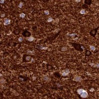
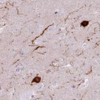
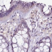
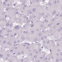


Comprehensive Human Protein Atlas IHC scoring per tissue (reliability: Approved — Low consistency between antibody staining and RNA expression data.). Rows are taken directly from the HPA tissue chart — click any row's HPA link to view the source.
| Tissue | Cell type | Level | Evidence | Source |
|---|---|---|---|---|
| Caudate | Neuronal cells | High | Protein (IHC) | HPA → |
| Cerebral cortex | Neuronal cells | High | Protein (IHC) | HPA → |
| Hippocampus | Neuronal cells | Medium | Protein (IHC) | HPA → |
| Nasopharynx | Respiratory epithelial cells | Medium | Protein (IHC) | HPA → |
| Tissue | Cell type | Level | Evidence | Source |
|---|---|---|---|---|
| Adipose tissue | Adipocytes | Not detected | Protein (IHC) | HPA → |
| Appendix | Glandular cells | Not detected | Protein (IHC) | HPA → |
| Bone marrow | Hematopoietic cells | Not detected | Protein (IHC) | HPA → |
| Breast | Adipocytes | Not detected | Protein (IHC) | HPA → |
| Cerebellum | Cells in granular layer | Not detected | Protein (IHC) | HPA → |
Use the catalog antibody’s paraffin section workflow as the starting point, then evaluate staining against BDKRB2 localisation and cell type evidence.
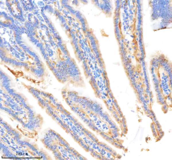
The IHC-validated antibody M03117 has paraffin-section images from human liver cancer and mouse small intestine; no IF/ICC image is supplied (M03117 IHC and IF image captions).
The M03117 card shows IHC staining of a paraffin-embedded human liver cancer section (M03117 card image caption). A second M03117 caption documents a paraffin-embedded mouse small intestine section (M03117 IHC image captions).
Which to pick: Choose M03117 for paraffin-section tissue IHC; its rabbit monoclonal antibody has documented human and mouse staining, and the fixative is unreported (catalog: host, clone and reactivity; M03117 IHC image captions). For work across those two species, M03117 is the catalog option (catalog: human and mouse reactivity; M03117 IHC image captions). No IF/ICC antibody choice is supported by this payload because M03117 has no listed IF application or IF image (catalog: applications and IF image captions).