This website uses cookies to ensure you get the best experience on our website.


- Table of Contents
Plan chromogenic BHMT IHC in paraffin sections using hepatocytes and kidney proximal tubules as positive tissue references (HPA tissue IHC). Score the observed cytoplasmic pattern (HPA tissue IHC), while recognizing that UniProt annotates a small nuclear pool that increases under oxidative stress (UniProt).

Expected localisation, validated protocols, controls and antibodies — the at-a-glance facts below, then the full design guide.
| Expected localisation | Cytoplasmic tissue staining; a small nuclear pool is annotated (HPA tissue IHC; UniProt) | |
| Staining pattern | Hepatocytes and proximal tubules show cytoplasmic staining (HPA tissue IHC) | |
| Antigen retrieval | EDTA pH 8.0 HIER, heat-mediated (datasheet A06712-2) | |
| Positive control | Kidney+1 more · see all | |
| Negative control | Adipose tissue+4 more · see all |
| Fixation | Keep tissue fixation consistent (standard IHC practice; not target-specific); Matched tissue-IHC evidence does not establish the fixation claim. Validate the specimen-specific method before use. (selected-SKU IHC image A06712-2) | |
| Caveat | Oxidative stress can shift BHMT into nuclei (UniProt) | |
| Regulation | Expression regulator not specified (UniProt) | |
| Isoform / epitope | No isoforms; one 1–406 chain with no extracellular domain (UniProt) |
Compare the catalog antibody’s IHC-P protocol with published BHMT staining methods for liver, bat brain, mouse ovary, and human brain sections (PMC11920202; PMC3871600; PMC9208444; PMC8112889).
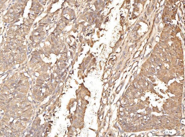
| Sample | Paraffin-embedded human bladder epithelial carcinoma tissue; fixative not specified (datasheet A06712-2) |
| Fixation | Image fixative and duration unreported (datasheet A06712-2); verify before use. |
| Sectioning | 4–5 µm sections on charged slides (standard) |
| Deparaffinisation | Xylene, graded ethanol series to water (standard) |
| Antigen retrieval | Heat retrieval: EDTA pH 8.0 (datasheet A06712-2); 20 min, 95–100 °C (standard) |
| Peroxidase block | 3% H2O2, 10 min, room temperature (standard) |
| Blocking | 10% goat serum (datasheet A06712-2) |
| Primary antibody | Rabbit anti-BHMT, 2-5 μg/ml (datasheet A06712-2) |
| Primary incubation | Overnight at 4 °C (datasheet A06712-2) |
| Detection | Streptavidin-biotin complex (SABC), DAB chromogen (datasheet A06712-2) |
| Counterstain | Hematoxylin, blue, dehydrate and mount (standard) |
| Expected result | BHMT-positive staining in proximal tubules (cell body) of kidney (HPA tissue IHC: High). HPA tissue profile: Selective cytoplasmic expression in hepatocytes and kidney tubules. No signal in the no-primary control. |
BHMT should show predominantly cytoplasmic staining in hepatocytes and kidney proximal tubules, where HPA reports high expression (HPA tissue IHC). A small nuclear fraction is possible, particularly with oxidative stress (UniProt Q93088). HPA rates its tissue staining reliability Enhanced, with high consistency between staining and RNA data (HPA tissue IHC). BHMT has no transmembrane segment, so a membrane-rim pattern is unexpected (UniProt Q93088 topology).
| Strong cytoplasmic signal in hepatocytes and proximal tubule cell bodies. | This matches the reported high expression and selective cytoplasmic pattern in liver and kidney (HPA tissue IHC). Assess signal in the relevant cells rather than treating every cell in a positive tissue as positive (HPA tissue IHC). |
| Predominantly membrane-rim or strong, widespread nuclear staining. | A membrane-rim pattern conflicts with BHMT's lack of a transmembrane segment (UniProt Q93088 topology). Widespread nuclear dominance also differs from its predominantly cytosolic distribution; a small nuclear fraction can occur, especially under oxidative stress (UniProt Q93088). |
| Strong staining in adipocytes or adrenal glandular cells. | These cells are reported as not detected (HPA tissue IHC). Treat a reproducible signal as unexpected and check antibody specificity and detection controls before assigning it to BHMT (standard IHC practice). |
| Diffuse color over many structures, including cells expected to be negative. | This lacks the selective cell and compartment pattern reported for BHMT (HPA tissue IHC). Review nonspecific antibody binding and detection background with appropriate controls (standard IHC practice). |
| No staining in hepatocytes or proximal tubules. | Both are high-expression reference cells (HPA tissue IHC). A blank result there cannot establish absence of BHMT in another sample; check staining controls and the assay workflow first (standard IHC practice). |
| Cell type and tissue | Liver hepatocytes and kidney proximal tubules provide high-signal reference cells; listed negative cells provide pattern checks (HPA tissue IHC). |
| Subcellular distribution | BHMT is predominantly cytosolic, with a small nuclear fraction that may increase under oxidative stress (UniProt Q93088; HPA subcellular ICC-IF). |
| Antibody validation | HPA038285 and HPA058310 each have Enhanced IHC validation; that supports their reported patterns without validating every antibody (HPA antibodies). |
| Antigen retrieval | Retrieval may be optimized using positive tissue and controls as a general paraffin-IHC step; BHMT-specific retrieval sensitivity is unreported in the supplied sources (standard IHC practice). |
| Detection background | Endogenous detection activity and nonspecific binding can mimic chromogenic staining; control slides help identify them (standard IHC practice). Neither establishes a BHMT-positive cell (HPA tissue IHC). |
| Situation | Likely cause | Next action |
|---|---|---|
| Hepatocytes and proximal tubules are both blank. | The result conflicts with their high reported expression (HPA tissue IHC); assay failure is possible (standard IHC practice). | Check the positive control, primary-antibody application, retrieval conditions, detection reagents, and counterstain before interpreting the sample (standard IHC practice). |
| A diffuse brown haze obscures cell boundaries. | Nonspecific binding or detection background can obscure a selective cytoplasmic pattern (standard IHC practice; HPA tissue IHC). | Compare a no-primary control; review blocking, washes, antibody concentration, and detection conditions (standard IHC practice). |
| Adipocytes or adrenal glandular cells stain strongly. | Both are reported as not detected, so the pattern is unexpected for BHMT (HPA tissue IHC). | Compare a known-positive liver or kidney section and a no-primary control; reassess specificity if unexpected staining persists (standard IHC practice; HPA tissue IHC). |
| Signal outlines membranes or fills most nuclei. | A membrane rim conflicts with BHMT topology; nuclear dominance differs from its usual distribution (UniProt Q93088). | Inspect morphology and control slides; allow for a small nuclear fraction, particularly with oxidative stress, before judging nuclear signal (UniProt Q93088; standard IHC practice). |
| Liver stains, but kidney proximal tubules do not. | Both cell types are reported high, so a tissue-dependent assay or specimen issue needs review (HPA tissue IHC; standard IHC practice). | Confirm proximal-tubule identity and tissue quality, then compare the two sections under the same staining conditions (standard IHC practice). |
| IF/ICC Q: Should a nuclear-only fluorescent pattern be accepted? | HPA reports approved cytosolic localization; UniProt allows a small nuclear fraction, especially with oxidative stress (HPA subcellular ICC-IF; UniProt Q93088). | A: Interpret nuclear-only signal cautiously and check staining controls and cell morphology; do not use it alone to assign BHMT localization (standard IF practice). |
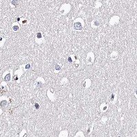
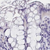


Comprehensive Human Protein Atlas IHC scoring per tissue (reliability: Enhanced — High consistency between antibody staining and RNA expression data.). Rows are taken directly from the HPA tissue chart — click any row's HPA link to view the source.
| Tissue | Cell type | Level | Evidence | Source |
|---|---|---|---|---|
| Kidney | Proximal tubules (cell body) | High | Protein (IHC) | HPA → |
| Liver | Hepatocytes | High | Protein (IHC) | HPA → |
| Tissue | Cell type | Level | Evidence | Source |
|---|---|---|---|---|
| Adipose tissue | Adipocytes | Not detected | Protein (IHC) | HPA → |
| Adrenal gland | Glandular cells | Not detected | Protein (IHC) | HPA → |
| Appendix | Glandular cells | Not detected | Protein (IHC) | HPA → |
| Bone marrow | Hematopoietic cells | Not detected | Protein (IHC) | HPA → |
| Breast | Adipocytes | Not detected | Protein (IHC) | HPA → |
Use BHMT’s expected cytoplasmic staining in hepatocytes and kidney tubules to guide IHC controls and interpretation (HPA tissue IHC).
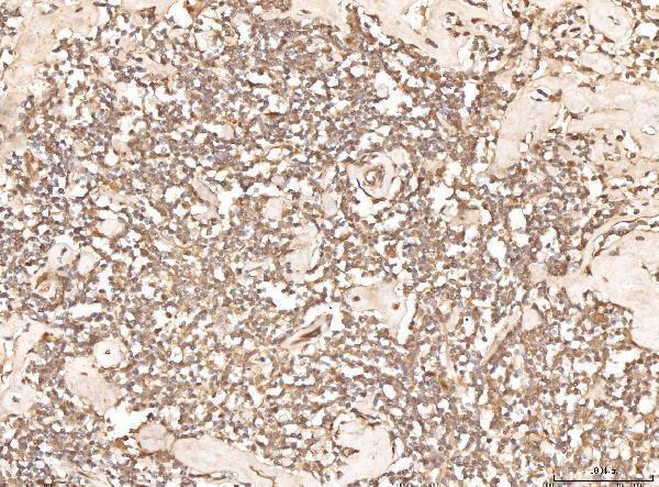
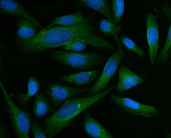
A06712-2 has human paraffin-section IHC images and a U20S-cell IF image (catalog image captions); its listed reactivity includes human, mouse, and rat (catalog applications/reactivity).
A06712-2 will render with its human bladder epithelial carcinoma paraffin-section IHC image; its catalog also lists IF/ICC and human, mouse, and rat reactivity (catalog image caption; applications/reactivity). M06712-2 is listed for human IHC, but has no supplied IHC image caption (catalog applications/reactivity; image alts).
Which to pick: Choose A06712-2 for tissue IHC when a documented paraffin-section example matters: its own image caption shows human bladder epithelial carcinoma stained at 2 μg/ml after EDTA retrieval, and does not report the fixative (A06712-2 IHC image caption). For IF/ICC or work involving mouse or rat, A06712-2 has the relevant applications and listed reactivity; its IF image documents U20S cells at 5 μg/ml (catalog applications/reactivity; A06712-2 IF image caption). M06712-2 offers a rabbit monoclonal option for human IHC, with no supplied IHC image or fixation details (catalog clone, applications/reactivity, image alts).