This website uses cookies to ensure you get the best experience on our website.


- Table of Contents
Plan paraffin BIN1 IHC around the high cytoplasmic staining reported in CNS and skeletal muscle (HPA tissue IHC). Use the staining pattern and controls to assess specificity, mindful of reported off-target binding (HPA tissue IHC).

Expected localisation, validated protocols, controls and antibodies — the at-a-glance facts below, then the full design guide.
| Expected localisation | Cytoplasm in CNS and skeletal muscle (HPA tissue IHC) | |
| Staining pattern | High cytoplasmic staining in CNS glia and skeletal myocytes (HPA tissue IHC) | |
| Antigen retrieval | EDTA pH 8.0 HIER, heat-mediated (datasheet A01551-1) | |
| Positive control | Caudate+4 more · see all | |
| Negative control | Esophagus+2 more · see all |
| Fixation | Keep fixation consistent across sections. (standard IHC practice; not target-specific) | |
| Caveat | Presumed off-target staining has been observed (HPA tissue IHC) | |
| Regulation | Highest expression in brain and muscle (UniProt) | |
| Isoform / epitope | 11 isoforms; epitope coverage is unspecified (UniProt) |
The catalog antibody has an IHC-P protocol (datasheet A01551-1). The published FFPE hepatocellular carcinoma method provides one comparison protocol (PMC10495885).
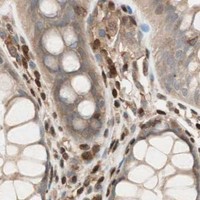
| Sample | Paraffin-embedded human appendiceal adenocarcinoma tissue; fixative not specified (datasheet A01551-1) |
| Fixation | Image fixative and duration unreported (datasheet A01551-1); verify before use. |
| Sectioning | 4–5 µm sections on charged slides (standard) |
| Deparaffinisation | Xylene, graded ethanol series to water (standard) |
| Antigen retrieval | Heat retrieval: EDTA pH 8.0 (datasheet A01551-1); 20 min, 95–100 °C (standard) |
| Peroxidase block | 3% H2O2, 10 min, room temperature (standard) |
| Blocking | 10% goat serum (datasheet A01551-1) |
| Primary antibody | Rabbit anti-BIN1, 2-5 μg/ml (datasheet A01551-1) |
| Primary incubation | Overnight at 4 °C (datasheet A01551-1) |
| Detection | Streptavidin-biotin complex (SABC), DAB chromogen (datasheet A01551-1) |
| Counterstain | Hematoxylin, blue, dehydrate and mount (standard) |
| Expected result | BIN1-positive staining in glial cells of caudate (HPA tissue IHC: High). HPA tissue profile: High cytoplasmic expression in CNS and skeletal muscle. No signal in the no-primary control. |
BIN1 staining should be predominantly cytoplasmic in CNS cells and skeletal myocytes (HPA: high cytoplasmic expression in CNS and skeletal muscle). BIN1 can also localise to the nucleus, endosomes, sarcolemma and T-tubules, and has no transmembrane segment (UniProt O00499: subcellular location and topology). Interpret tissue staining with caution: HPA rates its IHC profile Enhanced while reporting medium agreement with RNA data and presumed off-target staining that was disregarded (HPA: tissue IHC reliability).
| Strong cytoplasmic staining in skeletal myocytes or CNS glial cells (HPA: High in skeletal muscle myocytes and glial cells of caudate, cortex and hippocampus). | This fits the reported tissue pattern (HPA: high cytoplasmic expression in CNS and skeletal muscle). Judge positivity within the named cell population, since adjacent cells need not share its staining level (HPA: cell-specific tissue IHC results). |
| Predominantly nuclear staining, with little cytoplasmic signal in a known-positive cell population. | Review localisation and controls before scoring it as the expected pattern: HPA describes high cytoplasmic tissue expression, although UniProt also lists the nucleus as a possible location (HPA: tissue IHC profile; UniProt O00499: subcellular location). Nuclear staining alone does not establish an artefact. |
| Strong staining of esophageal, oral mucosal or vaginal squamous epithelium (HPA: Not detected in these squamous epithelial cells). | Treat this as discordant with the HPA tissue pattern and investigate antibody cross-reactivity or endogenous detection activity (HPA: tissue IHC results; standard chromogenic IHC practice). HPA reports presumed off-target binding in its reliability assessment (HPA: tissue IHC reliability). |
| Widespread, poorly localised colour across tissue structures and empty spaces. | A diffuse deposit is difficult to score as cell-specific BIN1 staining; assess background with the appropriate detection controls and inspect blocking and wash steps (standard chromogenic IHC practice). It does not reproduce the reported cell-specific cytoplasmic pattern (HPA: tissue IHC profile). |
| No staining in skeletal myocytes on a section intended as a positive reference (HPA: High in skeletal muscle myocytes). | First check tissue identity and whether the positive reference stained in the same run; then review retrieval, primary antibody, detection and counterstain conditions (standard chromogenic IHC practice). An absent signal in one run does not establish that BIN1 is absent from that tissue (HPA: tissue IHC result). |
| Cell population selected for interpretation | HPA reports High staining in skeletal myocytes, selected CNS cells and lung alveolar type I cells, but Not detected in specified squamous epithelial cells (HPA: tissue IHC results). Compare the same cell type when judging a positive or negative result. |
| Antibody validation and specificity | Three listed antibodies have Enhanced IHC validation, but HPA describes medium agreement with RNA data and disregarded presumed off-target binding (HPA: antibody validation; HPA: tissue IHC reliability). Concordant localisation and control results still matter when scoring an unfamiliar pattern. |
| Isoform and compartment context | UniProt lists 11 BIN1 isoforms and reports brain-restricted isoform IIA, while isoform BIN1 is widely expressed with its highest expression in skeletal muscle (UniProt O00499: isoforms and tissue specificity). HPA's tissue profile alone cannot assign staining to a particular isoform (HPA: tissue IHC profile). |
| Interpretation of membrane-associated signal | UniProt places BIN1 at the sarcolemma and T-tubules as well as in cytoplasm, endosomes and nucleus, despite listing no transmembrane segment (UniProt O00499: subcellular location and topology). Membrane-adjacent staining can be plausible, but assess it alongside the observed predominantly cytoplasmic IHC pattern (HPA: tissue IHC profile). |
| Retrieval and fixation evidence | The supplied HPA and UniProt records give no BIN1-specific fixation sensitivity or retrieval condition. Optimise retrieval against a known-positive tissue and matched controls as a general IHC workflow step (standard chromogenic IHC practice); do not infer a target-specific fixation effect from the tissue profile. |
| Situation | Likely cause | Next action |
|---|---|---|
| Known-positive skeletal muscle has no specific colour (HPA: High in skeletal muscle myocytes). | The run may have lost sensitivity through a retrieval, primary antibody or detection problem (standard chromogenic IHC practice). | Confirm tissue identity, include a same-run positive reference, and check the validated antibody's IHC conditions plus detection reagents before interpreting the sample as negative (standard chromogenic IHC practice). |
| Squamous epithelium stains strongly where HPA reports no detection (HPA: esophagus, oral mucosa and vagina). | Possible primary antibody cross-reactivity or endogenous detection activity; HPA also notes disregarded presumed off-target binding (HPA: tissue IHC reliability; standard chromogenic IHC practice). | Review a no-primary control and the relevant endogenous-activity block; compare localisation with an independently validated IHC antibody if available (standard chromogenic IHC practice; HPA: three Enhanced IHC antibodies). |
| Colour is diffuse or extends beyond recognisable cells. | Background from nonspecific reagent binding, inadequate washing or detection activity can obscure cell boundaries (standard chromogenic IHC practice). | Inspect no-primary and detection controls, washing and blocking, then score only clearly localised cellular staining (standard chromogenic IHC practice). |
| A section appears nuclear-only, unlike the expected cytoplasmic profile (HPA: high cytoplasmic expression). | UniProt permits nuclear localisation, so compartment alone cannot prove an artefact; the discrepancy may also reflect background or cell identification (UniProt O00499: subcellular location; standard IHC interpretation). | Identify the stained cell population, review counterstain and controls, and report the compartment explicitly rather than calling it the typical HPA tissue pattern (HPA: tissue IHC profile; standard IHC interpretation). |
| The sample stains weakly compared with a skeletal muscle reference (HPA: High in skeletal muscle myocytes). | Expression differs by cell type: HPA reports Low staining in cardiomyocytes, small-intestinal glandular cells, urothelial cells and breast adipocytes (HPA: tissue IHC results). | Compare like cell populations and confirm the reference works in the same run before changing the assay (HPA: cell-specific tissue IHC results; standard chromogenic IHC practice). |
| An IF/ICC image shows cytosolic BIN1 and seems different from a paraffin-section result. | HPA supports cytosolic localisation in ICC-IF, while its tissue IHC profile reports high cytoplasmic expression in CNS and skeletal muscle (HPA: subcellular ICC-IF summary; HPA: tissue IHC profile). | Interpret each result in its own preparation and named cell population; use the separate IF/ICC guide for its workflow (HPA: subcellular ICC-IF summary and tissue IHC results). |
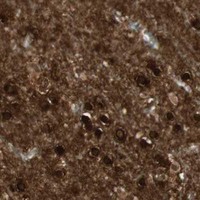


Comprehensive Human Protein Atlas IHC scoring per tissue (reliability: Enhanced — Medium consistency between antibody staining and RNA expression data. Presumed off target binding observed and disregarded.). Rows are taken directly from the HPA tissue chart — click any row's HPA link to view the source.
| Tissue | Cell type | Level | Evidence | Source |
|---|---|---|---|---|
| Caudate | Glial cells | High | Protein (IHC) | HPA → |
| Cerebellum | Granular cells - cytoplasm/membrane | High | Protein (IHC) | HPA → |
| Cerebral cortex | Glial cells | High | Protein (IHC) | HPA → |
| Hippocampus | Glial cells | High | Protein (IHC) | HPA → |
| Lung | Alveolar cells type I | High | Protein (IHC) | HPA → |
Troubleshoot BIN1 staining in paraffin sections by checking retrieval, compartment, cell type and controls before comparing chromogenic signal across samples.
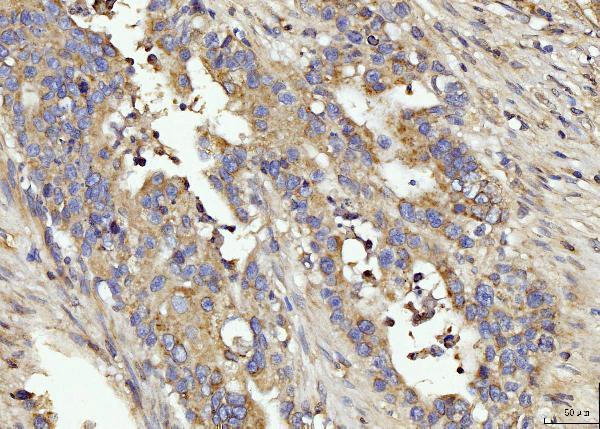
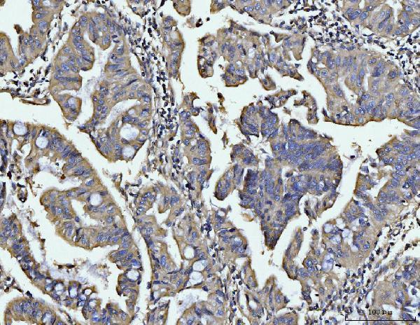
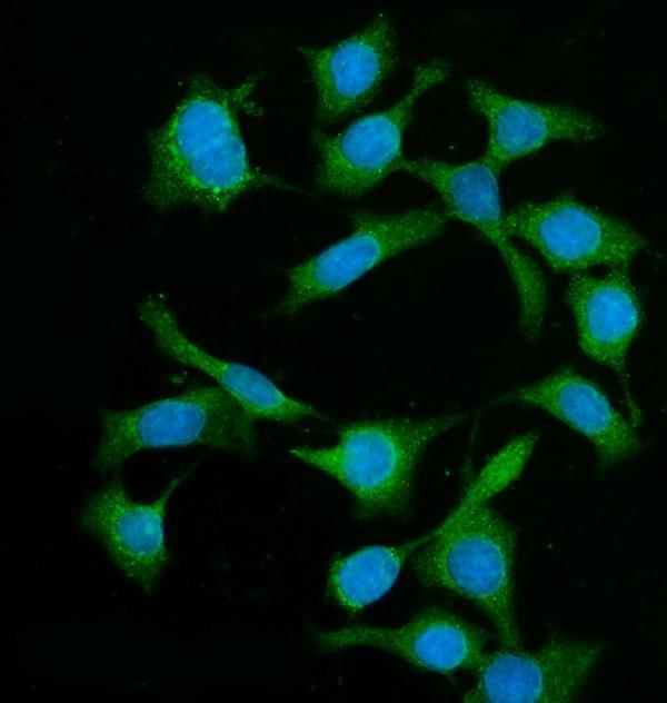
A01551-1 has IHC images from human paraffin sections and an IF image from Caco-2 cells (catalog image captions); its listed reactivity includes human, mouse and rat (catalog reactivity).
A01551-1 is the sole rendered card, with IHC images from human appendiceal adenocarcinoma, gastric adenocarcinoma, lung cancer and brain paraffin sections (catalog IHC captions). Its IF image uses Caco-2 cells, and its listed applications include IHC, IF and ICC (catalog IF caption; catalog applications).
Which to pick: For tissue IHC, choose A01551-1: its own caption documents human paraffin sections, EDTA retrieval at pH 8.0, and 2 μg/ml primary antibody; the fixative is unreported (A01551-1 IHC caption). For IF/ICC, A01551-1 has an IF image in Caco-2 cells using 5 μg/ml primary antibody (A01551-1 IF caption). For cross-species planning, A01551-1 lists human, mouse and rat reactivity, while rabbit monoclonals M01551-3 and M01551-4 list human/mouse and human reactivity, respectively; the supplied IHC and IF images for A01551-1 document human samples only (catalog reactivity; catalog image captions).