BLM / RecQ-like DNA helicase BLM · IHC design guide

Design Immunohistochemistry for BLM

Plan BLM paraffin IHC around the variable nuclear staining seen in most tissues (HPA tissue IHC). This guide covers fixation, detection, and scoring, using the catalog antibody at 1:100–1:300 (datasheet).

Evidence assembled Oct 2026 · For research use; verify linked source records and product datasheet before use
Immunohistochemistry protocol sheet for BLM (IHC for BLM): expected localisation Nuclear staining in most tissues (HPA tissue IHC), antibody A00724, validated IHC image, and IHC protocol steps
Printable BLM IHC protocol sheet — expected localisation Nuclear staining in most tissues (HPA tissue IHC), antibody A00724, controls and protocol steps. Open the full BLM IHC guide →

BLM Immunohistochemistry Experimental Design Guide

Expected localisation, validated protocols, controls and antibodies — the at-a-glance facts below, then the full design guide.

Must know before staining
Expected localisation Nuclear staining in most tissues (HPA tissue IHC)
Staining pattern Variable nuclear staining in most tissues (HPA tissue IHC)
Antigen retrieval Tris-EDTA pH 9.0 HIER, 95–98 °C, 20 min (rule: nuclear antigen)
Positive control ⓘ Adrenal gland+4 more · see all
Negative control ⓘ None in HPA (detected in all 44 tissues); use no-primary + isotype controls
Important caveats
Reasons your staining may differ from the expected pattern.
Fixation Keep fixation conditions consistent across sections. (standard IHC practice; not target-specific)
Caveat Staining and RNA expression show low consistency (HPA tissue IHC)
Regulation HU/CPT redistributes BLM into foci (UniProt)
Isoform / epitope No annotated isoforms; one 1–1417 chain (UniProt)
Section 1

Recommended BLM IHC & IF Protocols

The catalog antibody protocol and three published BLM IHC protocols provide starting points for paraffin sections.

Recommended immunohistochemistry (IHC-P) protocol parameters
SampleParaffin-embedded human lymph node tissue; fixative not specified (datasheet A00724)
FixationImage fixative and duration unreported (datasheet A00724); verify before use.
Sectioning4–5 µm sections on charged slides (standard)
DeparaffinisationXylene, graded ethanol series to water (standard)
Antigen retrievalHeat-induced epitope retrieval in Tris-EDTA buffer, pH 9.0, 20 min at 95–98 °C (standard rule: nuclear antigen)
Peroxidase block3% H2O2, 10 min, room temperature (standard)
Blocking10% normal serum of the secondary host, 30 min, room temperature (standard)
Primary antibodyRabbit anti-BLM, 1:100-1:300 (datasheet A00724)
Primary incubationOvernight at 4 °C (standard)
DetectionHRP-polymer secondary, DAB chromogen 5–10 min (standard)
CounterstainHematoxylin, blue, dehydrate and mount (standard)
Expected resultBLM-positive staining in glandular cells of adrenal gland (HPA tissue IHC: High). HPA tissue profile: Nuclear expression at varying levels in most tissues. No signal in the no-primary control.
💡Decision noteStart with Tris-EDTA pH 9.0 retrieval at 95–98 °C for 20 min (page retrieval rule); the colorectal study used pH 6 HIER (PMC4453952).
Section 2

What Is the Expected BLM Staining Pattern?

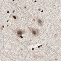
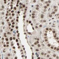
BLM is a nuclear DNA helicase with no transmembrane segment (UniProt P54132). In paraffin sections, expect nuclear staining at varying levels in most tissues (HPA: tissue IHC profile). Reported high-staining cells include glandular, respiratory epithelial, glial and endothelial cells (HPA: tissue IHC). Interpret intensity cautiously: HPA rates the tissue staining Approved but reports low consistency with RNA expression (HPA: reliability description).

What am I looking at on my slide?
Distinct nuclear staining in glandular cells of appendix or breast, with little staining outside nuclei.This fits the expected compartment (UniProt P54132: nucleus) and reported high staining in those cells (HPA: appendix and breast glandular cells, High). Compare cells within the same section before scoring intensity; HPA reports varying nuclear expression across most tissues (HPA: tissue IHC profile).
Predominantly membrane, extracellular or broad cytoplasmic staining, with weak nuclear staining.Treat this as a localization warning in IHC: BLM is nuclear and has no transmembrane segment (UniProt P54132). Check a staining control and detection background before calling it BLM. HPA also reports uncertain cytosolic localization in ICC-IF, so a minor cytosolic IF signal alone is inconclusive (HPA: subcellular).
Strong staining confined to an unexpected cell population while reported high-staining cells remain blank.Investigate antibody cross-reactivity or endogenous detection activity (general IHC practice). First verify cell identity and compare with the relevant HPA image: high staining is reported in several cell classes, not one universal positive cell type (HPA: tissue IHC). HPA supplies no negative tissue in this payload.
Uniform haze over nuclei, cytoplasm and tissue spaces obscures cell boundaries.A diffuse pattern cannot establish BLM localization; the expected tissue pattern is nuclear (HPA: tissue IHC profile; UniProt P54132). Review blocking, antibody concentration, washes and chromogen development with appropriate controls (general IHC practice). Do not assign intensity scores until individual nuclei can be assessed.
No nuclear signal in a section expected to contain HPA high-staining cells.Check tissue identity, section integrity and the assay's positive and detection controls before calling the sample BLM-negative (general IHC practice). HPA reports High staining in appendix glandular cells and bronchial respiratory epithelial cells, but its Approved result has low consistency with RNA expression (HPA: tissue IHC and reliability description).
💡Expected BLM appearanceCall a result positive when identifiable cells show nuclear staining at an intensity appropriate to the sampled tissue—for example, High in appendix glandular cells (HPA: tissue IHC)—and investigate dominant membrane or diffuse extracellular staining as a possible false positive (UniProt P54132: nucleus, no transmembrane segment; general IHC practice).
How each factor affects the staining
Compartment and topologyBLM is nuclear and lacks a transmembrane segment (UniProt P54132); score cellular nuclei rather than outlining membranes.
Tissue and cell variationHPA reports varying nuclear levels in most tissues, including Low in liver cholangiocytes and bone-marrow hematopoietic cells (HPA: tissue IHC).
Antibody evidenceHPA005689 is Approved for IHC, while HPA notes low staining–RNA consistency (HPA: antibody validation; tissue reliability).
DNA damage contextBLM can redistribute into nuclear foci after HU or CPT treatment (UniProt P54132); foci need not define a routine tissue positive.
Fixation evidenceTarget-specific fixation sensitivity is unreported in the supplied UniProt and HPA records; make no BLM-specific retrieval prediction.
Why is my staining missing, weak or wrong?
SituationLikely causeNext action
Reported high-staining cells show no nuclear signal.A failed staining or detection step is possible (general IHC practice); an individual HPA High result does not guarantee every specimen matches it (HPA: low consistency with RNA).Confirm tissue and cell identity; review run controls, retrieval and detection steps, then compare with the relevant HPA tissue image (general IHC practice; HPA: tissue IHC).
Most cells show uniform brown background.Non-specific antibody binding, incomplete blocking or endogenous detection activity can create background (general IHC practice).Review blocking and washes; use appropriate no-primary and detection controls before scoring nuclei (general IHC practice).
Signal is strongest on cell borders or outside cells.That distribution conflicts with nuclear BLM and its lack of a transmembrane segment (UniProt P54132).Check controls and staining specificity; score only convincing nuclear signal in identifiable cells (general IHC practice; HPA: nuclear tissue profile).
Bone marrow looks weaker than its tissue-enhanced RNA label suggests.RNA and IHC measure different readouts; HPA reports Low staining in hematopoietic cells and low staining–RNA consistency (HPA: tissue IHC).Interpret the observed nuclei against the HPA protein staining record, recording cell type and intensity separately from RNA specificity (HPA: tissue IHC).
Punctate nuclear staining appears in a damage-treated sample.BLM can accumulate in nuclear DNA damage-induced foci after HU or CPT treatment (UniProt P54132).Record treatment and assess whether puncta remain nuclear; compare with an appropriate untreated control before interpreting the change (UniProt P54132; general IHC practice).
IF/ICC: Where should BLM appear?HPA supports nucleoplasmic localization and lists additional cytosolic localization as uncertain (HPA: subcellular).Expect primarily nucleoplasmic signal; treat cytosolic signal cautiously. HPA005689 has Supported ICC validation (HPA: antibody validation and subcellular).

Sample controls for BLM IHC & IF

🧪Run breast first and score its glandular cells for nuclear staining (HPA: High in breast glandular cells; UniProt P54132: nucleus). HPA detects BLM in all 44 scored tissues, so use no-primary and isotype controls instead of a negative tissue; on the positive slide, cells without nuclear signal should show only background staining, but no cell type can be designated a confirmed internal negative (HPA: no negative tissue rows; HPA: BLM detected in all 44 scored tissues).
Positive control tissue: Adrenal gland (Glandular cells, HPA High)
Negative control tissue: None in HPA: BLM is detected in all 44 scored tissues. Use a no-primary (secondary-only) and an isotype control instead.
ICC-IF cell lines (HPA subcellular resource): HPA ICC-IF images show BLM in A-431, U-251MG, U2OS, with annotated localisation: Nucleoplasm (supported) (HPA subcellular).
Technical controls: Include a secondary-only slide without primary antibody and an isotype control matched to the primary antibody’s host species and clonality; a BLM knockout is a biological specificity control, while peptide blocking is documented for the catalog antibody in lymph node IHC (A00724 tissue-IHC caption: peptide block). Quench endogenous peroxidase for chromogenic detection, and block endogenous biotin if using biotin-based detection (standard IHC practice).
⚠️Feasibility: A target-specific fixation window and the effect of antigen retrieval are unreported in the supplied evidence; optimize retrieval on the selected paraffin sections (supplied target/application evidence: no fixation-window or retrieval comparison). The selected A00724 paraffin-section caption does not state a fixative (A00724 tissue-IHC caption: fixative unreported). The supplied evidence does not establish that frozen sections or IF are easier than paraffin IHC; for breast, check that stromal background does not obscure glandular-cell scoring (HPA: High in breast glandular cells; standard IHC practice).

HPA tissue IHC evidence for BLM

Comprehensive Human Protein Atlas IHC scoring per tissue (reliability: Approved — Low consistency between antibody staining and RNA expression data.). Rows are taken directly from the HPA tissue chart — click any row's HPA link to view the source.

Positive expression · recommended positive controls

TissueCell typeLevelEvidenceSource
Adrenal gland Glandular cells High Protein (IHC) HPA →
Appendix Glandular cells High Protein (IHC) HPA →
Breast Glandular cells High Protein (IHC) HPA →
Bronchus Respiratory epithelial cells High Protein (IHC) HPA →
Caudate Glial cells High Protein (IHC) HPA →

Undetected expression · recommended negative controls

TissueCell typeLevelEvidenceSource
None in HPA: BLM is detected in all 44 scored tissues. Use a no-primary (secondary-only) and an isotype control instead.
Section 3

Advanced BLM IHC Tips

Troubleshoot BLM staining in paraffin section IHC by checking retrieval, nuclear localisation, controls and scoring; use the IF guidance when planning multiplex imaging.

How should I retrieve BLM when nuclear staining is weak in paraffin sections?
Start with Tris-EDTA at pH 9.0, heated to 95–98 °C for 20 min (page retrieval rule: nuclear antigen). Keep section thickness, heating vessel and cooling procedure consistent across the test and control slides so differences in staining can be attributed to the retrieval change (standard IHC practice). If nuclear staining remains weak, compare a shorter heating interval or a citrate buffer at pH 6.0 on adjacent sections while monitoring morphology (standard IHC practice). Judge improvement by nuclear staining in an appropriate control, since BLM is nuclear and the supported HPA subcellular location is nucleoplasmic (UniProt P54132 localisation; HPA: supported nucleoplasm).
Could fixation explain absent BLM staining in my paraffin sections?
BLM-specific sensitivity to fixative type or fixation duration is unknown from the supplied evidence (selected A00724 caption: fixative not stated). The A00724 image documents staining in paraffin-embedded human lymph node and a synthesized-peptide block, but gives no fixative or fixation time (selected A00724 tissue-IHC caption). Record the actual fixative and processing history, then compare similarly processed control and test sections under one retrieval and detection setup (standard IHC practice). If fixation varies across specimens, interpret weak staining cautiously and assess nuclear morphology alongside the expected nuclear pattern; neither the tissue image nor BLM's topology establishes a fixation effect (UniProt P54132 localisation; selected A00724 caption).
Should BLM staining appear throughout the cell or mainly in nuclei?
Expect predominantly nuclear chromogenic staining: BLM is annotated in the nucleus, and HPA supports a mainly nucleoplasmic location (UniProt P54132 localisation; HPA: supported nucleoplasm). Score nuclear signal separately from cytoplasmic colour, because HPA lists its additional cytosolic location as uncertain (HPA: uncertain cytosol). If sections show discrete nuclear puncta, record their frequency and treatment context; BLM can redistribute with SPIDR into DNA damage induced nuclear foci after hydroxyurea or camptothecin exposure (UniProt P54132 localisation). Diffuse cytoplasmic staining without convincing nuclear signal warrants review of primary-antibody specificity, retrieval and detection controls before it is called BLM expression (UniProt P54132 localisation; standard IHC practice).
How can I assess whether the antibody epitope affects BLM IHC?
Check the catalog antibody's stated immunogen or epitope before interpreting a loss of staining, because the supplied record does not map that antibody's binding site (catalog evidence supplied: epitope not stated). BLM has 1,417 amino acids, no annotated isoforms in this record, and no transmembrane segment (UniProt P54132 record). Its annotated helicase ATP-binding, helicase C-terminal and HRDC regions occupy residues 676–851, 877–1024 and 1212–1292, respectively (UniProt P54132 domains). A peptide-blocked A00724 lymph-node image supports competition by the synthesized peptide, but does not establish recognition of every protein state or independently prove cellular specificity (selected A00724 tissue-IHC caption; standard IHC interpretation).
How should I adapt the BLM localisation check for multiplex IF?
Pair BLM with a validated marker for the cell population being assessed, and use a nuclear counterstain to confirm that candidate BLM signal falls within nuclei (UniProt P54132 localisation; standard IF practice). Choose spectrally separated fluorophores and inspect unstained tissue for autofluorescence before assigning a weak channel to BLM (standard IF practice). Because BLM is nuclear and has no transmembrane segment, use a compatible permeabilisation step to give antibody access across cellular and nuclear barriers, then verify that nuclear morphology remains intact (UniProt P54132 topology and localisation; standard IF practice). Confirm each channel with single-stain and secondary-only controls, especially when interpreting discrete puncta as possible damage-associated BLM foci (UniProt P54132 localisation; standard IF practice).
How can I distinguish weak nuclear BLM signal from chromogenic background?
Run a no-primary control through the same secondary reagent and chromogen steps to reveal detection-system background (standard IHC practice). Include a peroxidase block when using an HRP and DAB workflow, and compare pigment or endogenous enzyme deposits with the no-primary slide before scoring nuclei (standard IHC practice). Reduce nonspecific staining by checking antibody concentration, wash stringency and protein blocking on matched sections, while preserving the page retrieval condition during that comparison (standard IHC practice; page retrieval rule: nuclear antigen). The A00724 synthesized-peptide-blocked lymph-node image is a useful competition control, but background in a new specimen still needs its own controls (selected A00724 tissue-IHC caption; standard IHC practice).
What is a defensible way to quantify BLM staining across sections? ⚠ ANSWER MARKED FOR VERIFICATION
Define the tissue compartment and cell population before scoring, then report the percentage of nuclei positive for BLM alongside an intensity-based nuclear H-score (UniProt P54132 localisation; standard IHC scoring practice). An H-score can weight the percentages of nuclei at intensity grades 0–3, yielding a 0–300 scale when those percentages sum to 100% (standard IHC scoring practice). For spatial questions, report positive nuclei per mm² of viable, annotated tissue and normalise to the number of evaluable nuclei or tissue area specified in advance (standard IHC scoring practice). Use the same exposure-free chromogenic assessment, counterstain criteria and exclusion rules across slides; HPA reports varying nuclear expression across tissues and low staining–RNA consistency (HPA: tissue IHC profile and Approved reliability description; standard IHC practice).
When should an apparent BLM-positive area be treated as artefact?
A credible BLM-positive cell should show interpretable nuclear staining within intact tissue architecture, consistent with its nuclear annotation and HPA's supported nucleoplasmic location (UniProt P54132 localisation; HPA: supported nucleoplasm). Recheck isolated cytoplasmic colour, stained cut edges and necrotic regions against adjacent intact areas and the no-primary control before counting them (HPA: uncertain cytosol; standard IHC practice). In an HRP and DAB assay, residual endogenous peroxidase or pigment can imitate chromogen, so inspect the peroxidase-blocked control and tissue morphology (standard IHC practice). Interpret cell identity within its histological compartment: HPA reports high staining in several listed glandular populations but low staining in bone-marrow hematopoietic cells, with low staining–RNA consistency overall (HPA: tissue IHC profile and Approved reliability description).
Boster reagents

Best BLM / RecQ-like DNA helicase BLM IHC Antibodies

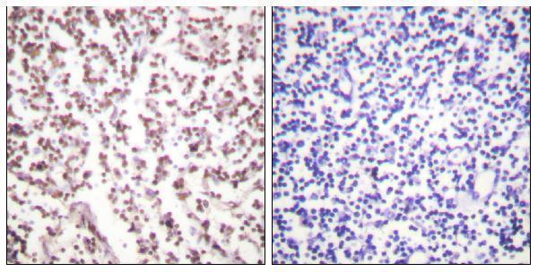
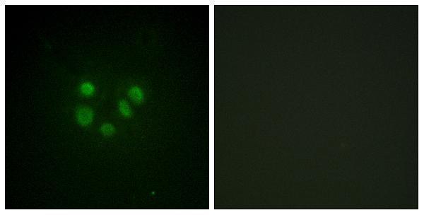
A00724 has IHC data from paraffin-embedded human lymph node and IF data from A549 cells (catalog image captions); its listed reactivity is human and mouse (catalog: reactivity).

Real IHC data Immunohistochemistry analysis of paraffin-embedded human lymph node tissue, using Bloom Syndrome Antibody. The picture on the right is blocked with the synthesized peptide.
Anti-Bloom syndrome protein BLM Antibody
Cat # A00724

A00724 will render with an IHC figure of paraffin-embedded human lymph node; the caption describes peptide blocking in a companion image (catalog: IHC image caption). A00724 also lists IF and shows A549-cell staining with a peptide-blocked companion image (catalog: applications; IF image caption).

Which to pick: For tissue IHC, choose A00724: its own figure shows paraffin-embedded human lymph node staining (catalog: IHC image caption); the fixative is unreported (catalog: IHC image caption). For IF/ICC, A00724 has an IF figure in A549 cells (catalog: IF image caption), while cross-species use is supported by listed human and mouse reactivity, with image data shown only for human tissue and A549 cells (catalog: reactivity; image captions). Clonality is unreported (catalog: clone field).

Each figure is that product's own IHC / IF validation image from its datasheet.

References

  1. UniProt Consortium. UniProt entry P54132 (BLM_HUMAN, RecQ-like DNA helicase BLM).
  2. Human Protein Atlas. BLM tissue IHC expression (reliability: Approved).
  3. Human Protein Atlas. BLM subcellular location (ICC-IF): Mainly localized to the nucleoplasm. In addition localized to the cytosol..
  4. Human Protein Atlas. BLM antibody validation summary (1 antibodies).
  5. Tailoring heated intraperitoneal mitomycin C for peritoneal metastases originating from colorectal carcinoma: a translational approach to improve survival. British journal of cancer 2015 — PMC4453952.
  6. Random-forest algorithm based biomarkers in predicting prognosis in the patients with hepatocellular carcinoma. Cancer cell international 2020 — PMC7302385.
  7. Sulforaphane attenuates pulmonary fibrosis by inhibiting the epithelial-mesenchymal transition. BMC pharmacology & toxicology 2018 — PMC5879815.
  8. Downregulation of BLM RecQ helicase inhibits proliferation, promotes the apoptosis and enhances the sensitivity of bladder cancer cells to cisplatin. Molecular medicine reports 2022 — PMC9437972.
  9. PubMed PMID:7585968 — UniProt-cited evidence.
  10. PubMed PMID:9388193 — UniProt-cited evidence.
  11. PubMed PMID:15489334 — UniProt-cited evidence.