This website uses cookies to ensure you get the best experience on our website.


- Table of Contents
Plan BLZF1 paraffin IHC using colon glandular cells as a high-staining reference (HPA tissue IHC). Start with the catalog antibody at 1:100–1:300 and include a peptide-absorbed negative control (datasheet A09997).

Expected localisation, validated protocols, controls and antibodies — the at-a-glance facts below, then the full design guide.
| Expected localisation | Nuclear and cytoplasmic tissue staining (HPA tissue IHC) | |
| Staining pattern | Colon glandular cells show nuclear and cytoplasmic staining (HPA tissue IHC) | |
| Antigen retrieval | Tris-EDTA pH 8.0 HIER, heat-mediated (datasheet A09997) | |
| Positive control | Cerebral cortex+4 more · see all | |
| Negative control | Adipose tissue |
| Fixation | Keep fixation consistent across sections (standard IHC practice; not target-specific); optimize empirically. | |
| Caveat | A discrete Golgi pattern may be hard to distinguish in tissue IHC (HPA tissue IHC; UniProt) | |
| Regulation | Low tissue specificity (HPA tissue RNA) | |
| Isoform / epitope | 2 isoforms; epitope impact is undetermined (UniProt) |
The catalog antibody’s IHC-P protocol (datasheet A09997) and one published BLZF1 IHC protocol (PMC13106729) provide starting conditions for paraffin sections.
| Sample | Paraffin-embedded Human brain tissue; fixative not specified (datasheet A09997) |
| Fixation | Image fixative and duration unreported (datasheet A09997); verify before use. |
| Sectioning | 4–5 µm sections on charged slides (standard) |
| Deparaffinisation | Xylene, graded ethanol series to water (standard) |
| Antigen retrieval | Heat retrieval: Tris-EDTA pH 8.0 (datasheet A09997); 20 min, 95–100 °C (standard) |
| Peroxidase block | 3% H2O2, 10 min, room temperature (standard) |
| Blocking | 10% normal serum of the secondary host, 30 min, room temperature (standard) |
| Primary antibody | Rabbit anti-BLZF1, 1:100 - 1:300 (datasheet A09997) |
| Primary incubation | Overnight at 4 °C (standard) |
| Detection | HRP-polymer secondary, DAB chromogen 5–10 min (standard) |
| Counterstain | Hematoxylin, blue, dehydrate and mount (standard) |
| Expected result | BLZF1-positive staining in neuropil of cerebral cortex (HPA tissue IHC: High). HPA tissue profile: General nuclear and cytoplasmic expression. No signal in the no-primary control. |
BLZF1 is mainly Golgi associated, with additional nucleoplasmic localization in ICC-IF (HPA: subcellular). Paraffin tissue IHC shows broader nuclear and cytoplasmic staining (HPA: tissue IHC). Expect staining in selected epithelial, tubular, alveolar, neural and germ cells listed below (HPA: tissue IHC). UniProt annotates a Golgi membrane association without a transmembrane segment (UniProt Q9H2G9 topology). HPA rates the tissue pattern Supported, with medium consistency between staining and RNA data (HPA: tissue IHC).
| Distinct staining in colon or duodenal glandular cells, kidney tubular cells, or esophageal squamous cells. | These are reported High IHC sites (HPA: tissue IHC). Score the named cell population against adjacent tissue and the negative control. A nuclear and cytoplasmic appearance fits the reported paraffin-section profile (HPA: tissue IHC); a resolved Golgi pattern is not required for an IHC-positive call. |
| Staining appears only in an unexpected compartment, with no convincing signal in the expected cells. | Check identity and specificity before interpreting it as BLZF1. Tissue IHC reports general nuclear and cytoplasmic expression (HPA: tissue IHC), while ICC-IF places BLZF1 mainly at the Golgi and additionally in nucleoplasm (HPA: subcellular). The two imaging methods offer different spatial detail. |
| Strong staining is confined to a cell population outside the reported positive populations, or adipocytes stain strongly. | Consider cross-reactivity or endogenous detection activity. Adipocytes are reported Not detected (HPA: adipose tissue IHC), whereas the named positive cells have tissue-specific observations (HPA: tissue IHC). Compare a no-primary control before assigning the unexpected signal to BLZF1 (general IHC practice). |
| Pale, diffuse chromogen covers many cells and the extracellular area, obscuring cell boundaries. | Treat this as background until controls establish a cellular pattern (general IHC practice). The HPA tissue profile describes nuclear and cytoplasmic expression, not an indiscriminate haze (HPA: tissue IHC). Review blocking, washes, detection reagent exposure and chromogen development using the same section conditions. |
| No interpretable staining appears in a section containing a reported High cell population. | A negative run in colon glandular cells or kidney tubular cells is inconclusive without a functioning positive control (HPA: tissue IHC; general IHC practice). Review section quality, the catalog antibody's IHC-P instructions, detection reagents and counterstain before concluding that the specimen lacks BLZF1. |
| Which tissue gives an informative comparison? | Colon and duodenal glandular cells, kidney tubular cells, and testis pachytene spermatocytes are reported High; adrenal glandular cells are Medium; adipocytes are Not detected (HPA: tissue IHC). Compare cell types within a section rather than treating an entire organ as uniformly positive or negative. |
| Does IF/ICC show the same level of detail? | ICC-IF places BLZF1 mainly at the Golgi apparatus and additionally in nucleoplasm (HPA: subcellular). Paraffin tissue IHC is summarized as general nuclear and cytoplasmic staining (HPA: tissue IHC). Use the separate IF/ICC guide for that application; a Golgi-resolved pattern is not a prerequisite for interpreting chromogenic IHC. |
| How strong is the antibody-pattern evidence? | HPA lists IHC as Supported for HPA025703 and HPA027331, and ICC as Supported for HPA067113 (HPA: antibodies). Its overall tissue reliability is Supported with medium staining-to-RNA consistency (HPA: tissue IHC). These ratings support comparison with the observed pattern; they do not establish specificity in every specimen. |
| Can topology or processing predict section behavior? | BLZF1 has no transmembrane segment, signal peptide, propeptide, or annotated glycosylation site; the annotated chain spans residues 1–400 (UniProt Q9H2G9). These facts do not identify the antibody epitope or establish antigen-retrieval needs or fixation sensitivity. Target-specific fixation sensitivity is unreported in the supplied sources. |
| Do variants or modifications change the expected stain? | UniProt lists two isoforms and modified residues including phosphoserines and a phosphothreonine (UniProt Q9H2G9). The supplied sources do not map the catalog antibody epitope to an isoform or modification. Do not attribute an unusual cellular pattern or a negative section to either without separate evidence. |
| Situation | Likely cause | Next action |
|---|---|---|
| Reported High cells are unstained. | The run may have failed, or the chosen section may lack an adequate internal positive population (HPA: tissue IHC; general IHC practice). | Check a section containing a named High population, such as colon glandular cells, and verify the catalog antibody's IHC-P conditions and detection controls (HPA: tissue IHC; general IHC practice). |
| All cell types carry similar weak chromogen. | Nonspecific reagent binding, excessive detection signal or insufficient washing can produce diffuse background (general IHC practice). | Compare the no-primary control; review blocking, washes and development time before scoring cell-specific staining (general IHC practice). |
| Adipocytes stain as strongly as reported positive cells. | Cross-reactivity or endogenous detection activity is possible; adipocytes are reported Not detected (HPA: adipose tissue IHC). | Check the no-primary and detection controls, then reassess whether staining tracks cell boundaries and the expected positive populations (general IHC practice; HPA: tissue IHC). |
| A nuclear or cytoplasmic IHC signal lacks a crisp Golgi shape. | HPA describes general nuclear and cytoplasmic tissue IHC, while its Golgi localization comes from ICC-IF (HPA: tissue IHC; HPA: subcellular). | Judge the paraffin section against the reported IHC cell populations and controls; use the separate IF/ICC guide if resolving the Golgi compartment is the experimental aim. |
| Only the counterstain is visible in both specimen and control. | A missing or inactive primary or detection step is possible (general IHC practice). | Verify reagent order and activity against a known-positive section before interpreting BLZF1 expression in the specimen (general IHC practice; HPA: tissue IHC). |
| The cell pattern differs between runs. | Variation in section selection, staining steps or scoring can change the apparent result (general IHC practice). HPA reports medium staining-to-RNA consistency (HPA: tissue IHC). | Repeat with matched controls and record the named cell type, compartment and intensity in each section; compare the same populations across runs (general IHC practice; HPA: tissue IHC). |
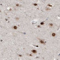
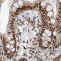
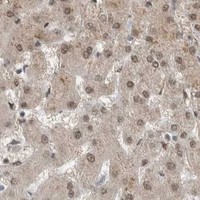
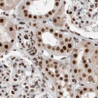
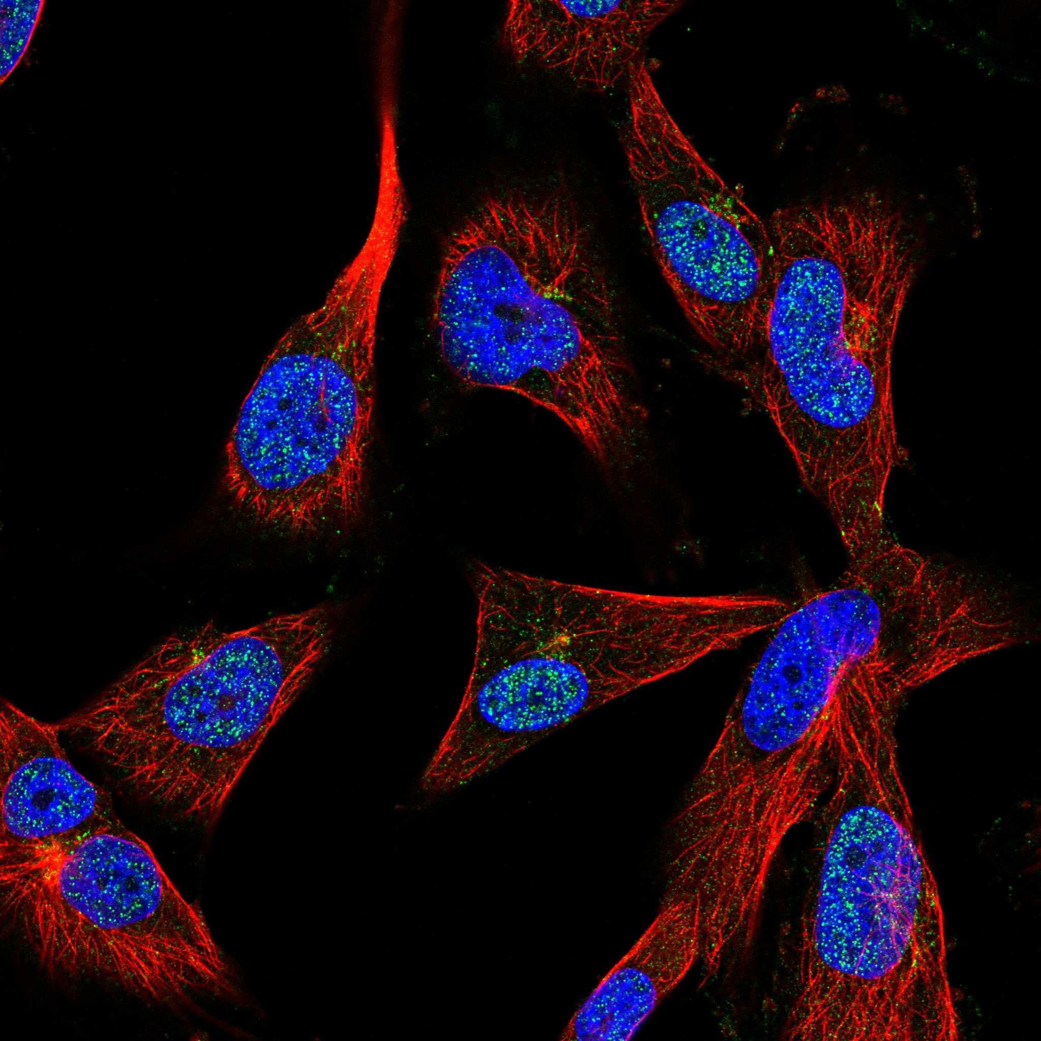
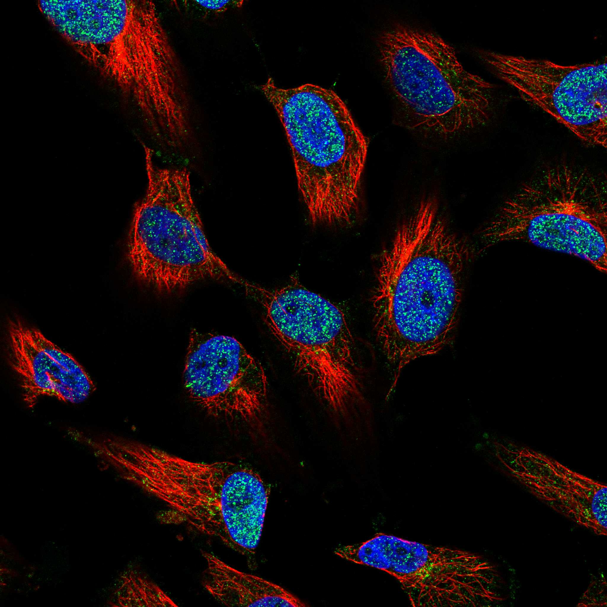
Comprehensive Human Protein Atlas IHC scoring per tissue (reliability: Supported — Medium consistency between antibody staining and RNA expression data.). Rows are taken directly from the HPA tissue chart — click any row's HPA link to view the source.
| Tissue | Cell type | Level | Evidence | Source |
|---|---|---|---|---|
| Cerebral cortex | Neuropil | High | Protein (IHC) | HPA → |
| Colon | Glandular cells | High | Protein (IHC) | HPA → |
| Duodenum | Glandular cells | High | Protein (IHC) | HPA → |
| Esophagus | Squamous epithelial cells | High | Protein (IHC) | HPA → |
| Kidney | Cells in tubules | High | Protein (IHC) | HPA → |
| Tissue | Cell type | Level | Evidence | Source |
|---|---|---|---|---|
| Adipose tissue | Adipocytes | Not detected | Protein (IHC) | HPA → |
Troubleshoot BLZF1 staining in paraffin sections by checking retrieval, tissue preservation, compartment pattern and controls before interpreting chromogenic signal.
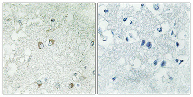
A09997 has an IHC image of a human brain paraffin section (A09997 IHC caption). The catalog lists human and mouse IHC/IF reactivity, plus rat reactivity for A09997-2 (catalog applications/reactivity).
The rendered SKU, A09997, has an IHC image from a human brain paraffin section (A09997 IHC caption). Its catalog lists IHC, IF and ICC applications for human and mouse samples; no IF image is supplied (A09997 catalog applications/reactivity; catalog image records).
Which to pick: For tissue IHC, start with A09997: its own image documents a human brain paraffin section at 1:100 with Tris-EDTA pH 8.0 retrieval (A09997 IHC caption). For IF/ICC, A09997 lists both applications for human and mouse, while A09997-2 lists IF but not ICC (catalog applications/reactivity). For rat samples, A09997-2 lists rat reactivity and IHC/IF applications (A09997-2 catalog applications/reactivity); the fixative in A09997’s paraffin-section image is unreported (A09997 IHC caption).