This website uses cookies to ensure you get the best experience on our website.


- Table of Contents
Plan chromogenic BMP1 IHC on paraffin sections using the reported cytoplasmic tissue pattern (HPA tissue IHC). Compare high staining in colon glandular cells with undetected staining in adipose tissue adipocytes, while considering BMP1 secretion when interpreting location (HPA tissue IHC; UniProt).

Expected localisation, validated protocols, controls and antibodies — the at-a-glance facts below, then the full design guide.
| Expected localisation | Cytoplasmic staining in tissue sections (HPA tissue IHC) | |
| Staining pattern | Cytoplasmic staining in most tissues (HPA tissue IHC) | |
| Antigen retrieval | Citrate pH 6.0 HIER, 95–98 °C, 20 min (rule: cytoplasmic / membrane antigen) | |
| Positive control | Colon+4 more · see all | |
| Negative control | Adipose tissue+4 more · see all |
| Fixation | Keep fixation consistent across sections. (standard IHC practice; not target-specific) | |
| Caveat | Secreted BMP1 may stain away from producer cells (HPA tissue IHC) | |
| Regulation | Low tissue specificity; no regulator specified (HPA tissue IHC) | |
| Isoform / epitope | 7 isoforms; precursor processing may affect epitope coverage (UniProt) |
The catalog antibody’s IHC-P protocol is followed by four published BMP1 IHC examples covering ovarian, lung, dental and keloid samples (PMC5326928; PMC8971496; PMC6635762; PMC5466890).
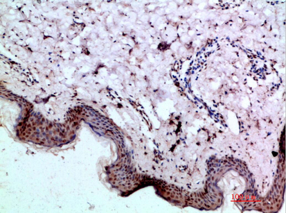
| Sample | Paraffin-embedded human skin tissue; fixative not specified (datasheet A02436) |
| Fixation | Image fixative and duration unreported (datasheet A02436); verify before use. |
| Sectioning | 4–5 µm sections on charged slides (standard) |
| Deparaffinisation | Xylene, graded ethanol series to water (standard) |
| Antigen retrieval | Heat-induced epitope retrieval in citrate buffer, pH 6.0, 20 min at 95–98 °C (standard rule: cytoplasmic / membrane antigen) |
| Peroxidase block | 3% H2O2, 10 min, room temperature (standard) |
| Blocking | 10% normal serum of the secondary host, 30 min, room temperature (standard) |
| Primary antibody | Rabbit anti-BMP1, 1:50 recommended; image 1:200 (datasheet A02436) |
| Primary incubation | Overnight at 4 °C (standard) |
| Detection | HRP-polymer secondary, DAB chromogen 5–10 min (standard) |
| Counterstain | Hematoxylin, blue, dehydrate and mount (standard) |
| Expected result | BMP1-positive staining in glandular cells of colon (HPA tissue IHC: High). HPA tissue profile: Cytoplasmic expression in most tissues. No signal in the no-primary control. |
BMP1 staining is predominantly cytoplasmic across tissues, with strong signals reported in intestinal glandular cells, cardiomyocytes and placental trophoblastic cells (HPA: tissue IHC, Enhanced reliability). BMP1 also localizes to the Golgi and is secreted into extracellular space and matrix; it has no transmembrane segment (UniProt P13497: subcellular location and topology). HPA notes only medium agreement between antibody staining and RNA expression because secreted protein can accumulate away from its source (HPA: reliability description).
| Strong cytoplasmic staining in colon, duodenum or small-intestinal glandular cells; cardiomyocytes or trophoblastic cells also stain. | These are the strongest listed tissue and cell-type benchmarks (High; HPA: tissue IHC). A Golgi-associated component or extracellular signal can fit BMP1 biology, but assess it alongside tissue architecture and controls (UniProt P13497: subcellular location). |
| Staining appears exclusively nuclear or sharply restricted to the plasma membrane. | That distribution does not match the reported cytoplasmic tissue pattern or Golgi, secreted and extracellular locations; BMP1 has no transmembrane segment (HPA: tissue IHC; UniProt P13497: subcellular location and topology). Check whether the same pattern persists in controls before interpreting it as BMP1. |
| Adipocytes or squamous epithelial cells stain strongly while expected glandular cells remain weak. | HPA reports BMP1 as not detected in adipocytes from adipose tissue and breast, and in squamous epithelial cells from cervix, esophagus and oral mucosa (HPA: tissue IHC). Consider cross-reactivity or detection activity; these cell-specific comparisons cannot exclude biological variation. |
| Diffuse chromogen covers stroma and cells without a readable cell-associated pattern. | BMP1 can enter extracellular matrix, so stromal signal alone is not proof of an artifact (UniProt P13497: subcellular location and POSTN interaction). Uniform staining that obscures architecture warrants a no-primary control and review of detection background (general IHC practice). |
| No signal is visible in colon or duodenal glandular cells processed with the study sections. | That conflicts with the reported High staining in those cells (HPA: tissue IHC). Review the control slide, antibody conditions, retrieval and detection before scoring study samples negative (general IHC practice). HPA's Enhanced rating still carries medium staining-to-RNA consistency for this secreted protein (HPA: reliability description). |
| Tissue and cell-type benchmark | HPA reports High staining in colon, duodenum and small-intestinal glandular cells, cardiomyocytes and trophoblastic cells; several other cell types are Medium, Low or not detected (HPA: tissue IHC). Compare the relevant cell population, rather than treating an entire tissue as uniformly positive. |
| Secretion and processing | BMP1 has a signal peptide at residues 1–22, a propeptide at 23–120 and a listed mature chain at 121–986 (UniProt P13497: processing). Its Golgi and extracellular locations mean intracellular and deposited staining need different spatial interpretations (UniProt P13497: subcellular location). |
| Epitope and isoform coverage | UniProt lists 7 BMP1 isoforms and proteolytic processing (UniProt P13497: isoforms and processing). The supplied antibody record gives no epitope, so which isoforms or processed forms contribute to staining is unresolved; avoid assigning a compartment to a particular form. |
| Antibody validation | The listed antibody HPA014572 has an Enhanced IHC assessment, meaning its pattern has support from independent antibodies or orthogonal data (HPA: antibody validation). HPA separately describes medium consistency between staining and RNA expression for secreted BMP1 (HPA: reliability description). |
| IF/ICC Q: Is a specific cellular pattern established? | A: No main location or cell-line ICC-IF images are supplied; HPA summarizes BMP1 as secreted (HPA: subcellular ICC-IF). The tissue IHC pattern should not be presented as an independently confirmed IF/ICC pattern. |
| Situation | Likely cause | Next action |
|---|---|---|
| The known-positive glandular control is blank. | The result conflicts with reported High glandular staining in colon and duodenum (HPA: tissue IHC); the failed step cannot be identified from appearance alone. | Check that the expected cells are present, then review antibody dilution, antigen retrieval, detection reagents and run controls in order (general IHC practice). |
| Signal is very weak in a cell type listed as Low. | Low staining is reported for several populations, including parathyroid glandular cells and bronchial respiratory epithelial cells (HPA: tissue IHC). Weak staining there is a limited sensitivity check. | Evaluate a listed High cell population in the same run before changing conditions (HPA: tissue IHC; general IHC practice). |
| Nuclear or membrane-edge staining dominates. | This distribution departs from the cytoplasmic HPA profile and BMP1's reported Golgi, secreted and extracellular locations (HPA: tissue IHC; UniProt P13497: subcellular location). | Compare the no-primary slide and expected positive cells; reassess specificity if the unusual pattern persists with the primary antibody (general IHC practice). |
| The slide has widespread brown haze, including cells expected to be negative. | Adipocytes and several squamous epithelial populations are listed as not detected (HPA: tissue IHC). Widespread color can also reflect detection background (general IHC practice). | Inspect a no-primary control; review blocking, washes and chromogen development if it also shows haze (general IHC practice). |
| Stromal deposits stain, but nearby cells show little signal. | BMP1 is secreted into extracellular matrix, and POSTN interaction promotes its matrix deposition (UniProt P13497: subcellular location and subunit). Protein location can therefore differ from RNA location (HPA: reliability description). | Record extracellular and cellular staining separately, then compare morphology and controls before assigning the deposit to BMP1 (general IHC practice). |
| Two sections give different intensities despite similar tissue labels. | HPA levels refer to specified cell types, and its BMP1 staining has medium agreement with RNA expression (HPA: tissue IHC and reliability description). A tissue label alone does not establish matching cell composition. | Score the same identifiable cell population in both sections and check run controls before attributing the difference to BMP1 abundance (general IHC practice). |


Comprehensive Human Protein Atlas IHC scoring per tissue (reliability: Enhanced — Medium consistency between antibody staining and RNA expression data. Secreted protein, tissue location of RNA and protein is expected to differ.). Rows are taken directly from the HPA tissue chart — click any row's HPA link to view the source.
| Tissue | Cell type | Level | Evidence | Source |
|---|---|---|---|---|
| Colon | Glandular cells | High | Protein (IHC) | HPA → |
| Duodenum | Glandular cells | High | Protein (IHC) | HPA → |
| Heart muscle | Cardiomyocytes | High | Protein (IHC) | HPA → |
| Placenta | Trophoblastic cells | High | Protein (IHC) | HPA → |
| Small intestine | Glandular cells | High | Protein (IHC) | HPA → |
| Tissue | Cell type | Level | Evidence | Source |
|---|---|---|---|---|
| Adipose tissue | Adipocytes | Not detected | Protein (IHC) | HPA → |
| Breast | Adipocytes | Not detected | Protein (IHC) | HPA → |
| Cervix | Squamous epithelial cells | Not detected | Protein (IHC) | HPA → |
| Esophagus | Squamous epithelial cells | Not detected | Protein (IHC) | HPA → |
| Liver | Cholangiocytes | Not detected | Protein (IHC) | HPA → |
Troubleshoot BMP1 staining in paraffin sections by checking retrieval, cellular and extracellular localisation, and controls before interpreting chromogenic signal (UniProt P13497; HPA tissue IHC).
A02436 has a paraffin-section IHC image from human skin (catalog: IHC image caption), lists IF without an IF image (catalog: applications and IF image alts), and lists human, mouse, and rat reactivity (catalog: reactivity).
A02436 will render with an IHC image of paraffin-embedded human skin, stained at 1:200 (catalog: IHC image caption). It is listed for IHC and IF in human, mouse, and rat, but has no supplied IF image (catalog: applications, reactivity, and IF image alts).
Which to pick: Choose A02436 for paraffin-section tissue IHC; its own image documents human skin, while the fixative is unreported (catalog: IHC image caption). For IF, A02436 is listed as applicable, but no IF image or ICC validation is supplied (catalog: applications and IF image alts). For work across human, mouse, and rat, A02436 is the listed polyclonal option; those species are catalog reactivity claims, while its supplied IHC image shows human tissue only (catalog: dilution_raw, reactivity, and IHC image caption).