BMP4 / Bone morphogenetic protein 4 · IHC design guide

Design Immunohistochemistry for BMP4

Plan paraffin BMP4 IHC using smooth muscle staining and placental extracellular positivity as reference patterns (HPA tissue IHC). This guide covers fixation consistency, the catalog antibody workflow, and interpretation of secreted protein staining (datasheet M00321; UniProt).

Evidence assembled Oct 2026 · For research use; verify linked source records and product datasheet before use
Immunohistochemistry protocol sheet for BMP4 (IHC for BMP4): expected localisation Extracellular in placenta (HPA tissue IHC), antibody M00321, validated IHC image, and IHC protocol steps
Printable BMP4 IHC protocol sheet — expected localisation Extracellular in placenta (HPA tissue IHC), antibody M00321, controls and protocol steps. Open the full BMP4 IHC guide →

BMP4 Immunohistochemistry Experimental Design Guide

Expected localisation, validated protocols, controls and antibodies — the at-a-glance facts below, then the full design guide.

Must know before staining
Expected localisation Extracellular in placenta (HPA tissue IHC)
Staining pattern Smooth muscle cells stain; placenta shows extracellular signal (HPA tissue IHC)
Antigen retrieval Citrate pH 6 HIER, heat-mediated (datasheet M00321)
Positive control ⓘ Adrenal gland+4 more · see all
Negative control ⓘ Adipose tissue+4 more · see all
Important caveats
Reasons your staining may differ from the expected pattern.
Fixation Keep fixation conditions consistent across sections. (standard IHC practice; not target-specific)
Caveat Secreted BMP4 may stain away from its source cells (UniProt)
Regulation Self-induction via MSX1 in dental cells (UniProt)
Isoform / epitope 0 isoforms; cleavage separates propeptide and mature BMP4 (UniProt)
Section 1

Recommended BMP4 IHC & IF Protocols

The catalog antibody’s IHC-P protocol is paired with four published BMP4 immunohistochemistry protocols (PMC3851443; PMC6337410; PMC10240375; PMC7140810).

Recommended immunohistochemistry (IHC-P) protocol parameters
SampleParaffin-embedded human breast cancer tissue; fixative not specified (datasheet M00321)
FixationImage fixative and duration unreported (datasheet M00321); verify before use.
Sectioning4–5 µm sections on charged slides (standard)
DeparaffinisationXylene, graded ethanol series to water (standard)
Antigen retrievalHeat retrieval: Citrate pH 6, 20 min (datasheet M00321)
Peroxidase block3% H2O2, 10 min, room temperature (standard)
Blocking10% goat serum (datasheet M00321)
Primary antibodyRabbit monoclonal (clone BBB-2) anti-BMP4, 1:50 recommended; image 1ug/ml (datasheet M00321)
Primary incubationOvernight at 4 °C (datasheet M00321)
DetectionStreptavidin-biotin complex (SABC), DAB chromogen (datasheet M00321)
CounterstainHematoxylin, blue, dehydrate and mount (standard)
Expected resultBMP4-positive staining in glandular cells of adrenal gland (HPA tissue IHC: Medium). HPA tissue profile: Smooth muscle expression. Extracellular positivity in placenta. No signal in the no-primary control.
💡Decision noteStart with citrate pH 6 heat retrieval for the catalog antibody (datasheet: M00321). The neuroblastic tumor study reports 20 min heat retrieval without specifying the solution (PMC7140810 methods).
Section 2

What Is the Expected BMP4 Staining Pattern?

BMP4 is secreted into extracellular space and matrix and has no transmembrane segment (UniProt P12644). Expect smooth muscle cell staining and extracellular positivity in placenta (HPA tissue IHC). HPA rates the tissue IHC pattern Uncertain, so interpret staining with controls (HPA tissue IHC; standard IHC practice).

What am I looking at on my slide?
Smooth muscle cells show medium staining (HPA tissue IHC).Consistent with the reported BMP4 tissue pattern (HPA tissue IHC).
Nuclear-only staining appears (UniProt P12644 localisation).Unexpected for secreted BMP4; assess nonspecific staining (UniProt P12644; standard IHC practice).
Adipocytes stain strongly (HPA tissue IHC: Not detected).Unexpected; assess cross-reactivity or endogenous detection activity (HPA tissue IHC; standard IHC practice).
Colour spreads uniformly across the section (standard IHC practice).Diffuse background obscures tissue-specific staining; check detection controls (standard IHC practice).
Smooth muscle cells show no signal (HPA tissue IHC: Medium).A reported positive cell type is unstained; check assay sensitivity and controls (standard IHC practice).
💡Expected BMP4 appearanceLook for medium smooth muscle cell staining and placental extracellular signal; nuclear-only staining is suspect (HPA tissue IHC; UniProt P12644).
How each factor affects the staining
Secretion and topology (UniProt P12644)Extracellular signal is plausible; a membrane-only pattern needs scrutiny (UniProt P12644).
Precursor processing (UniProt P12644)BMP4 is cleaved into propeptide and mature chain; the antibody epitope is unspecified (UniProt P12644).
Tissue-pattern confidence (HPA tissue IHC)Reliability is Uncertain because secreted protein and tissue RNA may differ (HPA tissue IHC).
Antibody validation (HPA antibodies)IHC status is Uncertain for HPA066235 and CAB006863 (HPA antibodies).
Why is my staining missing, weak or wrong?
SituationLikely causeNext action
No smooth muscle signal (HPA tissue IHC: Medium)The IHC assay may lack sensitivity (standard IHC practice).Check a positive section, then titrate antibody and retrieval (standard IHC practice).
Adipocytes stain (HPA tissue IHC: Not detected)Cross-reactivity or endogenous detection activity is possible (standard IHC practice).Check no-primary and endogenous enzyme controls (standard IHC practice).
Nuclear-only stain (UniProt P12644 localisation)This conflicts with secreted BMP4 localisation (UniProt P12644).Compare with negative controls and extracellular staining (standard IHC practice).
Uniform diffuse colour (standard IHC practice)Background may reflect inadequate blocking or detection washout (standard IHC practice).Review blocking, washes and detection controls (standard IHC practice).
Placental extracellular signal is overlooked (HPA tissue IHC)Cell-only scoring can miss extracellular positivity (HPA tissue IHC; standard IHC practice).Score extracellular staining separately (standard IHC practice).
What should IF/ICC show? (HPA subcellular)HPA says Secreted but provides no ICC-IF images or main location (HPA subcellular).Assess extracellular signal with IF controls; treat localisation as provisional (UniProt P12644; standard IF practice).

Sample controls for BMP4 IHC & IF

🧪Run small intestine first: glandular cells should stain (HPA: Medium in small-intestine glandular cells). Use appendix glandular cells as the negative tissue (HPA: Not detected in appendix glandular cells); on the positive slide, score cells without specific staining against local background, allowing for extracellular BMP4 signal (UniProt P12644: secreted, extracellular matrix).
Positive control tissue: Adrenal gland (Glandular cells, HPA Medium)
Negative control tissue: Adipose tissue (HPA Not detected)
ICC-IF cell lines (HPA subcellular resource): HPA carries no ICC-IF cell line for BMP4; derive a cell-line control from the positive tissue's cell type (Glandular cells) and confirm it by RNA or western blot first.
Technical controls: Include a no-primary secondary-only slide, a control IgG matched to the primary antibody’s host species and clonality, and BMP4-knockout material as a biological negative. For DAB detection, block endogenous peroxidase and check for endogenous biotin signal when using the caption’s avidin–biotin detection system (selected-SKU IHC caption: SABC with DAB).
⚠️Feasibility: No target-specific fixation window or fixation effect is reported in the supplied evidence, and the selected-SKU paraffin-section caption does not state a fixative (selected-SKU IHC caption: fixative not stated). Use heat retrieval in citrate buffer at pH 6 for 20 minutes as a starting condition, then verify staining in the chosen tissues (selected-SKU IHC caption: citrate retrieval, 20 minutes). The evidence does not establish that frozen sections or IF are easier; with avidin–biotin chromogenic detection, check small-intestine sections for endogenous biotin background (selected-SKU IHC caption: SABC with DAB).

HPA tissue IHC evidence for BMP4

Comprehensive Human Protein Atlas IHC scoring per tissue (reliability: Uncertain — At least one protein variant secreted, tissue location of RNA and protein might differ and correlation is complex.). Rows are taken directly from the HPA tissue chart — click any row's HPA link to view the source.

Positive expression · recommended positive controls

TissueCell typeLevelEvidenceSource
Adrenal gland Glandular cells Medium Protein (IHC) HPA →
Heart muscle Cardiomyocytes Medium Protein (IHC) HPA →
Skeletal muscle Myocytes Medium Protein (IHC) HPA →
Small intestine Glandular cells Medium Protein (IHC) HPA →
Smooth muscle Smooth muscle cells Medium Protein (IHC) HPA →

Undetected expression · recommended negative controls

TissueCell typeLevelEvidenceSource
Adipose tissue Adipocytes Not detected Protein (IHC) HPA →
Appendix Glandular cells Not detected Protein (IHC) HPA →
Bone marrow Hematopoietic cells Not detected Protein (IHC) HPA →
Bronchus Respiratory epithelial cells Not detected Protein (IHC) HPA →
Caudate Glial cells Not detected Protein (IHC) HPA →
Section 3

Advanced BMP4 IHC Tips

Troubleshoot BMP4 staining in paraffin sections using the catalog antibody’s tissue IHC conditions and BMP4’s secreted, extracellular localisation (datasheet M00321; UniProt P12644).

What retrieval condition should I try first for weak BMP4 staining?
Use heat-mediated citrate buffer at pH 6 for 20 minutes as the first retrieval condition for paraffin sections (datasheet M00321). Run a known positive section alongside the test section, keeping section thickness, heating, and cooling consistent so staining differences are interpretable (standard IHC practice). If signal remains weak, optimise heating time in a small series before trying another retrieval buffer, and inspect tissue integrity after each condition (standard IHC practice). The caption reports staining of paraffin-embedded human breast cancer under the stated citrate condition, but gives no fixation method; retrieval performance after other processing remains unknown (datasheet M00321).
Could fixation explain inconsistent BMP4 staining between paraffin blocks?
The selected tissue IHC caption does not state a fixative, so BMP4-specific sensitivity to fixation cannot be determined from this evidence (datasheet M00321). Record the fixative, fixation duration, processing schedule, and storage history for each block before comparing staining intensity (standard IHC practice). Test sections from blocks with different histories in the same staining run, using the documented citrate pH 6, 20-minute retrieval condition as a shared starting point (datasheet M00321; standard IHC practice). Include a positive control section and review morphology; a difference between blocks warrants controlled optimisation but does not by itself establish a BMP4-specific fixation effect (standard IHC practice).
Should BMP4 staining appear inside cells or in surrounding matrix?
Assess both cell-associated and extracellular staining because BMP4 is secreted and is annotated in extracellular space and extracellular matrix (UniProt P12644). Its precursor has a signal peptide at residues 1–19 and no transmembrane segment, so a continuous membrane rim is not a required staining pattern (UniProt P12644). Compare any extracellular deposit with nearby intact cells and tissue structure, using the same exposure and scoring rules across sections (standard IHC practice). HPA reports smooth muscle expression and extracellular positivity in placenta, but labels its tissue IHC reliability uncertain because a secreted protein’s RNA and protein locations may differ (HPA tissue IHC).
How might BMP4 processing affect which structures the antibody detects?
BMP4 is annotated as a 408-amino-acid precursor with a propeptide at residues 20–292 and a mature chain at 293–408 (UniProt P12644). Four glycosylation sites are annotated at residues 143, 208, 350, and 365, and no isoforms are listed (UniProt P12644). The supplied caption does not identify the antibody epitope, so staining cannot be assigned specifically to the propeptide or mature chain from this evidence (datasheet M00321). If localisation changes across samples, compare staining with a separately characterised epitope reagent or an appropriate specificity control before attributing the change to cleavage or glycosylation (standard IHC practice; UniProt P12644).
How should I assess BMP4 localisation in a multiplex IF experiment?
For the secondary IF application, pair BMP4 with a marker of the cell population being assessed, such as a validated smooth muscle marker when examining the HPA-reported smooth muscle pattern (HPA tissue IHC; standard IF practice). Choose a fluorophore channel with low tissue autofluorescence, and collect single-label controls to assess bleed-through before interpreting overlap (standard IF practice). BMP4 has a signal peptide, lacks a transmembrane segment, and is secreted; choose permeabilisation according to whether the characterised antibody epitope requires access to intracellular precursor or extracellular protein (UniProt P12644; standard IF practice). The supplied catalog evidence documents chromogenic staining of paraffin sections, while the epitope and an IF-specific method are not supplied (datasheet M00321).
How can I distinguish BMP4 signal from diffuse DAB background?
Start with the documented 1 µg/ml primary antibody incubation overnight at 4 °C, then compare a primary-omission control processed through the same detection steps (datasheet M00321; standard IHC practice). The caption uses 10% goat serum blocking, a biotinylated secondary antibody, an avidin–biotin detection complex, and DAB; assess background from those steps independently (datasheet M00321; standard IHC practice). Apply an endogenous peroxidase block and assess endogenous biotin where relevant to the tissue and detection system (standard IHC practice). Diffuse extracellular staining can be plausible for secreted BMP4, but staining concentrated at section edges, folds, or damaged tissue calls for review against controls and morphology (UniProt P12644; standard IHC practice).
How should I score BMP4 when staining is both cellular and extracellular? ⚠ ANSWER MARKED FOR VERIFICATION
Define cellular and extracellular regions before scoring because BMP4 is secreted and is annotated in extracellular matrix (UniProt P12644; standard IHC practice). For cellular staining, report the percentage of positive cells and, when intensity categories are reproducible, an H-score calculated from the percentage at each intensity (standard IHC practice). For extracellular staining, report DAB-positive area or optical density per mm² of viable tissue, with the threshold set using matched controls (standard IHC practice). Normalise cellular counts to the relevant intact cell population and extracellular measurements to viable tissue area; exclude folds, edges, and necrotic areas by a prespecified rule (standard IHC practice).
When should an apparent BMP4-positive result be treated as artefact?
Interpret signal in extracellular space or matrix as anatomically plausible because BMP4 is secreted, while treating an exclusive sharp membrane rim cautiously because no transmembrane segment is annotated (UniProt P12644). Compare the stained cell population with the tissue context: HPA reports smooth muscle expression and extracellular positivity in placenta, but assigns its tissue IHC results uncertain reliability (HPA tissue IHC). Staining restricted to cut edges, folds, necrosis, or the primary-omission control suggests a processing or detection artefact requiring investigation (standard IHC practice). Check endogenous peroxidase and biotin contributions when using the caption’s avidin–biotin and DAB workflow, and confirm disputed patterns with an independent specificity control (datasheet M00321; standard IHC practice).
Boster reagents

Best BMP4 / Bone morphogenetic protein 4 IHC Antibodies

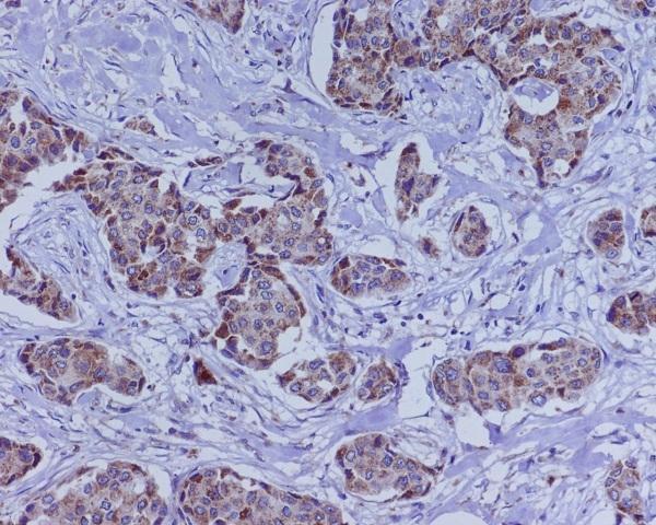
M00321 is listed for human IHC and IF/ICC (catalog: applications, reactivity). Its real IHC image shows paraffin-embedded human breast cancer (M00321 image caption).

Real IHC data Immunohistochemical analysis of paraffin-embedded human breast cancer, using BMP4 Antibody(M00321) BMP4 was detected in paraffin-embedded tissue section. Heat mediated antigen retrieval was performed in citrate buffer (pH6, epitope retrieval solution) for 20 mins. The tissue section was blocked with 10% goat serum. The tissue section was then incubated with 1ug/ml rabbit anti-BMP4 Antibody (M00321)overnight at 4°C. Biotinylated goat anti-rabbit IgG was used as secondary antibody and incubated for 30 minutes at 37°C. The tissue section was developed using Strepavidin-Biotin-Complex (SABC)(Catalog # SA1022) with DAB as the chromogen.
Anti-BMP4 Rabbit Monoclonal Antibody
Cat # M00321

M00321 is the only SKU supplied and is listed for human IHC and IF/ICC (catalog: SKU, applications, reactivity). Its IHC image shows paraffin-embedded human breast cancer (M00321 image caption); no IF image is supplied (catalog: if_image_alts).

Which to pick: Choose M00321 for human paraffin-section IHC: it is a rabbit monoclonal listed for human IHC (catalog: host, clone, applications, reactivity), with a paraffin-section image (M00321 image caption); the fixative is unreported (M00321 image caption). For IF/ICC, M00321 is listed at 1:50 (catalog: applications, if_dilution), but has no supplied IF image (catalog: if_image_alts). No cross-species option is supported: M00321 lists human reactivity only (catalog: reactivity).

Each figure is that product's own IHC / IF validation image from its datasheet.

References

  1. UniProt Consortium. UniProt entry P12644 (BMP4_HUMAN, Bone morphogenetic protein 4).
  2. Human Protein Atlas. BMP4 tissue IHC expression (reliability: Uncertain).
  3. Human Protein Atlas. BMP4 subcellular location (ICC-IF): Secreted.
  4. Human Protein Atlas. BMP4 antibody validation summary (2 antibodies).
  5. BMP4, a new prognostic factor for glioma. World journal of surgical oncology 2013 — PMC3851443.
  6. Myelination in Multiple Sclerosis Lesions Is Associated with Regulation of Bone Morphogenetic Protein 4 and Its Antagonist Noggin. International journal of molecular sciences 2019 — PMC6337410.
  7. Transcriptome identification of genes associated with uterus-vagina junction epithelial folds formation in chicken hens. Poultry science 2023 — PMC10240375.
  8. A Wnt-BMP4 Signaling Axis Induces MSX and NOTCH Proteins and Promotes Growth Suppression and Differentiation in Neuroblastoma. Cells 2020 — PMC7140810.
  9. PubMed PMID:3201241 — UniProt-cited evidence.
  10. PubMed PMID:9701626 — UniProt-cited evidence.
  11. PubMed PMID:15489334 — UniProt-cited evidence.