This website uses cookies to ensure you get the best experience on our website.


- Table of Contents
Plan BMPR1A IHC in paraffin sections using skeletal myocytes as a high-staining reference (HPA tissue IHC) and the catalog antibody's 2–5 μg/ml range (datasheet A01581-1). Assess observed cytoplasmic staining (HPA tissue IHC) alongside expected cell-surface localisation (UniProt), accounting for low antibody–RNA consistency (HPA tissue IHC).

Expected localisation, validated protocols, controls and antibodies — the at-a-glance facts below, then the full design guide.
| Expected localisation | Cytoplasmic staining (HPA tissue IHC); cell surface expected (UniProt) | |
| Staining pattern | Cytoplasmic staining in most tissues (HPA tissue IHC) | |
| Antigen retrieval | EDTA pH 8.0 HIER, heat-mediated (datasheet A01581-1) | |
| Positive control | Gallbladder+4 more · see all | |
| Negative control | Adipose tissue+4 more · see all |
| Fixation | Keep fixation consistent across sections (standard IHC practice; not target-specific) | |
| Caveat | Antibody staining and RNA show low consistency (HPA tissue IHC) | |
| Regulation | Expression regulation unreported (UniProt) | |
| Isoform / epitope | No isoforms listed; extracellular vs cytoplasmic epitope matters (UniProt) |
Compare the catalog antibody’s IHC-P protocol (datasheet A01581-1) with published rat testis (PMC9748027) and mouse skeletal section methods (PMC2694443).
| Sample | Paraffin-embedded human breast cancer tissue; fixative not specified (datasheet A01581-1) |
| Fixation | Image fixative and duration unreported (datasheet A01581-1); verify before use. |
| Sectioning | 4–5 µm sections on charged slides (standard) |
| Deparaffinisation | Xylene, graded ethanol series to water (standard) |
| Antigen retrieval | Heat retrieval: EDTA pH 8.0 (datasheet A01581-1); 20 min, 95–100 °C (standard) |
| Peroxidase block | 3% H2O2, 10 min, room temperature (standard) |
| Blocking | 10% goat serum (datasheet A01581-1) |
| Primary antibody | Rabbit anti-BMPR1A, 2-5 μg/ml (datasheet A01581-1) |
| Primary incubation | Overnight at 4 °C (datasheet A01581-1) |
| Detection | HRP-conjugated secondary, DAB chromogen (datasheet A01581-1) |
| Counterstain | Hematoxylin, blue, dehydrate and mount (standard) |
| Expected result | BMPR1A-positive staining in glandular cells of gallbladder (HPA tissue IHC: High). HPA tissue profile: Cytoplasmic expression in most tissues. No signal in the no-primary control. |
BMPR1A is a cell-surface receptor with one transmembrane segment (UniProt P36894 topology). In paraffin-section IHC, HPA reports mainly cytoplasmic staining across tissues, including high staining in skeletal myocytes and several glandular cell populations (HPA tissue IHC). Treat that observed pattern as a reference, not a definitive distribution: HPA rates the IHC antibody Approved but reports low consistency between staining and RNA expression (HPA tissue IHC).
| Strong cytoplasmic staining in skeletal myocytes, pancreatic exocrine glandular cells, or gallbladder glandular cells. | These are high-staining cell populations in HPA tissue IHC. Record which cells stain and how intensely relative to adjacent cells (HPA tissue IHC; general IHC practice). Skeletal muscle also has high BMPR1A expression reported by UniProt, making it a useful reference tissue (UniProt P36894 tissue specificity). |
| Staining outlines cells at the surface, with or without cytoplasmic signal. | A surface component is compatible with BMPR1A's annotated cell-membrane location and extracellular, transmembrane, and cytoplasmic regions (UniProt P36894 topology). HPA's tissue-IHC summary emphasizes cytoplasmic staining, so compare the full pattern with a positive reference tissue before interpreting an exclusively membranous result (HPA tissue IHC; general IHC practice). |
| Predominantly nuclear staining, or staining confined to an unexpected compartment. | Nuclear localization is not the reported reference pattern: UniProt places BMPR1A at the cell surface, while HPA describes cytoplasmic tissue-IHC staining and approved cytosolic ICC-IF localization (UniProt P36894 subcellular location; HPA tissue IHC; HPA subcellular). Check the counterstain and detection controls, then reassess the result as a possible artifact (general IHC practice). |
| Strong staining in a cell population listed as not detected, such as adipocytes or esophageal squamous epithelial cells. | That conflicts with HPA's observations for those specific cell populations (HPA tissue IHC). Check whether the stained cells were identified correctly, then use detection controls to investigate nonspecific antibody binding or endogenous chromogen-generating activity (general IHC practice). A listed negative population does not establish that every cell in its tissue is negative. |
| Diffuse staining across cells and tissue spaces, including a nominally negative area. | A broad haze that obscures cell boundaries is difficult to score as BMPR1A staining (general IHC practice). Compare a primary-antibody-omission control and an HPA high-staining reference population; background in the omission control points to the detection system or tissue activity (HPA tissue IHC; general IHC practice). |
| Compartment and antibody interpretation | BMPR1A has an extracellular region at residues 24–152, a transmembrane segment at 153–176, and a cytoplasmic region at 177–532 (UniProt P36894 topology). The supplied record does not identify the antibody epitope, so these coordinates cannot predict its staining intensity or a required retrieval condition. |
| Choice of reference cells | HPA reports high staining in gallbladder and stomach glandular cells, pancreatic exocrine cells, placental trophoblasts, and skeletal and smooth muscle cells (HPA tissue IHC). It reports no detection in adipocytes and selected glial and epithelial populations (HPA tissue IHC). Score the named cell population, not an entire tissue as uniformly positive or negative. |
| Strength of evidence | The HPA tissue-IHC antibody is Approved, but staining has low consistency with RNA expression; the supplied validation status is Approved for IHC and ICC, not Enhanced (HPA tissue IHC; HPA antibody validation). Interpret a surprising distribution alongside morphology and controls rather than treating one stain as conclusive (general IHC practice). |
| IF/ICC: should its pattern match paraffin-section IHC? | HPA calls cytosol the approved ICC-IF location and lists images from A-431, U-251MG, and U2OS; the tissue-IHC summary also describes cytoplasmic expression (HPA subcellular; HPA tissue IHC). These observations provide a compartment comparison, but image availability does not establish staining intensity or a protocol for those cell lines. |
| Situation | Likely cause | Next action |
|---|---|---|
| No visible signal in a skeletal-muscle section expected to contain myocytes. | HPA reports high myocyte staining and UniProt reports high skeletal-muscle expression, so absent signal merits a technical check; neither source establishes a BMPR1A-specific fixation effect (HPA tissue IHC; UniProt P36894 tissue specificity). | Confirm myocytes are present and the chromogenic detection run worked. Review the antibody's IHC validation and the run's retrieval and dilution records; optimize those conditions using routine IHC controls without assuming a target-specific cause (HPA antibody validation; general IHC practice). |
| Signal is strong in adipocytes but weak in nearby expected-positive cells. | HPA lists adipocytes as not detected, so the distribution differs from its reference observations; nonspecific staining or tissue-associated detection activity is possible (HPA tissue IHC; general IHC practice). | Recheck cell identification, compare a primary-antibody-omission control, and repeat with appropriately controlled antibody and detection conditions before assigning the signal to BMPR1A (general IHC practice). |
| Brown precipitate appears in the primary-antibody-omission control. | Signal without primary antibody implicates the chromogenic detection workflow or endogenous tissue activity rather than specific primary-antibody binding (general IHC practice). | Review the applicable endogenous-activity blocking step, detection reagents, and wash quality; rerun the omission control with the test section (general IHC practice). |
| The entire section has diffuse brown haze that hides cellular boundaries. | Excess background can arise from detection or antibody conditions, and it prevents reliable comparison with HPA's named cell populations (general IHC practice; HPA tissue IHC). | Compare omission and positive-reference controls, then adjust routine blocking, washes, and antibody or detection conditions as indicated by those controls (general IHC practice). |
| Nuclei dominate the staining pattern while cytoplasm and cell borders are faint. | Dominant nuclear staining diverges from UniProt's cell-surface annotation, HPA's cytoplasmic tissue-IHC profile, and HPA's approved cytosolic ICC-IF location (UniProt P36894 subcellular location; HPA tissue IHC; HPA subcellular). | Check the counterstain and omission control, inspect whether pigment or precipitate overlaps nuclei, and reassess specificity before scoring the nuclei as BMPR1A-positive (general IHC practice). |
| A reference tissue stains, but intensity varies among cases or cell populations. | HPA reports low tissue RNA specificity and low consistency between antibody staining and RNA data; its high, medium, low, and not-detected calls refer to specified cell populations (HPA tissue IHC). | Score location, fraction, and intensity within the same identified cell population, include a comparable reference section in each run, and report deviations from the HPA pattern rather than imposing one intensity threshold on every tissue (general IHC practice; HPA tissue IHC). |



Comprehensive Human Protein Atlas IHC scoring per tissue (reliability: Approved — Low consistency between antibody staining and RNA expression data.). Rows are taken directly from the HPA tissue chart — click any row's HPA link to view the source.
| Tissue | Cell type | Level | Evidence | Source |
|---|---|---|---|---|
| Gallbladder | Glandular cells | High | Protein (IHC) | HPA → |
| Pancreas | Exocrine glandular cells | High | Protein (IHC) | HPA → |
| Placenta | Trophoblastic cells | High | Protein (IHC) | HPA → |
| Skeletal muscle | Myocytes | High | Protein (IHC) | HPA → |
| Smooth muscle | Smooth muscle cells | High | Protein (IHC) | HPA → |
| Tissue | Cell type | Level | Evidence | Source |
|---|---|---|---|---|
| Adipose tissue | Adipocytes | Not detected | Protein (IHC) | HPA → |
| Caudate | Glial cells | Not detected | Protein (IHC) | HPA → |
| Cerebellum | Cells in granular layer | Not detected | Protein (IHC) | HPA → |
| Esophagus | Squamous epithelial cells | Not detected | Protein (IHC) | HPA → |
| Fallopian tube | Glandular cells | Not detected | Protein (IHC) | HPA → |
Use the catalog antibody’s paraffin-section conditions as an IHC starting point, then check staining against BMPR1A topology and cell-specific reference patterns.
Two rabbit polyclonal anti-BMPR1A antibodies have human tissue IHC images (catalog; image captions). Catalog reactivity also includes mouse or rat, depending on SKU (catalog). Neither has IF/ICC validation listed (catalog).
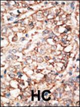
A01581 has an IHC-P image of formalin-fixed, paraffin-embedded human cancer tissue, including breast carcinoma and hepatocarcinoma (A01581 image caption). A01581-1 has an IHC image of a paraffin-embedded human breast cancer section; its fixative is unreported (A01581-1 image caption).
Which to pick: For human tissue IHC, choose A01581 for documented paraffin-section, paraffin-embedded tissue (A01581 image caption; datasheet: 1:50–1:100), or A01581-1 for a documented paraffin-section workflow whose fixative is unreported (A01581-1 image caption; datasheet: 2–5 μg/ml). Neither SKU lists IF/ICC validation, so there is no catalog-supported IF/ICC pick (catalog applications). Both are rabbit polyclonals (catalog); for cross-species planning, A01581 lists human and mouse reactivity, while A01581-1 lists human and rat reactivity, with the pictured IHC examples limited to human tissue (catalog reactivity; image captions). The selected A01581-1 tissue-IHC caption documents paraffin sections, but does not specify the fixative (selected-SKU IHC image A01581-1).