BNIP3L / BCL2/adenovirus E1B 19 kDa protein-interacting protein 3-like · IHC design guide

Design Immunohistochemistry for BNIP3L

Plan chromogenic BNIP3L IHC in paraffin sections using the IHC-validated antibody (datasheet M03107). This guide covers controls and scoring for the often granular cytoplasmic tissue pattern (HPA tissue IHC).

Evidence assembled Oct 2026 · For research use; verify linked source records and product datasheet before use
Immunohistochemistry protocol sheet for BNIP3L (IHC for BNIP3L): expected localisation Cytoplasm observed (HPA tissue IHC); outer mitochondrial membrane expected (UniProt), antibody M03107, validated IHC image, and IHC protocol steps
Printable BNIP3L IHC protocol sheet — expected localisation Cytoplasm observed (HPA tissue IHC); outer mitochondrial membrane expected (UniProt), antibody M03107, controls and protocol steps. Open the full BNIP3L IHC guide →

BNIP3L Immunohistochemistry Experimental Design Guide

Expected localisation, validated protocols, controls and antibodies — the at-a-glance facts below, then the full design guide.

Must know before staining
Expected localisation Cytoplasm observed (HPA tissue IHC); outer mitochondrial membrane expected (UniProt)
Staining pattern Ubiquitous, often granular cytoplasmic staining (HPA tissue IHC)
Antigen retrieval EDTA pH 8.0 HIER, heat-mediated (datasheet M03107)
Positive control ⓘ Breast+4 more · see all
Negative control ⓘ Soft tissue
Important caveats
Reasons your staining may differ from the expected pattern.
Fixation Keep fixation consistent across sections (standard IHC practice; not target-specific)
Caveat Staining has medium consistency with RNA data (HPA tissue IHC)
Regulation PPTC7 interaction promotes degradation (UniProt)
Isoform / epitope 2 isoforms; epitope differences are unknown (UniProt)
Section 1

Recommended BNIP3L IHC & IF Protocols

Compare the catalog antibody’s IHC-P protocol (datasheet M03107) with four published BNIP3L IHC protocols (PMC8258758; PMC2360231; PMC11872638; PMC5432493).

Recommended immunohistochemistry (IHC-P) protocol parameters
SampleParaffin-embedded human liver cancer tissue; fixative not specified (datasheet M03107)
FixationImage fixative and duration unreported (datasheet M03107); verify before use.
Sectioning4–5 µm sections on charged slides (standard)
DeparaffinisationXylene, graded ethanol series to water (standard)
Antigen retrievalHeat retrieval: EDTA pH 8.0 (datasheet M03107); 20 min, 95–100 °C (standard)
Peroxidase block3% H2O2, 10 min, room temperature (standard)
Blocking10% goat serum (datasheet M03107)
Primary antibodyRabbit monoclonal (clone ABIB-2) anti-BNIP3L, 1:50 (datasheet M03107)
Primary incubationOvernight at 4 °C (datasheet M03107)
DetectionHRP-conjugated secondary, DAB chromogen (datasheet M03107)
CounterstainHematoxylin, blue, dehydrate and mount (standard)
Expected resultBNIP3L-positive staining in myoepithelial cells of breast (HPA tissue IHC: High). HPA tissue profile: Ubiquitous cytoplasmic expression, which often had a granular pattern. No signal in the no-primary control.
💡Decision noteStart with heat-mediated EDTA retrieval at pH 8.0 (datasheet M03107). Published alternatives used citrate at pH 6.0 (PMC2360231) or EDTA at pH 9.0 (PMC11872638).
Section 2

What Is the Expected BNIP3L Staining Pattern?

BNIP3L should appear mainly as cytoplasmic, often granular staining in paraffin sections (HPA tissue IHC: Enhanced reliability). Strong staining is reported in kidney tubular cells, lung type II alveolar cells, and several glandular populations (HPA tissue IHC: High). Its mitochondrial outer membrane, endoplasmic reticulum, and nuclear envelope locations are annotated separately (UniProt O60238: subcellular location); residues 188–208 form a transmembrane segment (UniProt O60238: topology).

What am I looking at on my slide?
Granular cytoplasmic staining in kidney tubular cells or lung type II alveolar cells (HPA tissue IHC: High).This matches the reported tissue pattern (HPA tissue IHC: ubiquitous cytoplasmic expression, often granular). Score the relevant cells and their intensity, while checking that the signal is distinct from background (general IHC practice).
Predominantly strong nuclear staining, with little cytoplasmic signal (HPA tissue IHC: cytoplasmic profile).This does not match the reported tissue IHC profile (HPA tissue IHC: cytoplasmic expression). UniProt also annotates the nuclear envelope, so review a nuclear-rim pattern separately from staining that fills the nucleus (UniProt O60238: subcellular location).
Strong staining in chondrocytes from soft tissue (HPA tissue IHC: Not detected).This conflicts with the reported cell-level result and raises concern about nonspecific binding or detection activity (HPA tissue IHC: chondrocytes Not detected; general IHC practice). Recheck morphology and controls before assigning it to BNIP3L.
Diffuse color across tissue and empty spaces, obscuring cell borders (general IHC practice).Treat this as background until controls show a cell-associated pattern (general IHC practice). The expected HPA observation is cytoplasmic and often granular, so an even haze cannot establish BNIP3L localization (HPA tissue IHC: profile).
No visible staining in kidney tubular cells despite an interpretable section (HPA tissue IHC: High).A failed known-positive reference is a warning about assay performance, not evidence that BNIP3L is absent from the test specimen (HPA tissue IHC: kidney tubular cells High; general IHC practice). Review the stain run and controls first.
💡Expected BNIP3L appearanceCall positive when the relevant cells show distinct cytoplasmic, often granular staining—potentially strong in kidney tubules—above background; broad haze or dominant nuclear fill is suspect (HPA tissue IHC: profile and kidney High; general IHC practice).
How each factor affects the staining
Tissue and cell context (HPA tissue IHC: cell-level results).High staining is reported in breast myoepithelial cells, endometrial and epididymal glandular cells, kidney tubules, lung type II alveolar cells, ovarian follicle cells, and prostate glandular cells (HPA tissue IHC: High). Chondrocytes offer a reported unstained comparator (HPA tissue IHC: Not detected).
Interpretation confidence (HPA tissue IHC: Enhanced reliability).The HPA profile reports medium consistency between antibody staining and RNA expression data (HPA tissue IHC: reliability description). Compare the observed compartment and named cell population with controls before interpreting an unexpected pattern (general IHC practice).
Antibody evidence (HPA antibodies: IHC Enhanced).HPA lists two antibodies with Enhanced IHC validation, HPA015652 and CAB025371 (HPA antibodies: IHC Enhanced). That supports the reported pattern but does not establish that a different catalog antibody will give an identical stain (general IHC practice).
Membrane location and transcript forms (UniProt O60238: topology and isoforms).BNIP3L has one annotated transmembrane segment at residues 188–208 and two listed isoforms (UniProt O60238: topology, isoforms). These facts alone do not identify the epitope recognized by a particular antibody or predict its staining strength.
IF/ICC Q&A: Where should fluorescence appear? (HPA subcellular ICC-IF).Mainly at mitochondria, with an additional nuclear-speckle location reported (HPA subcellular ICC-IF: mitochondria enhanced; nuclear speckles supported). This is ICC-IF evidence; assess paraffin IHC against the separate tissue IHC profile (HPA tissue IHC: cytoplasmic, often granular).
Why is my staining missing, weak or wrong?
SituationLikely causeNext action
No staining in the test section and kidney positive control (HPA tissue IHC: kidney tubules High).A run-level detection or staining failure is possible when the expected positive control is also blank (general IHC practice).Check that the positive-control section and reagent steps were included and that detection developed; repeat the run after resolving the failed control (general IHC practice).
Weak or absent signal only in the test specimen (general IHC practice).A negative specimen may be genuine, but tissue composition or a technical difference can also explain the result (general IHC practice).Confirm the intended cell population is present, compare it with the positive control, and record the result by cell type (general IHC practice; HPA tissue IHC: cell-level results).
Uniform brown haze masks the expected granular cytoplasm (HPA tissue IHC: often granular).Background from nonspecific staining or detection can obscure a cell-associated signal (general IHC practice).Review the no-primary control and blocking, washing, and detection steps; adjust the workflow only after locating the source of background (general IHC practice).
Signal appears to fill nuclei instead of marking cytoplasm (HPA tissue IHC: cytoplasmic profile).A dominant nuclear-fill pattern is discordant with tissue IHC, although UniProt annotates the nuclear envelope (HPA tissue IHC: profile; UniProt O60238: subcellular location).Check focus and counterstain, then compare the nuclear rim and cytoplasm in a positive reference section before accepting the result (general IHC practice).
Chondrocytes stain strongly in a soft-tissue section (HPA tissue IHC: Not detected).This conflicts with the HPA cell-level observation; nonspecific antibody binding or endogenous detection activity is possible (HPA tissue IHC: chondrocytes Not detected; general IHC practice).Inspect the no-primary control and cell morphology, and verify that expected positive cells stain appropriately before interpreting this signal (general IHC practice).
Chromogen accumulates near section edges or in damaged areas (general IHC practice).Uneven reagent coverage or local tissue damage can create a pattern unrelated to cell localization (general IHC practice).Assess intact areas, check reagent coverage and washes, and compare with the control section before scoring BNIP3L (general IHC practice).

Sample controls for BNIP3L IHC & IF

🧪Run breast first and expect staining in myoepithelial cells (HPA: High in breast myoepithelial cells). Use soft tissue chondrocytes as the negative tissue (HPA: Not detected in chondrocytes); on the breast slide, assess cells outside the myoepithelial compartment for low background without assuming they are BNIP3L-negative (HPA: breast myoepithelial cells are High).
Positive control tissue: Breast (Myoepithelial cells, HPA High)
Negative control tissue: Soft tissue (HPA Not detected)
ICC-IF cell lines (HPA subcellular resource): HPA ICC-IF images show BNIP3L in A-431, U-251MG, U2OS, with annotated localisation: Mitochondria (enhanced) (HPA subcellular).
Technical controls: Include no-primary (secondary-only) and host-species- and immunoglobulin-class-matched isotype controls, plus a BNIP3L knockout specimen as a biological negative (standard IHC practice; caption: rabbit primary antibody). Block endogenous peroxidase and check for background DAB signal in breast sections (standard chromogenic IHC practice; caption: peroxidase detection with DAB).
⚠️Feasibility: A target-specific fixation window and fixation effect are unreported, and the selected M03107 tissue-IHC caption does not state the fixative (M03107 caption: fixative not stated). The demonstrated paraffin-section procedure used heat retrieval in EDTA at pH 8.0, but the caption does not establish that retrieval is required (M03107 caption: heat-mediated EDTA retrieval). No supplied evidence establishes whether frozen sections or IF would be easier; endogenous peroxidase is a potential chromogenic artefact to control in the breast section (standard IHC practice; M03107 caption: peroxidase detection with DAB).

HPA tissue IHC evidence for BNIP3L

Comprehensive Human Protein Atlas IHC scoring per tissue (reliability: Enhanced — Medium consistency between antibody staining and RNA expression data.). Rows are taken directly from the HPA tissue chart — click any row's HPA link to view the source.

Positive expression · recommended positive controls

TissueCell typeLevelEvidenceSource
Breast Myoepithelial cells High Protein (IHC) HPA →
Endometrium Glandular cells High Protein (IHC) HPA →
Epididymis Glandular cells High Protein (IHC) HPA →
Fallopian tube Glandular cells High Protein (IHC) HPA →
Kidney Cells in tubules High Protein (IHC) HPA →

Undetected expression · recommended negative controls

TissueCell typeLevelEvidenceSource
Soft tissue Chondrocytes Not detected Protein (IHC) HPA →
Section 3

Advanced BNIP3L IHC Tips

Troubleshoot BNIP3L staining by checking retrieval, controls, compartment, and cell identity before comparing chromogenic IHC results across sections.

What retrieval should I use when BNIP3L staining is weak in paraffin sections?
Start with heat-mediated retrieval in EDTA at pH 8.0 for this antibody (datasheet M03107). The selected paraffin-section image used that retrieval before overnight incubation with primary antibody at 4°C (datasheet M03107). If staining remains weak, compare retrieval heating and cooling consistency across slides while keeping antibody concentration and detection conditions fixed (standard IHC practice). Record the heating duration, section retention, background, and cytoplasmic granular staining together; a stronger DAB signal alone does not establish more specific BNIP3L detection (HPA: ubiquitous, often granular cytoplasmic staining; standard IHC practice).
How should I assess whether fixation is limiting BNIP3L detection?
The selected paraffin-section caption does not state its fixative, so BNIP3L-specific sensitivity to fixation is unknown for this antibody (datasheet M03107). Document the specimen’s actual fixative and fixation interval, then compare sections processed under those conditions using the same pH 8.0 EDTA retrieval and detection settings (datasheet M03107; standard IHC practice). Include a tissue area with interpretable morphology and an independent positive control in each run to distinguish lost antigen signal from a failed staining run (standard IHC practice). Assess tissue preservation and staining together; do not infer a preferred fixation condition from the protein’s membrane topology or tissue expression pattern (UniProt O60238 topology; HPA tissue IHC; standard IHC practice).
Is diffuse cytoplasmic BNIP3L staining plausible when mitochondrial staining is expected?
Evaluate cytoplasmic signal in context: tissue IHC describes ubiquitous expression that often appears granular, while subcellular imaging places BNIP3L mainly at mitochondria (HPA tissue IHC; HPA subcellular). BNIP3L also has annotated outer mitochondrial membrane, endoplasmic reticulum, and nuclear-envelope locations, with a transmembrane segment at residues 188–208 (UniProt O60238 localisation and topology). Chromogenic IHC may show a cytoplasmic granular pattern without resolving individual mitochondria, so compare cell morphology and the distribution of granules before assigning an organelle (HPA tissue IHC; standard IHC practice). Treat isolated nuclear DAB signal cautiously because the reported nuclear-speckle location comes from subcellular imaging, whereas the tissue IHC profile is cytoplasmic (HPA subcellular; HPA tissue IHC).
Can isoforms or epitope accessibility explain inconsistent BNIP3L staining?
BNIP3L has 2 annotated isoforms and a transmembrane segment at residues 188–208, so document the antibody’s mapped epitope before interpreting isoform coverage (UniProt O60238 isoforms and topology; standard IHC practice). The supplied caption gives an antibody dilution but no epitope map or isoform validation, leaving recognition of each isoform unresolved (datasheet M03107). Several phosphoserines are annotated, including residues 62, 117, 118, 120, and 166; their effect on this antibody’s staining is unknown (UniProt O60238 modified residues; datasheet M03107). If the expected pattern is absent, compare independently validated antibody epitopes and keep retrieval, detection, and tissue processing matched (standard IHC practice).
How can IF help assess a questionable chromogenic BNIP3L pattern?
Use IF as a separate localisation check alongside chromogenic IHC, whose selected evidence is a paraffin-section image with an unreported fixative (datasheet M03107; standard IHC practice). Multiplex BNIP3L with a validated marker of the cell type under examination and a mitochondrial marker, then inspect whether signals occupy the expected cells and compartments (HPA subcellular: mainly mitochondrial; standard IF practice). Choose fluorophores after measuring tissue autofluorescence and include single-label controls to assess spectral bleed-through (standard IF practice). Because the annotated transmembrane segment spans residues 188–208, determine which side contains the antibody epitope before selecting permeabilisation conditions; the supplied evidence does not map that epitope (UniProt O60238 topology; datasheet M03107; standard IF practice).
What should I check when BNIP3L DAB staining looks widespread?
Widespread staining is not automatically background: tissue IHC describes ubiquitous, often granular cytoplasmic BNIP3L expression, although signal varies among cell types (HPA tissue IHC). Run a no-primary control, check endogenous peroxidase blocking, and examine whether DAB deposits follow tissue edges or damaged regions (standard IHC practice). The selected image used 10% goat-serum blocking, primary antibody at 1:50 overnight at 4°C, and peroxidase-based DAB detection (datasheet M03107). If background obscures cell boundaries, adjust blocking, washing, or antibody concentration one variable at a time and judge changes against preserved granular staining in an interpretable control (HPA tissue IHC; standard IHC practice).
How should I score heterogeneous BNIP3L staining across tissue compartments? ⚠ ANSWER MARKED FOR VERIFICATION
Define the cell population and region before scoring, since HPA reports different staining levels across cell types within its tissue IHC profile (HPA tissue IHC; standard IHC practice). For chromogenic sections, report the percentage of positive cells and an H-score using the same intensity categories and threshold across slides (standard IHC practice). If counting discrete positive cells, report density per mm² of viable, evaluable tissue and exclude folds, necrosis, and section edges by a preset rule (standard IHC practice). Normalise each measurement to the relevant cell population or evaluable tissue area, and keep acquisition, DAB development, and counterstaining consistent between samples (standard IHC practice).
How do I distinguish true BNIP3L staining from artefact?
A credible result should show interpretable cellular staining, with cytoplasmic granularity consistent with tissue IHC and the mainly mitochondrial localisation reported by subcellular imaging (HPA tissue IHC; HPA subcellular). Check cell identity rather than tissue name alone: HPA reports high staining in kidney tubular cells and low staining in liver cholangiocytes (HPA tissue IHC). Signal restricted to torn edges, necrotic areas, or no-primary controls warrants investigation of section artefact or endogenous enzyme activity before biological interpretation (standard IHC practice). Finally, assess staining against the matched control and morphology; the selected liver-cancer image establishes staining under its stated conditions but does not establish tumour-specific expression (datasheet M03107; standard IHC practice).
Boster reagents

Best BNIP3L / BCL2/adenovirus E1B 19 kDa protein-interacting protein 3-like IHC Antibodies

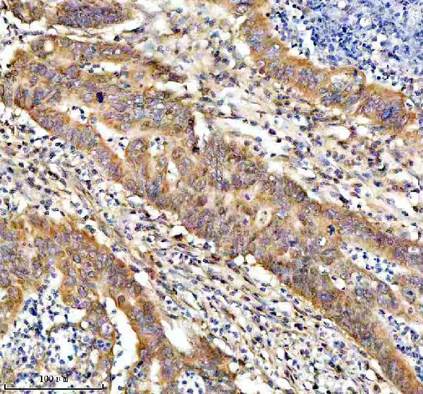
Anti-BNIP3L antibodies have IHC images from human liver cancer and kidney tissue and an IF/ICC image from U2OS cells (catalog image captions); catalog reactivity includes human, mouse and rat (catalog reactivity).

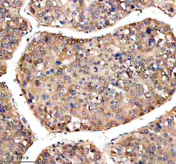
Real IHC data IHC analysis of BNIP3L using anti-BNIP3L antibody (M03107). BNIP3L was detected in a paraffin-embedded section of human liver cancer tissue. Heat mediated antigen retrieval was performed in EDTA buffer (pH 8.0, epitope retrieval solution). The tissue section was blocked with 10% goat serum. The tissue section was then incubated with 1:50 rabbit anti-BNIP3L Antibody (M03107) overnight at 4°C. Peroxidase Conjugated Goat Anti-rabbit IgG was used as secondary antibody and incubated for 30 minutes at 37°C. The tissue section was developed using HRP Conjugated Rabbit IgG Super Vision Assay Kit (Catalog # SV0002) with DAB as the chromogen.
Anti-BNIP3L/Nix Rabbit Monoclonal Antibody
Cat # M03107
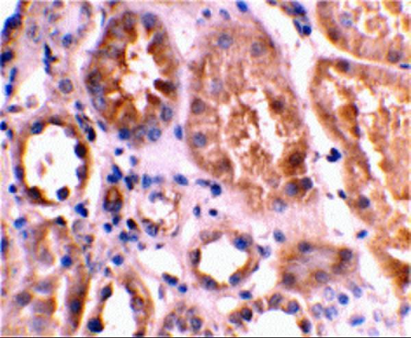
Real IHC data Immunohistochemical staining of human kidney tissue using Bnip3L antibody at 2 μg/mL.
Anti-Bnip3L Antibody
Cat # A03107
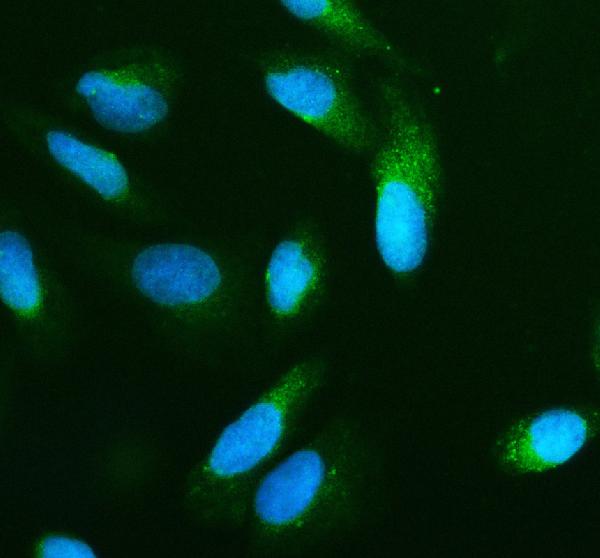
Real IF data IF analysis of BNIP3L using anti-BNIP3L antibody (A03107-3). BNIP3L was detected in an immunocytochemical section of U2OS cells. Enzyme antigen retrieval was performed using IHC enzyme antigen retrieval reagent (AR0022) for 15 mins. The cells were blocked with 10% goat serum. And then incubated with 5 μg/mL rabbit anti-BNIP3L Antibody (A03107-3) overnight at 4°C. DyLight®488 Conjugated Goat Anti-Rabbit IgG (BA1127) was used as secondary antibody at 1:500 dilution and incubated for 30 minutes at 37°C. The section was counterstained with DAPI. Visualize using a fluorescence microscope and filter sets appropriate for the label used.
Anti-BNIP3L Antibody ®
Cat # A03107-3

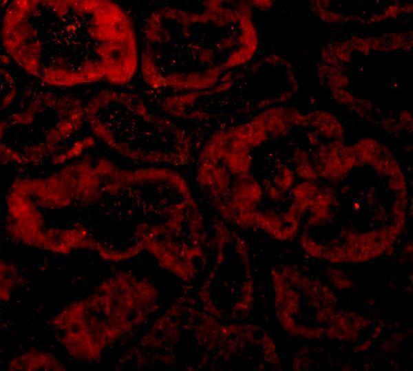
M03107 has IHC data from paraffin-embedded human liver cancer tissue, and A03107 has IHC data from human kidney tissue (catalog image captions). A03107-3 has IF/ICC data from U2OS cells; A03107 also has IF data from human kidney tissue (catalog image captions).

Which to pick: For tissue IHC, choose M03107 when a paraffin-section example is useful: it is a rabbit monoclonal with listed human, mouse and rat reactivity, and its IHC caption uses EDTA retrieval at pH 8.0 and primary antibody at 1:50; the fixative is unreported (catalog applications, reactivity and M03107 IHC caption). A03107 is another human tissue IHC option at 2 μg/mL in the kidney image, with no processing method reported in that caption (A03107 IHC caption). For IF/ICC, choose A03107-3 for the U2OS cell example at 5 μg/mL, or A03107 for human kidney IF at 10 μg/mL; M03107 has the broader listed species reactivity for IHC, while A03107 lists human only (catalog applications, reactivity and image captions).

Each figure is that product's own IHC / IF validation image from its datasheet.

References

  1. UniProt Consortium. UniProt entry O60238 (BNI3L_HUMAN, BCL2/adenovirus E1B 19 kDa protein-interacting protein 3-like).
  2. Human Protein Atlas. BNIP3L tissue IHC expression (reliability: Enhanced).
  3. Human Protein Atlas. BNIP3L subcellular location (ICC-IF): Mainly localized to the mitochondria. In addition localized to the nuclear speckles..
  4. Human Protein Atlas. BNIP3L antibody validation summary (2 antibodies).
  5. Expression and Clinical Significance of BCL2 Interacting Protein 3 Like in Multiple Myeloma. Technology in cancer research & treatment 2021 — PMC8258758.
  6. Gene expression profiling of liver metastases and tumour invasion in pancreatic cancer using an orthotopic SCID mouse model. British journal of cancer 2007 — PMC2360231.
  7. EPAS1 induction drives myocardial degeneration in desmoplakin-cardiomyopathy. iScience 2025 — PMC11872638.
  8. BNIP3L promotes cardiac fibrosis in cardiac fibroblasts through [Ca(2+)](i)-TGF-β-Smad2/3 pathway. Scientific reports 2017 — PMC5432493.
  9. PubMed PMID:9523198 — UniProt-cited evidence.
  10. PubMed PMID:9867803 — UniProt-cited evidence.
  11. PubMed PMID:10381623 — UniProt-cited evidence.