This website uses cookies to ensure you get the best experience on our website.


- Table of Contents
Plan BORA staining in paraffin sections using the cytoplasmic tissue pattern reported by HPA (HPA tissue IHC). Compare candidate positive tissues with adipocytes, where staining was not detected, and start the catalog antibody at 2.5 μg/mL (HPA tissue IHC; datasheet).

Expected localisation, validated protocols, controls and antibodies — the at-a-glance facts below, then the full design guide.
| Expected localisation | Cytoplasmic staining in most tissues (HPA tissue IHC) | |
| Staining pattern | Cytoplasmic staining across most tissues (HPA tissue IHC) | |
| Antigen retrieval | Citrate pH 6.0 HIER, 95–98 °C, 20 min (rule: cytoplasmic / membrane antigen) | |
| Positive control | Adrenal gland+4 more · see all | |
| Negative control | Adipose tissue |
| Fixation | Keep formalin fixation consistent across sections. (standard IHC practice; not target-specific) | |
| Caveat | Adipocytes may be unstained despite the broad profile (HPA tissue IHC) | |
| Regulation | Expression regulation is unannotated (UniProt) | |
| Isoform / epitope | 2 isoforms; epitope coverage is unspecified (UniProt; datasheet) |
The catalog antibody’s IHC-P protocol is accompanied by one published BORA IHC protocol using breast cancer tissue microarray slides (PMC5546444).
| Sample | Tissue sections; selected-image fixative not specified (standard IHC workflow) |
| Fixation | Image fixative and duration unreported (datasheet A02087); verify before use. |
| Sectioning | 4–5 µm sections on charged slides (standard) |
| Deparaffinisation | Xylene, graded ethanol series to water (standard) |
| Antigen retrieval | Heat-induced epitope retrieval in citrate buffer, pH 6.0, 20 min at 95–98 °C (standard rule: cytoplasmic / membrane antigen) |
| Peroxidase block | 3% H2O2, 10 min, room temperature (standard) |
| Blocking | 10% normal serum of the secondary host, 30 min, room temperature (standard) |
| Primary antibody | Rabbit anti-BORA, 2.5 μg/mL (datasheet A02087) |
| Primary incubation | Overnight at 4 °C (standard) |
| Detection | HRP-polymer secondary, DAB chromogen 5–10 min (standard) |
| Counterstain | Hematoxylin, blue, dehydrate and mount (standard) |
| Expected result | BORA-positive staining in glandular cells of adrenal gland (HPA tissue IHC: High). HPA tissue profile: Cytoplasmic expression in most tissues. No signal in the no-primary control. |
In paraffin-section IHC, expect BORA staining mainly in the cytoplasm across many tissues, including glandular cells, hematopoietic cells and selected other cell populations (HPA tissue IHC: cytoplasmic expression in most tissues; listed High cells). BORA has no annotated transmembrane segment (UniProt Q6PGQ7 topology). Treat the pattern as a comparison point: HPA rates its tissue IHC Approved, with medium staining–RNA consistency and external verification pending (HPA tissue IHC reliability).
| Cytoplasmic staining in adrenal or appendix glandular cells (HPA tissue IHC: High in both). | This matches the reported IHC compartment and cell types (HPA tissue IHC). Compare cells within the section: a High designation identifies a reported positive population, but does not require every cell to stain equally (HPA tissue IHC: High). |
| Predominantly nuclear staining with little cytoplasmic signal in a paraffin section (HPA tissue IHC: cytoplasmic profile). | Investigate a compartment mismatch before scoring it as expected IHC. HPA separately reports nucleoplasm and nucleoli fibrillar-center signal by ICC-IF, with additional cytosol signal; that IF observation does not establish a nuclear IHC pattern (HPA subcellular ICC-IF; HPA tissue IHC). |
| Strong signal in adipocytes while the reported positive cells remain faint (HPA tissue IHC: adipocytes Not detected). | This conflicts with the HPA cell-type comparison and raises concern for nonspecific staining or endogenous detection activity (HPA tissue IHC; general IHC practice). Check the no-primary control and inspect other cells in the same section before assigning BORA positivity (general IHC practice). |
| Broad, fairly uniform color over cells and tissue spaces, without a discernible cytoplasmic pattern (HPA tissue IHC: cytoplasmic profile). | Treat this as background until controls clarify it; diffuse chromogen alone does not identify BORA-positive cells (general IHC practice). Review blocking, washing and detection controls, then reassess whether staining follows the reported cellular compartment (general IHC practice; HPA tissue IHC). |
| No detectable signal in adrenal or appendix glandular cells on the test run (HPA tissue IHC: High in both). | A negative result in a reported High population calls for a technical check before interpreting the study tissue as negative (HPA tissue IHC; general IHC practice). Review the antibody's IHC-P instructions, retrieval, detection and a positive control processed in the same run (general IHC practice). |
| Tissue and cell selection (HPA tissue IHC). | HPA reports High staining in adrenal, appendix, duodenal and endometrial glandular cells; bone-marrow hematopoietic cells; cerebellar granular-layer cells; colonic endothelial cells; and esophageal squamous epithelial cells (HPA tissue IHC). Adipocytes are Not detected, while caudate glial and ovarian follicle cells are Low (HPA tissue IHC). Select controls by cell type within the tissue (general IHC practice). |
| Compartment by application (HPA tissue IHC; HPA subcellular ICC-IF). | IHC has a cytoplasmic tissue profile, whereas ICC-IF reports approved nucleoplasm and nucleoli fibrillar-center locations plus cytosol (HPA tissue IHC; HPA subcellular ICC-IF). IF/ICC Q&A: What should IF show? Those reported nuclear locations and additional cytosol; the separate IF/ICC guide should govern its workflow (HPA subcellular ICC-IF). |
| Evidence and antibody validation (HPA tissue IHC; HPA antibodies). | The supplied HPA antibody HPA040866 is Approved for IHC and ICC; the tissue profile has medium staining–RNA consistency and awaits external verification (HPA antibodies; HPA tissue IHC reliability). Use concordant controls to support a local interpretation without treating approval as proof that every stained cell is specific (general IHC practice). |
| Topology, processing and variants (UniProt Q6PGQ7). | BORA has no annotated transmembrane segment, signal peptide or propeptide, and UniProt lists two isoforms (UniProt Q6PGQ7 topology, processing, isoforms). Those entries give no basis for expecting a membrane rim or secreted deposit. Isoform recognition by the staining antibody is unspecified in the supplied evidence (UniProt Q6PGQ7; HPA antibodies). |
| Phosphorylation and target-specific retrieval (UniProt Q6PGQ7). | UniProt lists six modified residues, including phosphoserines, but the supplied evidence gives no antibody epitope or BORA-specific retrieval response (UniProt Q6PGQ7 modified residues; HPA antibodies). Do not infer that phosphorylation or fixation explains a weak or absent stain; optimize retrieval using the IHC antibody instructions and controls (general IHC practice). |
| Situation | Likely cause | Next action |
|---|---|---|
| Positive-control glandular cells are blank (HPA tissue IHC: adrenal and appendix High). | The run may have failed at retrieval, primary-antibody incubation or detection (general IHC practice). | Check the IHC-P antibody instructions and reagent sequence; repeat with a reported High tissue and a same-run detection control (general IHC practice; HPA tissue IHC). |
| Every compartment appears faintly brown, including spaces between cells (general IHC practice). | Diffuse background can obscure the reported cytoplasmic pattern (general IHC practice; HPA tissue IHC). | Inspect a no-primary control; review blocking, wash steps and chromogen development, then rescore identifiable cells (general IHC practice). |
| Adipocytes stain as strongly as reported High cells (HPA tissue IHC: adipocytes Not detected). | Nonspecific binding or endogenous detection activity is possible (general IHC practice). | Compare no-primary and detection controls, then reassess staining in the named cell populations (general IHC practice; HPA tissue IHC). |
| Only nuclei stain in IHC-P sections (HPA tissue IHC: cytoplasmic profile). | The result disagrees with the tissue IHC profile; nuclear ICC-IF localization alone cannot validate it as IHC (HPA tissue IHC; HPA subcellular ICC-IF). | Check the positive-control compartment, no-primary control and morphology before reporting nuclear IHC positivity (general IHC practice). |
| A Low population is scored negative despite a working positive control (HPA tissue IHC: caudate glia and ovarian follicle cells Low). | Weak reported staining may fall below the run's visual detection threshold (HPA tissue IHC; general IHC practice). | Record the detection limit and avoid using a Low population alone to judge run failure (general IHC practice; HPA tissue IHC). |
| Strong staining appears outside the reported cell population in an otherwise positive tissue (HPA tissue IHC: cell-specific entries). | The tissue name alone does not establish that each stained cell expresses BORA (HPA tissue IHC; general IHC practice). | Identify cells by morphology, compare the HPA cell-type entry, and check no-primary staining before scoring them (HPA tissue IHC; general IHC practice). |


Comprehensive Human Protein Atlas IHC scoring per tissue (reliability: Approved — Medium consistency between antibody staining and RNA expression data. Pending external verification.). Rows are taken directly from the HPA tissue chart — click any row's HPA link to view the source.
| Tissue | Cell type | Level | Evidence | Source |
|---|---|---|---|---|
| Adrenal gland | Glandular cells | High | Protein (IHC) | HPA → |
| Appendix | Glandular cells | High | Protein (IHC) | HPA → |
| Bone marrow | Hematopoietic cells | High | Protein (IHC) | HPA → |
| Cerebellum | Cells in granular layer | High | Protein (IHC) | HPA → |
| Colon | Endothelial cells | High | Protein (IHC) | HPA → |
| Tissue | Cell type | Level | Evidence | Source |
|---|---|---|---|---|
| Adipose tissue | Adipocytes | Not detected | Protein (IHC) | HPA → |
Use compartment, cell type, and matched controls to troubleshoot BORA staining in chromogenic tissue IHC (HPA tissue IHC; HPA subcellular).
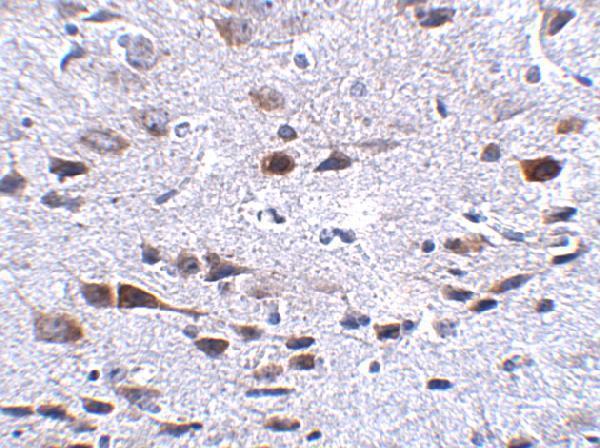
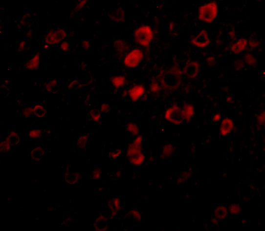
A02087 has IHC data in mouse brain tissue and IF data in mouse brain cells (catalog image captions); Human, Mouse and Rat reactivity is listed (catalog reactivity).
A02087 will render with an IHC image of mouse brain tissue at 2.5 μg/mL (IHC image caption). The same SKU has an IF image of mouse brain cells at 20 μg/mL (IF image caption).
Which to pick: Choose A02087 for tissue IHC: its mouse brain tissue image uses 2.5 μg/mL (IHC image caption), and IHC-P is listed (catalog applications); the fixative is unreported (IHC image caption). For IF/ICC, A02087 has an IF image in mouse brain cells at 20 μg/mL (IF image caption). For other species, A02087 lists Human, Mouse and Rat reactivity (catalog reactivity), while the supplied IHC and IF images show mouse samples (catalog image captions).