This website uses cookies to ensure you get the best experience on our website.
- Table of Contents
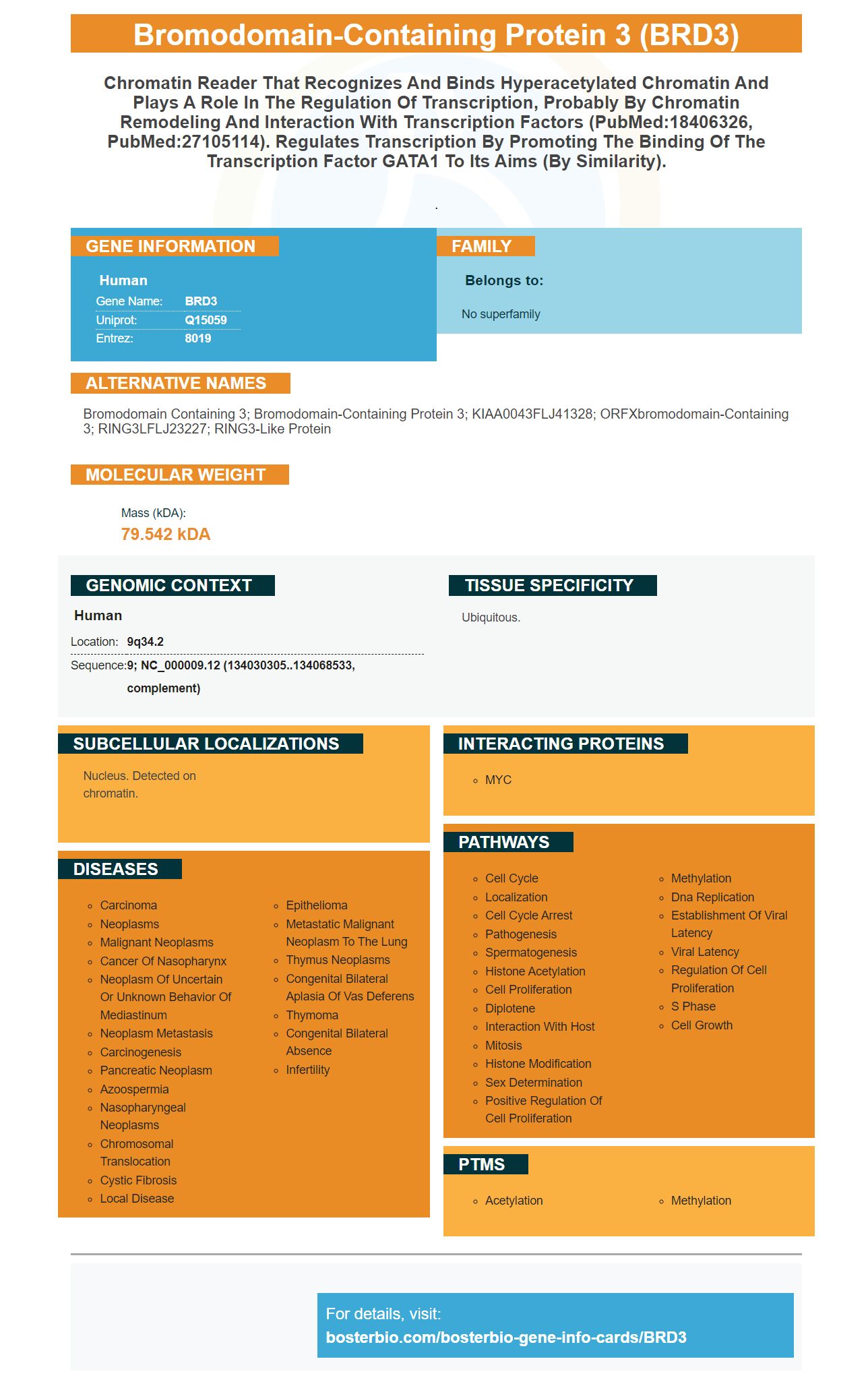
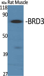
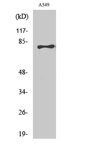
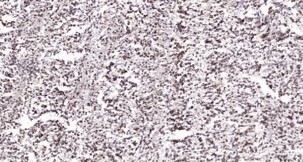
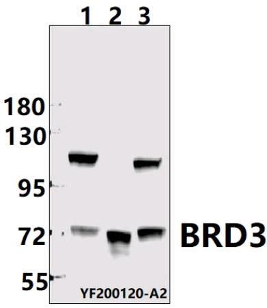
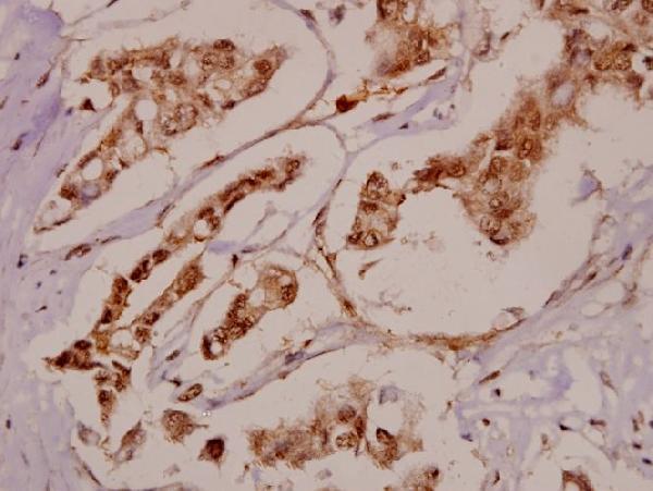
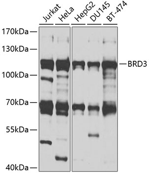
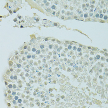
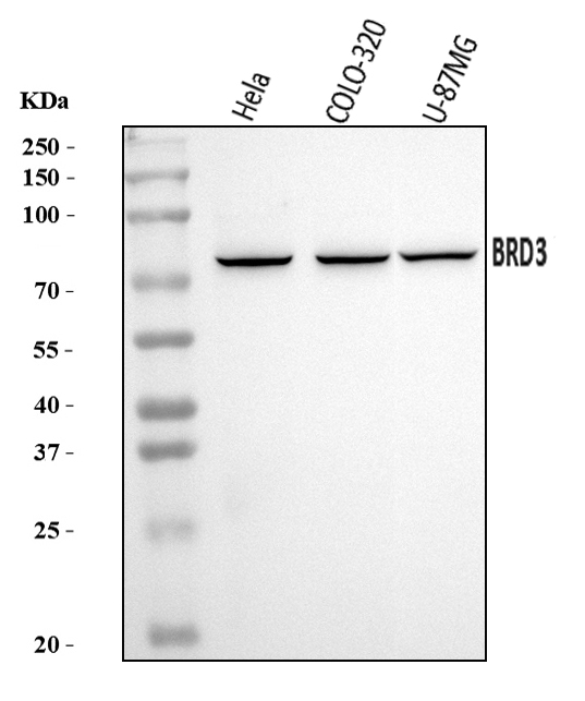
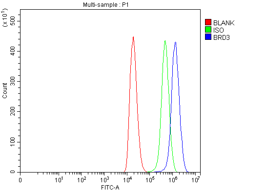
Facts about Bromodomain-containing protein 3.
.
Human | |
---|---|
Gene Name: | BRD3 |
Uniprot: | Q15059 |
Entrez: | 8019 |
Belongs to: |
---|
No superfamily |
bromodomain containing 3; bromodomain-containing protein 3; KIAA0043FLJ41328; ORFXbromodomain-containing 3; RING3LFLJ23227; RING3-like protein
Mass (kDA):
79.542 kDA
Human | |
---|---|
Location: | 9q34.2 |
Sequence: | 9; NC_000009.12 (134030305..134068533, complement) |
Ubiquitous.
Nucleus. Detected on chromatin.
There are many benefits to using the BRD3 Marker. This marker can also be used to identify special samples and species. Researchers can also use it to identify mycoplasma contamination in cell lines. It is also a highly sensitive and easy-to-use assay kit. Scientists from all parts of the world can benefit from the BRD3 Marker. Continue reading for more information.
Flowcytometry has many applications in science and can be used by researchers to study cells or particles. Boster Bio supplies high-affinity primary antibodies. These antibodies are monoclonal or polyclonal. They have been cited for more than 25 years in scientific journals. Their high-affinity antibody are used for ELISA, protein purification, as well as labeling. They are widely used in a variety of applications, including research into cancer.
Boster Bio's ELISA kit and antibodies can be used by researchers to identify biomarkers for cancer, neurogenesis, and inflammation. The high-affinity antibodies of the company can be detected down to picogram levels. Boster Bio's subsidiary tebubio offers ELISA kits. These products are produced to meet stringent quality standards. Boster Bio has developed a wide range secondary antibodies by using trade secrets.
Using a streptavidin-biosensor, four biotinylated antibodies bind to the same epitope. These antibodies have a dissociation coefficient of 0.7-10x10-4s-1. They are therefore very easy to integrate. The affinity is usually in the range of 0.6 to 45nM. Another two antibodies bind to ricin, but do not fall within this correlation trend.
Dual-label technology has many benefits. You can ask more detailed questions and gain more context for your research by using both primary antibodies as well as secondary antibodies. A combination of a secondary and primary antibody allows you to measure the antigen in two different samples, and determine which antibody binds best to the antigen. This allows you to measure the amount of a specific antigen and purify it before further testing it.
The recombinant antibodies used in research are highly specific and stable, and are free of cell-line drift or lot-to-lot variation. This ensures that your research will be successful. Recombinant antibodies are less susceptible to cross-reactivity between different batches of antibody. This allows researchers to maximize their research potential by obtaining high quality antibodies at a lower price and with minimal risk.
You can further test the effectiveness of high-affinity primary antibody by comparing them with a variety of antigens to detect pathogens in different environments. High-affinity antibodies are a vital tool for research and development, as they are more specific. With their increased specificity, these antibodies are the most effective choice for your research. Boster Bio is the right company if you are looking for a high-quality, high-affinity prim antibody.
The BRD3 gene has become a very desirable target for research. BRD3 gene expression is associated in a number of cancers, and other diseases. Recently, the BRD3 gene was recognized as a biomarker for prostate cancer. To target drugs that inhibit tumor growth, it is important to recognize BRD3 gene expression in these conditions.
Gene-specific antigens allow for the identification of several strains. BRD3 is a good example of such an antigen. This antibody binds to BRD3 in a variety of host tissues, including human serum. This antibody-based test is more specific than other immunoassays.
A new Boster kit, which is sensitive, high-throughput, easy to use, and provides a method for BRD3 measurement, is available for studies that use the BRD3 marker. Researchers from the National Institute of General Medical Sciences have published recent results that show the utility of BRD3 immuneassays kits in diagnosing cancer. Researchers can now identify BRD3 within various cancers in a wide array of tissues and cancer types thanks to the new kit.
The BRD3 marker, which is a tumor specific mutation, means that a certain population of subjects will be affected at least once by one of these mutations. In this example, at minimum 6.7% of subjects will carry any combination listed in Table 8.
This assay is convenient, lysate-free, sensitive, and high-throughput for researchers who are conducting clinical trials. The assay employs a proprietary peptide immunegen with numerous modifications that increase its invivo halflife and cellular targeting. The peptide can be further modified by polysialylation or recombinant PEG micrometics, as well as Fc-fusion.
One of the most important tools in the study of biological processes is cell culture. Mycoplasmas can infect cell cultures via airborne microscopic aerosols that are released during routine pipetting. This can easily be prevented using good aseptic techniques, routine testing, and clean to dirty work practices. In order to ensure your cell culture is mycoplasma-free, here are three steps to take:
Mycoplasma contamination is detectable in C3A or Tc cell lines. This test uses Tc cells as positive controls. Mycoplasma contamination can be detected in monoclonal monoclonal human-mouse cell lines by day. C3A cells can also be used as positive control. Despite their small size, mycoplasma infection can cause cell growth changes and altered transfection rates. It is important to ensure that your cell culture is clean from mycoplasma contamination.
Mycoplasma contamination can be very damaging to cell culture techniques. Mycoplasma contamination can affect cell proliferation by competing with nutrients in the culture medium. Other signs include cell aggregation, changes in morphology, and poor transfection rates. Independent laboratories should be able to provide results if your cell culture has been infected by mycoplasmas. They can even test your Boster Bio cell lines in-house to ensure that they are free of mycoplasma contamination.
The testing procedure involves DNA sequencing followed by PCR to identify Mycoplasma infection. The American Type Culture Collection provided cells for this study. These cells are derived directly from Hep G2 cells. They produce high levels albumin. DNA sequencing confirmed the contamination. The cells were then grown to adhere in Biofil plastic plates. The cells were cultured in Eagle's minimum necessary medium, which contained 20% heat-inactivated foetus serum. They were kept at 37°C with 90% relative humidity. The culture medium contained no additional supplements.
52 samples were used for the analysis to determine protein expression differences between mycoplasma and uninfected cell line. The three other cell lines only had one post-treatment sample that was infected and the other two were uninfected. There were no significant difference between infected or uninfected cell types. These cell line were not infected following treatment. In fact, a large part of their protein expression was very similar to their post-treatment counterparts.
InvivoGen San Diego, CA is the only place that offers PlasmocinTM (tm) treatment. PlasmocinTM Treatment is a DNA sequencing method that detects more 60 mycoplasma species. These include Acholeplasma laidlawii. Spiroplasma. and Ureaplasma. The technique uses universal primers that target the 16S rRNA genes and a unique thermostable, DNA polymerase. The PCR method has passed all standards of European Pharmacopoea 2.6.7.
These results are generally reliable. Below are the most commonly found mycoplasma. If you suspect contamination, it is best to get your samples tested by a professional. If you are concerned about contamination with a particular species, however, it is recommended that you obtain an infected sample culture. It is very fast and sensitive, and you can easily confirm contamination.
PMID: 16008511 by Ishii H., et al. Differentially expressed genes in endothelial differentiation.
PMID: 9373153 by Thorpe K.L., et al. Chromosomal localization, gene structure and transcription pattern of the ORFX gene, a homologue of the MHC-linked RING3 gene.